User login
Healing chronic venous ulcers? Compression, compression, compression
ESTES PARK, COLO. – Meg A. Lemon, MD, said at a conference on internal medicine sponsored by the University of Colorado at Denver, Aurora.
“The top nine ways of treating venous leg ulcers are compression,” explained Dr. Lemon, a Denver-area dermatologist in private practice. “You generally don’t have an active patient who’s moving around [and] allowing the calf to squeeze venous blood back into the circulation, so you have to compress externally. I spend a lot of time with patients saying, ‘If we don’t wear the compression stockings and we don’t elevate the legs, the wound will never heal.’ ”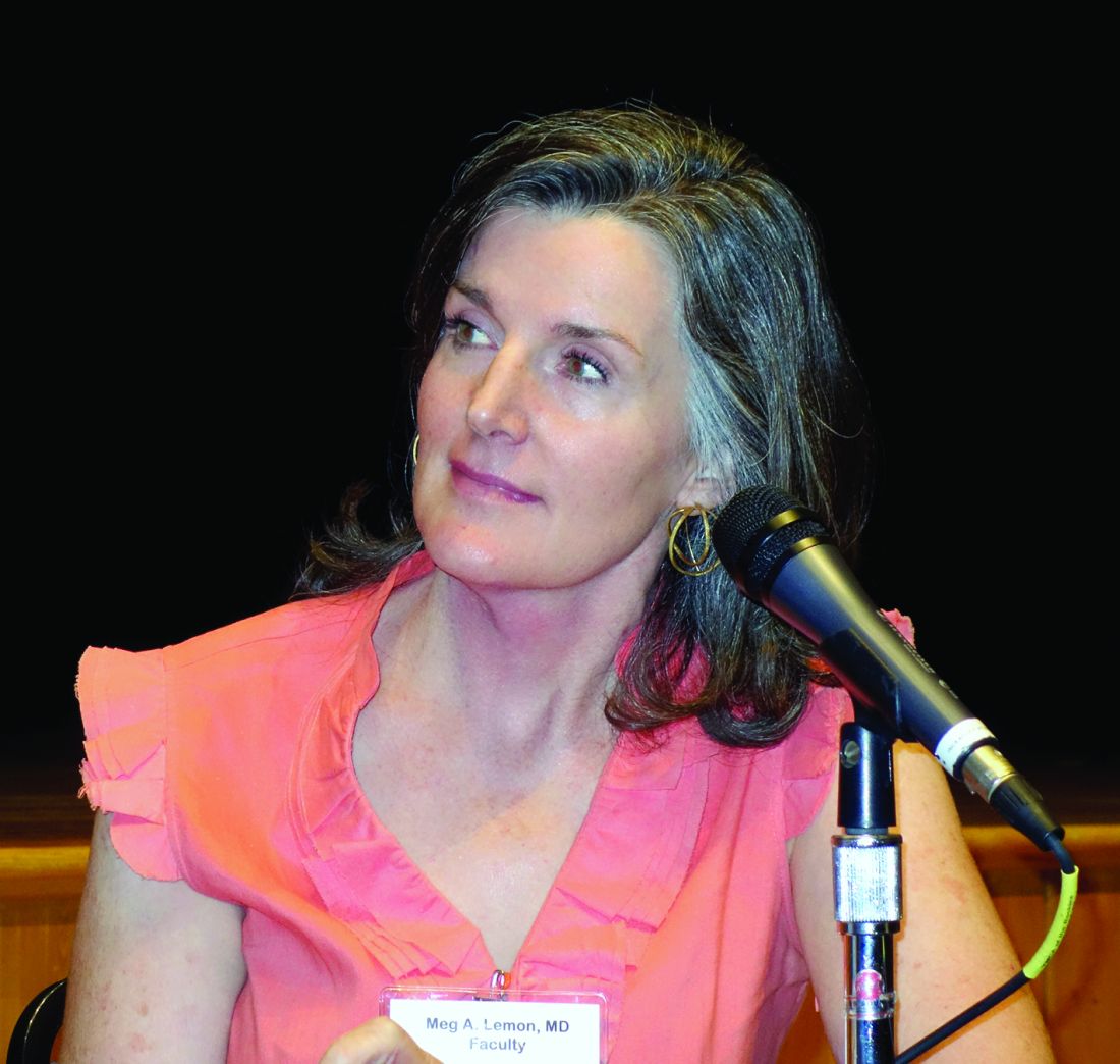
Compression stockings are no longer frumpy – they have gotten hip. Endurance athletes have embraced them as a recovery aid, and they now come in a multitude of colorful styles. It’s best to start patients off at 15-20 mm Hg of compression so they don’t get discouraged by the initial challenges of getting the stockings on and off, Dr. Lemon explained, then work up to at least 30 mm Hg. Application devices that function much like a shoehorn can assist patients in getting the stockings on if they report difficulties.
As soon as patients get out of bed in the morning, their venous pressure skyrockets, and inflammatory compounds start accumulating in their legs: “I tell patients they need to put the stockings on before getting out of bed to pee in the morning,” the dermatologist said.
A meta-analysis has shown that adequate compression, coupled with wound debridement when indicated, resulted in the healing of 57% of longstanding chronic venous lower-extremity ulcers within 10 weeks and of 75% within 16 weeks, she reported.
Elevating the legs has therapeutic benefit as well, but it has to be done right. The legs must be above the heart for hours at a time, such as while sleeping or while laying on a couch to read or watch television. Putting three bed pillows under the entire calf – rather than under the feet – does the job. Simply sticking the pillows under the feet can result in painful knee hyperextension, which can lead to poor compliance.
“We’re all basically large columns of fluid,” Dr. Lemon said. “We stand around or sit all the time, and the fluid pools in our legs because the valves in the veins stop closing properly as we age. The increased pressure in the vein causes the vein to leak inflammatory compounds into the surrounding tissue, producing edema and chronic inflammation. The chronicity of the disease leads to really profound changes in the skin that are extraordinarily helpful diagnostically.”
Diagnostic tips
These cutaneous changes include a dark brown discoloration – reminiscent of rust – because of deposition of hemosiderin in tissues. The skin becomes fibrous, hard, and ulcerated. The end stage of chronic venous insufficiency is lipodermatosclerosis, in which the skin becomes almost immobile and the lower leg takes on a champagne bottle shape because of hardening of the skin close to the ankle.
Venography, long preferred in definitively diagnosing venous lower-extremity ulcers, is giving way to venous duplex ultrasound, which is not quite as accurate but spares patients the pain associated with the older procedure.
Roughly 70% of chronic lower-extremity ulcers are of venous origin, Dr. Lemon noted.
She encounters quite a few patients who are hospitalized for what is mistakenly diagnosed as bilateral cellulitis, when their true problem is chronic venous insufficiency. The patient’s response to two questions makes it easy to differentiate the two disorders. One is, “Do your legs hurt more or itch more?”
“If the legs itch more, the patient doesn’t have cellulitis,” Dr. Lemon explained. “Cellulitis doesn’t itch. Also, ask the patient, ‘When did your legs last look normal?’ They usually say it was years ago. That’s not bilateral cellulitis – an acute problem requiring hospitalization. It’s a chronic problem requiring extensive chronic wound care.”
If the lower leg wound site looks like cellulitis, with redness, swelling, and warmth to touch, but it itches rather than hurts, she noted, it’s probably stasis dermatitis, which is very common in patients with venous lower-extremity ulcers. The treatment is a potent topical steroid.
“Don’t be afraid of potent steroids,” Dr. Lemon said. “There is so much inflammation in those tissues, they need a potent steroid on the leg. I usually prescribe fluocinonide 0.05% because it’s reliably beneficial.”
An exudative venous wound should receive a moist dressing.
“Forget what your granny told you, and stop telling patients their wounds need to breathe,” she cautioned. “Wounds generally need to be suffocated and covered with an ointment like Vaseline. A dry wound heals about six times slower than a moist one; that’s been extensively studied in the dermatology, surgery, and burn literature.”
If, after 5-6 months of Dr. Lemon’s efforts, a patient’s wound still isn’t healing, the dermatologist will obtain a venous surgical consultation. In some reported series, surgical treatment of insufficiency – venous stripping – has resulted in improved healing time and fewer recurrent ulcers.
Dr. Lemon reported having no financial conflicts regarding her presentation.
EXPERT ANALYSIS FROM THE ANNUAL INTERNAL MEDICINE PROGRAM
Are those hemorrhoids? Get to know the anorectal imitators
ESTES PARK, COLO. – As a colorectal surgeon, Michelle Cowan, MD, sees a steady parade of primary care referrals for surgical evaluation of hemorrhoids.
The thing is, most of the time, the referred patients don’t have hemorrhoids. They have one of the other common anorectal disorders, including anal fissure, anoperineal abscess, fistula-in-ano, or an anorectal sexually transmitted infection, according to Dr. Cowan.
The diagnostic challenge stems from the fact that most common anorectal diseases – whether benign or malignant – present with the same constellation of symptoms: pain, bleeding, itching or burning, swelling, and leakage.
The quality and intensity of the pain “down under” provides a useful clue in differentiating the disorders.
“Hemorrhoids rarely cause legit pain,” said Dr. Cowan, who practices at the University of Colorado at Denver, Aurora. “Excruciating pain, where the patient will only sit on one side, that’s typically an abscess, a fissure, or an STI.”
The exceptions in the hemorrhoid realm are external thrombosed hemorrhoids, which are exceedingly painful but also readily identifiable, and incarcerated hemorrhoids, which are quite rare.
The pain associated with an anal fissure is distinct from that of an abscess or thrombosed hemorrhoid – it’s a throbbing pain lasting minutes to hours per episode.
“These are the people who won’t sit down in your office,” Dr. Cowan said.
Anal fissure is a common condition in young and middle-aged adults, and especially in peripartum women. The pathophysiology involves microtrauma, typically either because of passing rock-hard stools, diarrhea, or the rigors of childbirth, any of which can cause a break in the anal mucosa. That break causes the internal sphincter muscle to go into spasm, temporarily choking off the blood supply to the area of the fissure. Those wounds won’t heal on their own. Close to 90% of the fissures are located in the posterior midline; if the fissure is ectopic, it’s time to consider Crohn’s disease, HIV infection, tuberculosis, cancer, and other possibilities.
The patient with an anoperineal abscess presents with extreme pain, a sensation of fullness in the anus and rectum, erythema, fullness of the perineum, drainage, and sometimes fever.
“This is legit pain, like with a fissure or thrombosed hemorrhoids,” she explained. “Patients with any of these conditions can tell you exactly when they went from feeling normal to when the pain started.”
The abscess is caused by an infected anal gland. The location is most commonly perianal or ischioanal. If that’s not the suppuration site, the abscess is intersphincteric or supralevator, in which case a confirmatory CT scan is called for before proceeding with treatment.
Regardless of the suspected cause of a patient’s anorectal symptoms, any GI bleeding needs to be taken seriously. Young adults are the only segment of the population in whom the incidence of colorectal cancer is going up. In response, the American Society for Gastrointestinal Endoscopy and other groups now recommend colonoscopy for all patients older than age 40 years with GI bleeding, even if their family histories for colorectal cancer are negative and they lack other high-risk factors. For those younger than age 40 years, flexible sigmoidoscopy is recommended, even if it’s obvious that the patient has external thrombosed hemorrhoids that are bleeding.
“I tell people that I will not do hemorrhoid surgery until they have the scope,” Dr. Cowan said.
Office-based treatment of common anorectal disorders
Nonoperative treatment of anal fissures and internal hemorrhoids is all about encouraging patient adherence.
“Patient expectations are often overlooked,” according to the surgeon. “It’s rare that these patients actually need to go to surgery, but they oftentimes don’t do what we tell them to do, which is why they end up in my office.”
With anal fissure, the goal is to relax the spastic sphincter muscle, allowing the fissure to heal. That can be accomplished medically or surgically.
Medically, treatment consists of increased water intake, incorporation of more fiber in the diet, undertaking warm sitz baths a couple times a day, and application of a pea-sized amount of topical 2% diltiazem three times daily on the outside of the anus for 6-8 weeks.
“Compliance is huge. This whole thing is about consistency. Oftentimes, the reason treatment fails is people can’t do this. They feel good after about a week, so they stop before the fissure is completely healed,” she said.
The topical diltiazem must be prepared at a compounding pharmacy. It’s usually covered by insurance. Even if it’s not, an 8-week prescription costs only about $25. The drug is effective in up to 95% of patients who follow the instructions.
Topical 0.2% nitroglycerin, an alternative treatment, is less attractive because 30% of patients experience often-disabling headaches as a side effect. Topical diltiazem has a much better side effect profile, Dr. Cowan noted. If a patient shows a partial response to 6-8 weeks of topical diltiazem, it’s worth prescribing a second round. If the fissure still hasn’t healed after that, it’s time for referral to a surgeon. The options are onabotulinumtoxinA (Botox) and lateral internal sphincterotomy.
Botox is effective in 60%-80% of patients, she explained, providing temporary benefit lasting up to 3 months with a much lower risk of incontinence than with lateral internal sphincterotomy. Open and closed sphincterotomy techniques yield a similar success rate, with healing in 93% of cases.
For internal hemorrhoids, stool softeners, 25-30 g of fiber supplements per day, warm sitz baths, avoiding straining during defecation, and not loitering on the toilet are key elements in achieving symptomatic control nonoperatively.
Patients who don’t have a bathtub in which to take sitz baths can accomplish the same thing using an easily removable, commercially available device that fits over a toilet bowl.
Disposable baby wipes for adults have become the No. 1 cause of anal itching and are to be shunned by patients with internal hemorrhoids or other anorectal disorders.
“Patients often engage in excessive wiping because of the poor consistency of their bowel movements,” Dr. Cowan explained. “If they’re pasty and not coming out in one fell swoop, it leads to residue that patients appropriately feel they need to wipe multiple times to keep clean. The majority of these dipe wipes for adults are alcohol based, and even though on your exam you may see nothing, the dipe wipes cause microexcoriations of the skin. The patient itches and doesn’t know why.”
Primary care physicians can readily learn to do mucosal banding for grade II and III prolapsing hemorrhoids in the office, she noted. However, banding should never be attempted on external thrombosed hemorrhoids, though.
Surgical excisional hemorrhoidectomy is a lasting solution for such hemorrhoids, but patients need to understand that even though it’s only a 10- to 15-minute procedure performed in an outpatient setting, it’s excruciatingly painful for a week – and that’s not the end of the story.
“I tell patients to take a week off work,” the surgeon said. “And don’t sit on a donut; it pulls on the suture line. Pillows are okay. But it takes 6-8 weeks to heal, so even though they’re only in excruciating pain for about a week, they have to poop past the suture line, so they’ve got to avoid rock-hard stools.”
With an anoperineal abscess, first-line treatment is incision of the abscess as close as possible to the anus, followed by placement of a drain to be left in place for 7-10 days. Prophylactic antibiotics are reserved for immunosuppressed patients.
Patients need to understand up front that, 30%-50% of the time, a fistula can develop after drainage of an abscess. Indeed, abscessed anoperineal fistula is one of the most common conditions Dr. Cowan sees in the emergency department and clinic. The telltale symptoms are recurrent abscess and/or persistent drainage. Those patients need referral to a colorectal surgeon.
“Fistula-in-ano is a frustrating disease for the patient and the surgeon. As surgeons, we like to fix – and there’s really no good option,” according to Dr. Cowan.
Among the surgical treatment options are debridement followed by fibrin glue injection, an anal fistula plug, an endorectal flap closure, and ligation of the intersphincteric fistula tract, or LIFT, procedure.
Dr. Cowan reported serving as a consultant to Applied Medical.
EXPERT ANALYSIS FROM THE ANNUAL INTERNAL MEDICINE PROGRAM
For interstitial cystitis, restrictive diet pays off
ESTES PARK, CO. – When patients with interstitial cystitis (IC) learn that first-line therapy is a rigorous diet designed to eliminate common bladder irritants, they tend to react in one of two ways, according to Julie A. Chacko, MD, a urologist in private practice in Santa Barbara, Calif.
Some “are just so grateful that they’re not crazy, which is what they’ve been told after 15 negative urine cultures. (Others) “look at the diet and think I’m sentencing them to death,” she said.
The sole medication approved by the Food and Drug Administration for IC is pentosan polysulfate sodium (Elmiron), and it should be reserved for the minority of patients who don’t experience significant improvement after giving the diet a reasonable shot, Dr. Chako advised. “When Elmiron works it’s great, but it’s not usually my go-to agent because it’s very expensive, you have to take it for 3-6 months to know for sure if it’s efficacious, and it has to be taken on an empty stomach. It’s a difficult medication.”
She advises patients to work with the diet. “Over time, they’re going to be able to find what I call their island – a point where they know very well their limitations and become quite comfortable with them,” she said at a conference on internal medicine sponsored by the University of Colorado.
A poorly understood yet common disorder, IC has a prevalence estimated at 0.5%-4% in women, less in men. Although typically diagnosed in the fourth decade or later, IC occurs at all ages. In some studies, the delay from first appearance of symptoms to arrival at a diagnosis is up to 8 years.
Interstitial cystitis is increasingly being called bladder pain syndrome in the literature, said Dr. Chako, who added, “I personally don’t love bladder pain syndrome as a description for this process. This syndrome has variable symptoms, and patients can have no pain at all.”
The mechanisms that result in IC are a mystery. The leading theory is that a bladder permeability problem allows urinary irritants to reach the interstitium. Nearly 80% of patients with IC can, with coaxing, identify dietary triggers for their symptoms, thereby basically establishing the diagnosis.
Other proposed mechanisms include an infectious agent that’s yet to be identified, allergic reaction, and neuromodulatory dysfunction. Common triggers other than foods include menses, copulation, emotional distress, and bladder trauma, including transvaginal ultrasound.
Conditions commonly associated with IC include fibromyalgia, irritable bowel syndrome, chronic fatigue, vulvodynia, migraines, depression, and anxiety.
The most common symptoms of IC are urinary urgency and frequency. Many affected patients have dysuria. Some have pain, which is typically suprapubic. However, pain can be present anywhere in a band circumscribing the whole central section of the torso, including the lower back, lower abdomen, urethra, vagina, and vulva. Patients describe a range of pain – burning, aching, stabbing, itching, buzzing, or a feeling of pressure.
“Most women who come in with IC are married to the idea that they’re having recurrent UTIs. They’re going to get antibiotics any way they can for their UTIs: over the phone, at urgent care. You need to get them to buy into the idea that even though UTIs are common, maybe not all of their flares are infections. They ask, ‘Then why do I feel better when I’m on antibiotics for recurrent UTI even though the cultures are negative?’ I say, ‘You feel less stress and anxiety because you think you’re on effective treatment,” Dr. Chacko said.
The diagnosis of IC is one of exclusions. Diagnoses to rule out before arriving at IC include recurrent UTI; overactive bladder, which should present with pure urge frequency and respond to medications for that condition; kidney stone disease present at the end of the ureter where it enters the bladder; gastrointestinal pathology; bladder cancer; and ovarian or uterine pathology.
Referral to a urologist for cystoscopy and cytology is appropriate in patients with microscopic hematuria, a significant smoking history predisposing to bladder cancer, or severe pain with severe frequency, which raises the possibility of Hunner’s ulcers, considered pathognomic for IC, respond “beautifully” to fulguration, she said.
Otherwise, IC can readily be managed by interested primary care physicians. The IC diet initially calls for 2 weeks of strict avoidance of all high-risk foods, most of which are acidic foods. These include fruits and fruit juices, especially citrus and cranberry juices; tomatoes and tomato products, including ketchup; yogurt; chocolate; coffee and tea, including decaf; vinegar; spicy foods; and carbonated beverages, water included.
These foods can later be added back one at a time to the diet while watching for IC flares, which typically occur within hours to several days of re-introducing the food. The return to coffee consumption, if that’s something important to the patient, should be with low-acid coffee. If that triggers an IC flare, try decaf. In time, many patients find they can consume some trigger foods in modest amounts.
“I tell patients it will take 12-18 months to get a good handle on their IC,” Dr. Chacko noted.
The use of OTC alkalizing agents such as Prelief may diffuse dietary triggers. A teaspoon of baking soda in water is also effective.
Second-line treatments include oral hydroxyzine 10-20 mg at bedtime; amitriptyline 10-20 mg at bedtime, mainly for patients with predominant pain symptoms; cimeditine; and pentosan polysulfate at 100 mg TID.
For IC patients with pelvic muscle tightness on pelvic examination, referral to a physical therapist adept at pelvic floor trigger point release can work wonders, she added.
One second-line option is bladder instillations of dimethyl sulfoxide weekly for 6 weeks, cutting back to once monthly maintenance therapy if the more intensive regimen is effective. Instillation of “heparin with lidocaine is a rescue solution. If it’s going to work, it kicks in within a few hours and usually lasts for 24-72 hours. It gets patients through a weekend, a wedding, or a funeral. A response can help make the IC diagnosis, too,” Dr. Chacko said.
She reported having no financial conflicts of interest regarding her presentation.
bjancin@frontlinemedcom.com
EXPERT ANALYSIS FROM THE ANNUAL INTERNAL MEDICINE PROGRAM
Opioid use disorder: Simplifying diagnosis and treatment in primary care
ESTES PARK, COLO. – Have a low threshold for diagnosing opioid use disorder in chronic pain patients, Joshua Blum, MD, advised at a conference on internal medicine sponsored by the University of Colorado.
Be alert to so-called ‘chemical copers’ as they skate through your practice, he said. “Maybe they call in here and there for an early refill; maybe they go to the ER and get a few pills here and there. But they never really surface to the level where we recognize them as an opioid use disorder patient.
“Some of them cross that line where they go from use and maybe intermittent misuse to meeting the criteria for opioid use disorder. I think we underdiagnose this in our chronic pain patients. I spend a lot of time trying to convince patients who tell me I just need to take their pain seriously that, yes, you’re in pain, and you have a pain diagnosis, but you also have an opioid use disorder. Medicalizing it makes things a lot easier for them; it helps take away the blame,” said Dr. Blum, program coordinator for the HIV primary care clinic at Denver Health. 
Chemical copers, a well-established term in addiction medicine, are not the glaringly obvious substance abusers or addicts. “They’re the ones in your practice who are on four or five different psychoactive drugs. They’ve never met a psychoactive drug they didn’t like. When they hurt their back, they’re on Flexoril [cyclobenzaprine]; they’re on an antidepressant, a neuropathic pain agent; they’re on a sedative or sleeping agent or trazadone. If you took the five pills they’re on, you’d be knocked out for 2 days. And they always go to the maximum dose,” he said.
As newly described in the the Diagnostic and Statistical Manual of Mental Disorders, DSM-5, the diagnosis of opioid use disorder (OUD) requires that any 2 of the following 11 criteria be met:
- Taking more opioids than intended.
- Unsuccessful efforts to control opioid use.
- Spending a great deal of time in activities aimed at obtaining, using, or recovering from the effects of opioids.
- Craving opioids.
- Failure to fulfill major work, school, or home obligations because of opioid use.
- Worsening interpersonal problems related to opioid use.
- Giving up or reducing involvement in important social, recreational, or occupational activities because of opioid use.
- Recurrent use in situations where it’s physically hazardous, such as driving under the influence.
- Continued use despite physical or psychological problems stemming from opioid use.
- And finally, two special criteria applicable only if the opioid wasn’t prescribed and therefore isn’t being used under medical supervision: tolerance for opioids and withdrawal symptoms when they aren’t taken.
“Even if you can’t remember all these criteria, all you basically have to remember is ‘control.’ Many of these criteria describe situations where the patient is losing control of the drug. When they’re not controlling their drug use, the drug use is controlling them – and that’s addiction. All you need is for a patient to tell you ‘I tried to cut back on these drugs and I couldn’t,’ and that they’re experiencing some health consequences related to use yet still want to stay on the drugs, and, boom, they meet criteria for at least mild opioid use disorder,” he explained.
The standard treatment for OUD is opioid replacement therapy using methadone or buprenorphine (Subutex).
This approach is evidence-based therapy, Dr. Blum said, citing a recent meta-analysis of studies totaling nearly 123,000 opioid-dependent patients treated long-term with methadone and 16,000 treated with buprenorphine. The risk of all-cause mortality dropped by two-thirds when patients went on methadone, from 36.1% while out of treatment to 11.3% while on treatment. Similarly, all-cause mortality was 4.3% in patients on buprenorphine, compared with 9.5% in those out of treatment (BMJ. 2017 Apr 26;357:j1550. doi: 10.1136/bmj.j1550).
Access to methadone for treatment of OUD is available only through authorized methadone clinics, typically found only in big cities. But buprenorphine is a very useful alternative to methodone, Dr. Blum said.
“Buprenorphine is a schedule III drug that’s safe to prescribe in an office-based setting. It’s a partial mu opioid agonist with a ceiling effect, so people using buprenorphine can’t die from taking excessive amounts of it. You can even write refills on the prescription. And in head-to-head studies, it looks about as effective as moderate-dose methodone at 60 mg/day,” he said.
Opioid replacement therapy reduces euphoria and extinguishes craving. When methadone or buprenorphine is on board, saturating the opioid receptors, patients can use prescription or illicit opioids, but they won’t get high.
“People can really get their brains back online again,” Dr. Blum said.
Opioid maintenance therapy is consistent with the principles of harm reduction, a philosophy Dr. Blum said he embraces. Harm reduction can be summarized as “a set of practical strategies that reduce negative consequences of drug use, incorporating a spectrum of strategies from safer use, to managed use, to abstinence,” according to the Harm Reduction Coalition.
“Primary care doctors are very used to meeting patients where they are. We don’t require perfectionism from our patients. We don’t withhold insulin from diabetic patients because they’re not exercising, for example. In the case of drug use, there is complete abstinence on one end and really severe misuse on the other, and there’s a whole lot of life that happens in the middle. We’re addressing that part in the middle,” Dr. Blum said.
Dr. Blum reported having no financial conflicts of interest regarding his presentation.
ESTES PARK, COLO. – Have a low threshold for diagnosing opioid use disorder in chronic pain patients, Joshua Blum, MD, advised at a conference on internal medicine sponsored by the University of Colorado.
Be alert to so-called ‘chemical copers’ as they skate through your practice, he said. “Maybe they call in here and there for an early refill; maybe they go to the ER and get a few pills here and there. But they never really surface to the level where we recognize them as an opioid use disorder patient.
“Some of them cross that line where they go from use and maybe intermittent misuse to meeting the criteria for opioid use disorder. I think we underdiagnose this in our chronic pain patients. I spend a lot of time trying to convince patients who tell me I just need to take their pain seriously that, yes, you’re in pain, and you have a pain diagnosis, but you also have an opioid use disorder. Medicalizing it makes things a lot easier for them; it helps take away the blame,” said Dr. Blum, program coordinator for the HIV primary care clinic at Denver Health.
Chemical copers, a well-established term in addiction medicine, are not the glaringly obvious substance abusers or addicts. “They’re the ones in your practice who are on four or five different psychoactive drugs. They’ve never met a psychoactive drug they didn’t like. When they hurt their back, they’re on Flexoril [cyclobenzaprine]; they’re on an antidepressant, a neuropathic pain agent; they’re on a sedative or sleeping agent or trazadone. If you took the five pills they’re on, you’d be knocked out for 2 days. And they always go to the maximum dose,” he said.
As newly described in the the Diagnostic and Statistical Manual of Mental Disorders, DSM-5, the diagnosis of opioid use disorder (OUD) requires that any 2 of the following 11 criteria be met:
- Taking more opioids than intended.
- Unsuccessful efforts to control opioid use.
- Spending a great deal of time in activities aimed at obtaining, using, or recovering from the effects of opioids.
- Craving opioids.
- Failure to fulfill major work, school, or home obligations because of opioid use.
- Worsening interpersonal problems related to opioid use.
- Giving up or reducing involvement in important social, recreational, or occupational activities because of opioid use.
- Recurrent use in situations where it’s physically hazardous, such as driving under the influence.
- Continued use despite physical or psychological problems stemming from opioid use.
- And finally, two special criteria applicable only if the opioid wasn’t prescribed and therefore isn’t being used under medical supervision: tolerance for opioids and withdrawal symptoms when they aren’t taken.
“Even if you can’t remember all these criteria, all you basically have to remember is ‘control.’ Many of these criteria describe situations where the patient is losing control of the drug. When they’re not controlling their drug use, the drug use is controlling them – and that’s addiction. All you need is for a patient to tell you ‘I tried to cut back on these drugs and I couldn’t,’ and that they’re experiencing some health consequences related to use yet still want to stay on the drugs, and, boom, they meet criteria for at least mild opioid use disorder,” he explained.
The standard treatment for OUD is opioid replacement therapy using methadone or buprenorphine (Subutex).
This approach is evidence-based therapy, Dr. Blum said, citing a recent meta-analysis of studies totaling nearly 123,000 opioid-dependent patients treated long-term with methadone and 16,000 treated with buprenorphine. The risk of all-cause mortality dropped by two-thirds when patients went on methadone, from 36.1% while out of treatment to 11.3% while on treatment. Similarly, all-cause mortality was 4.3% in patients on buprenorphine, compared with 9.5% in those out of treatment (BMJ. 2017 Apr 26;357:j1550. doi: 10.1136/bmj.j1550).
Access to methadone for treatment of OUD is available only through authorized methadone clinics, typically found only in big cities. But buprenorphine is a very useful alternative to methodone, Dr. Blum said.
“Buprenorphine is a schedule III drug that’s safe to prescribe in an office-based setting. It’s a partial mu opioid agonist with a ceiling effect, so people using buprenorphine can’t die from taking excessive amounts of it. You can even write refills on the prescription. And in head-to-head studies, it looks about as effective as moderate-dose methodone at 60 mg/day,” he said.
Opioid replacement therapy reduces euphoria and extinguishes craving. When methadone or buprenorphine is on board, saturating the opioid receptors, patients can use prescription or illicit opioids, but they won’t get high.
“People can really get their brains back online again,” Dr. Blum said.
Opioid maintenance therapy is consistent with the principles of harm reduction, a philosophy Dr. Blum said he embraces. Harm reduction can be summarized as “a set of practical strategies that reduce negative consequences of drug use, incorporating a spectrum of strategies from safer use, to managed use, to abstinence,” according to the Harm Reduction Coalition.
“Primary care doctors are very used to meeting patients where they are. We don’t require perfectionism from our patients. We don’t withhold insulin from diabetic patients because they’re not exercising, for example. In the case of drug use, there is complete abstinence on one end and really severe misuse on the other, and there’s a whole lot of life that happens in the middle. We’re addressing that part in the middle,” Dr. Blum said.
Dr. Blum reported having no financial conflicts of interest regarding his presentation.
EXPERT ANALYSIS AT THE ANNUAL INTERNAL MEDICINE PROGRAM
Don’t omit extragenital gonorrhea, chlamydia testing
ESTES PARK, COLO. – Close to 80% of men who have sex with men who had gonorrhea or chlamydia in a recent study were infected only at extragenital sites – and therein lies a tale for primary care physicians.
“Five or six years ago my infectious diseases colleagues were pushing extragenital testing in MSM, and I thought then it was a little over the top and excessive. But I now think this is something we should be doing. Two studies from last year highlight this. I think we’re probably missing a lot of infections if we’re only doing genitourinary testing,” John Koeppe, MD, said at a conference on internal medicine sponsored by the University of Colorado.
“It takes labs quite a while to get certified for extragenital testing. Many of my colleagues were sending samples to noncertified labs as urethral samples even though they were actually from the rectum or pharynx. The results were probably reliable. I’ll let you decide if that’s ok,” said Dr. Koeppe, an internist and infectious diseases specialist at the university.
He highlighted one recent potentially practice-changing study in which University of Pittsburgh investigators tested 224 MSM and 175 women with a history of receptive anal intercourse for genitourinary, rectal, and oral gonorrhea and chlamydia. A total of 22.8% of men and 3.4% of women had gonorrhea, while 21.9% of men and 12.6% of women had chlamydia. The major finding: 79.6% of the chlamydia infections and 76.5% of the gonorrhea infections in men were detected by NAAT only in the pharynx or rectum. So were 18.2% of chlamydia and 16.7% of gonorrhea infections in women (Sex Transm Dis. 2016 Feb;43[2]:105-9).
“So in gay men we’d be potentially missing more than three-quarters of infections by only doing genitourinary testing. And in women, it would be more than 16%,” Dr. Koeppe observed.
Moreover, in a national cross-sectional study of 1,071 MSM and bisexual men known as the One Thousand Strong Panel, the prevalence of gonorrhea and chlamydia in urine testing was 0.5% and 1.4%, respectively, whereas in rectal samples the rates were more than threefold higher at 1.8% for gonorrhea and 4.4% for chlamydia.
“Our finding that insertive CAS [condomless anal sex acts] was associated with rectal GC/CT highlights that providers should screen patients for GC/CT [gonococcus/Chlamydia trachomatis] via a full range of transmission routes, lest GC/CT go undiagnosed” the investigators concluded (Sex Transm Dis. 2016 Mar;43[3]:165-71).
Dr. Koeppe noted that major guidelines are in discord regarding chlamydia and gonorrhea screening in men. The U.S. Preventive Services Task Force and American Academy of Family Physicians don’t recommend the practice, while the Centers for Disease Control and Prevention and the Canadian STD guidelines do. The Canadian guidelines even include a series of specific questions to ask men to determine if they are at increased risk. If any of the answers raise a concern, then the guidelines urge testing, since chlamydia and gonorrhea are often asymptomatic.
Dr. Koeppe believes the CDC and the Canadians got it right.
“I think it makes sense to screen men. The CDC’s STD surveillance data indicate the incidence of chlamydia infection in U.S. women is twice as high as in men. That probably has a lot to do with the fact that all of the guidelines recommend screening sexually active women under age 25. I don’t think women are getting most of their chlamydia from other women, they’re probably getting it from men who we’re not screening,” said Dr. Koeppe.
He reported having no financial conflicts regarding his presentation.
EXPERT ANALYSIS FROM THE ANNUAL INTERNAL MEDICINE PROGRAM
Cancer screening in elderly: When to just say no
ESTES PARK, COLO. – A simple walking speed measurement over a 20-foot distance is an invaluable guide to physiologic age as part of individualized decision making about when to stop cancer screening in elderly patients, according to Jeff Wallace, MD, professor of geriatric medicine at the University of Colorado at Denver.
“If you have one measurement to assess ‘am I aging well?’ it’s your gait speed. A lot of us in geriatrics are advocating evaluation of gait speed in all patients as a fifth vital sign. It’s probably more useful than blood pressure in some of the older adults coming into our clinics,” he said at a conference on internal medicine sponsored by the University of Colorado.
Dr. Wallace also gave a shout-out to the ePrognosis cancer-screening decision tool, available free at www.eprognosis.org, as an aid in shared decision-making conversations regarding when to stop cancer screening. This tool, developed by researchers at the University of California, San Francisco, allows physicians to plug key individual patient characteristics into its model, including comorbid conditions, functional status, and body mass index, and then spits out data-driven estimated benefits and harms a patient can expect from advanced-age screening for colon or breast cancer.
Of course, guidelines as to when to stop screening for various cancers are available from the U.S. Preventive Services Task Force, the American Cancer Society, and specialty societies. However, it’s important that nongeriatricians understand the serious limitations of those guidelines.
“We’re not guidelines followers in the geriatrics world because the guidelines don’t apply to most of our patients,” he explained. “We hate guidelines in geriatrics because few studies – and no lung cancer or breast cancer trials – enroll patients over age 75 with comorbid conditions. Also, most of these guidelines do not incorporate patient preferences, which probably should be a primary goal. So we’re left extrapolating.“
Regrettably, though, “it turns out most Americans are drinking the Kool-Aid when it comes to patient preferences. It’s amazing how much cancer screening is going on in this country. We’re doing a lot more than we should,” said Dr. Wallace.
All of that is clearly overscreening. Experts unanimously agree that if someone is not going to live for 10 years, that person is not likely to benefit from cancer screening. The one exception is lung cancer screening of high-risk patients, where there are data to show that annual low-dose CT screening is beneficial in those with even a 5-year life expectancy.
As part of the Choosing Wisely program, the American Geriatric Society has advocated that physicians “don’t recommend screening for breast, colorectal, prostate, or lung cancer without considering life expectancy and the risks of testing, overdiagnosis, and overtreatment.”
That’s where gait speed and ePrognosis come in handy in discussions with patients regarding what they can realistically expect from cancer screening at an advanced age.
The importance of gait speed was highlighted in a pooled analysis of nine cohort studies totaling more than 34,000 community-dwelling adults aged 65 years and older with 6-21 years of follow-up. Investigators at the University of Pittsburgh identified a strong relationship between gait speed and survival. Every 0.1-m/sec made a significant difference (JAMA. 2011 Jan 5;305[1]:50-8).
A gait speed evaluation is simple: The patient is asked to walk 20 feet at a normal speed, not racing. For men age 75, the Pittsburgh investigators found, gait speed predicted 10-year survival across a range of 19%-87%. The median speed was 0.8 m/sec, or about 1.8 mph, so a middle-of-the-pack walker ought to stop all cancer screening by age 75. A fast-walking older man won’t reach a 10-year remaining life expectancy until he’s in his early to mid-80s; a slow walker reaches that life expectancy as early as his late 60s, depending upon just how slow he walks. A woman at age 80 with an average gait speed has roughly 10 years of remaining life, factoring in plus or minus 5 years from that landmark depending upon whether she is a faster- or slower-than-average walker, Dr. Wallace explained.
The U.S. Preventive Services Task Force currently recommends colon cancer screening routinely for 50- to 75-year-olds, declaring in accord with other groups that this strategy has a high certainty of substantial net benefit. But the USPSTF also recommends selective screening for those aged 76-85, with a weaker C recommendation (JAMA. 2016 Jun 21;315[23]:2564-75).
What are the practical implications of that recommendation for selective screening after age 75?
Investigators at Harvard Medical School and the University of Oslo recently took a closer look. Their population-based, prospective, observational study included 1,355,692 Medicare beneficiaries aged 70-79 years at average risk for colorectal cancer who had not had a colonoscopy within the previous 5 years.
The investigators demonstrated that the benefit of screening colonoscopy decreased with age. For patients aged 70-74, the 8-year risk of colorectal cancer was 2.19% in those who were screened, compared with 2.62% in those who weren’t, for an absolute 0.43% difference. The number needed to be screened to detect one additional case of colorectal cancer was 283. Among those aged 75-79, the number needed to be screened climbed to 714 (Ann Intern Med. 2017 Jan 3;166[1]18-26).
Moreover, the risk of colonoscopy-related adverse events also climbed with age. These included perforations, falls while racing to the bathroom during the preprocedural bowel prep, and the humiliation of fecal incontinence. The excess 30-day risk for any adverse event in the colonoscopy group was 5.6 events per 1,000 patients aged 70-74 and 10.3 per 1,000 in 75- to 79-year-olds.
In a similar vein, Mara A. Schonberg, MD, of Harvard Medical School, Boston, has shed light on the risks and benefits of biannual mammographic screening for breast cancer in 70- to 79-year-olds, a practice recommended in American Cancer Society guidelines for women who are in overall good health and have at least a 10-year life expectancy.
She estimated that 2 women per 1,000 screened would avoid death due to breast cancer, for a number needed to screen of 500. But roughly 200 of those 1,000 women would experience a false-positive mammogram, and 20-40 of those false-positive imaging studies would result in a breast biopsy. Also, roughly 30% of the screen-detected cancers would not otherwise become apparent in an older woman’s lifetime, yet nearly all of the malignancies would undergo breast cancer therapy (J Am Geriatr Soc. 2016 Dec;64[12]:2413-8).
Dr. Schonberg’s research speaks to Dr. Wallace.
“It’s breast cancer therapy: It’s procedures; it’s medicalizing the patient’s whole life and creating a high degree of angst when she’s 75 or 80,” he said.
As to when to ‘just say no’ to cancer screening, Dr. Wallace said his answer is after age 65 for cervical cancer screening in women with at least two normal screens in the past 10 years or a prior total hysterectomy for a benign indication. All of the guidelines agree on that, although the American Congress of Obstetricians and Gynecologists recommends in addition that women with cervical intraepithelial neoplasia 2 be screened for the next 20 years.
For prostate cancer, Dr. Wallace recommends his colleagues just say no to screening at age 70 and above because harm is more likely than benefit to ensue.
“I don’t know about you, but I have a ton of patients over age 70 asking me for PSAs. That’s one place I won’t do any screening. I tell them I know you’re in great shape for 76 and you think it’s a good idea, but I think it’s bad medicine and I won’t do it. Even the American Urological Association says don’t do it after age 70,” he said.
For prostate cancer screening at age 55-69, however, patient preference rules the day, he added.
He draws the line at any cancer screening in patients aged 90 or over. Mean survival at age 90 is another 4-5 years. Only 11% of 90-year-old women will reach 100.
“Everybody has to die eventually,” he mused.
Dr. Wallace reported having no financial conflicts regarding his presentation.
EXPERT ANALYSIS FROM THE ANNUAL INTERNAL MEDICINE PROGRAM
Helpful schedules ease task of tapering opioids
ESTES PARK, COLO. – Now that the opioid epidemic has formally been declared a national emergency, physicians can expect to encounter growing pressure to taper opioids in their chronic pain patients, Sunny Linnebur, PharmD, predicted at a conference on internal medicine sponsored by the University of Colorado.
As an example of what physicians around the country might expect, she added, Colorado state health officials recently announced that coverage of opioid therapy for Medicaid patients will be reduced. State health officials recommended that physicians taper down their patients’ opioids.
Fortunately, helpful tools for doing so are just a few mouse clicks away, according to Dr. Linnebur, professor of clinical pharmacy at the University of Colorado, Aurora.
Indications for opioid tapering as described in a guide provided by the Centers for Disease Control and Prevention include lack of a sustained or clinically meaningful improvement in pain and functioning as defined, for example, by at least a 30% improvement on the three-item PEG scale; use of opioids at a daily dosage of 50 morphine equivalent doses or more without evidence of benefit; signs of a substance use disorder other than tobacco dependence; warning signs of harms, such as drowsiness, slurred speech, or difficulty controlling use of the medication; patient request; and any situation where the physician deems that the benefits no longer outweigh the risks (www.cdc.gov/drugoverdose/pdf/clinical_pocket_guide_tapering-a.pdf).
General principles of tapering opioids as outlined by the CDC include reducing the dosage by about 10% a week at a time – although if a patient has been on opioids for years, then at a slower rate, perhaps 10% per month, may be more appropriate. If a patient has been using a 12.5 mcg/hour fentanyl patch, a switch to an oral opioid is recommended to complete the taper. When the smallest dosage has been reached, the interval between doses can be stretched; and once the medication is being taken less than once per day, it can be stopped.
She highlighted an opioid tapering schedule form developed by experts at the Washington State Health Care Authority as being particularly useful.
“If you type in a patient’s opioid medication and dose, it will give you a week-to-week calendar schedule for tapering,” she explained. “We know that getting patients on the safest dose of opioid is important, but it’s also difficult. This is an objective taper schedule that will prevent the patient from withdrawing from their opioid and hopefully will help in tolerating the reduction.”
Dr. Linnebur reported having no financial conflicts of interest regarding her presentation.
EXPERT ANALYSIS FROM THE ANNUAL INTERNAL MEDICINE PROGRAM
How to rule out secondary causes of osteoporosis
ESTES PARK, COLO. – Everyone diagnosed with osteoporosis deserves a laboratory assessment to rule out unsuspected secondary causes, according to Sterling West, MD. And he’s got a doozy of a workup he recommends to primary care physicians as “incredibly cost effective.”
“With this workup you’ll identify 98% of abnormalities at a mean cost of $366 per diagnosis. That’s incredibly cost effective. You’re going to get a lot of information with actually not very much outlay at all,” he said at a conference on internal medicine sponsored by the University of Colorado.
The tests he advocates that primary care physicians order in all their patients with osteoporosis include a complete blood count, a complete metabolic panel, a 24-hour urine calcium/sodium/creatinine, a serum 25-hydroxyvitamin D level, and a serum phosphorus. In addition, men with osteoporosis should have their serum testosterone measured. A thyroid-stimulating hormone level should be obtained in patients who are taking thyroxine or if they look clinically hyperthyroid.
A measurement of parathyroid hormone is warranted as part of the screen in patients with an abnormal serum calcium. If the parathyroid hormone is normal, hyperparathyroidism can be ruled out.
Ordering a serum protein electrophoresis to check for multiple myeloma is appropriate in osteoporotic patients over age 50 years with an abnormal complete blood count.
This basic laboratory workup will identify patients with the relatively common secondary causes of low bone mineral density which account for 98% of all cases. These causes include vitamin D deficiency, malabsorption, hypogonadism, hypercalciuria, and myeloma.
“Leave the other 2% to me,” the rheumatologist suggested.
Special laboratory tests Dr. Sterling recommended that are best left to bone disease specialists include bone turnover markers, a serum tryptase/urine N-methylhistamine to screen for systemic mastocytosis, antitransglutaminase antibodies for celiac disease, a 24-hour urinary free cortisol and/or overnight dexamethasone suppression test to identify patients with Cushing syndrome, and bone biopsy.
Who should be referred to a bone specialist for a more extensive workup?
“If somebody is losing bone or fracturing and they’re on appropriate therapy and you believe they’re taking their medication, that’s for sure somebody that we should see. Also, a premenopausal woman with a high Z score who has had a fracture that’s atypical. And patients with stage 4 or 5 chronic kidney disease; those are some of the toughest cases and are best referred to a bone expert,” Dr. Sterling said.
On the other hand, if an osteoporotic patient simply can’t tolerate guideline-recommended initial therapy with an oral bisphosphonate such as alendronate (Fosamax) or risedronate (Actonel), there’s no need to bring in a specialist. Simply switch the patient to denosumab (Prolia), a monoclonal antibody against receptor activator of nuclear factor kappa-B ligand, administered by subcutaneous injection once every 6 months. The cost is about $2,200 per year, but the drug is covered by Medicare Part B. Clinical trials have demonstrated that denosumab boosts bone mineral density by 6%-9%, with an absolute 5% reduction in fractures and a 40%-68% relative risk reduction, he noted.
Dr. West reported having no financial conflicts of interest regarding his presentation.
EXPERT ANALYSIS FROM THE ANNUAL INTERNAL MEDICINE PROGRAM
Is that thyroid nodule malignant?
ESTES PARK, COLO. – Before ordering any tests at all, an astute clinician can have a pretty good idea as to whether a palpable thyroid nodule is at higher or lower risk for being malignant based upon the patient’s history and physical examination, Michael T. McDermott, MD, said at a conference on internal medicine sponsored by the University of Colorado.
“The possibility of cancer is what scares people, and that’s what people want to know,” observed Dr. McDermott, professor of medicine and director of endocrinology and diabetes practice at University of Colorado Hospital.
This is a common clinical problem.
“If you felt the neck of every patient that you saw, 4%-7% would have a palpable thyroid nodule,” the endocrinologist said. 
Moreover, upon imaging the thyroid by ultrasound, CT, or MRI, the prevalence of a thyroid nodule is close to 40%. And because the prevalence increases with age, thyroid nodules are found at autopsy in 50%-60% of individuals.
Ninety percent of thyroid nodules are benign. And even when they are malignant, for the most part the prognosis is very good. Ten-year survival is in excess of 90% in patients with papillary, follicular, or Hurthle-cell thyroid cancer and greater than 60% in the 2%-5% of thyroid cancers classified as medullary. The one bad actor is anaplastic thyroid cancer, with a 1-year survival rate of less than 5%; however, only 2%-5% of thyroid cancers are of the anaplastic type.
The cancer risk of a palpable thyroid nodule is increased if it is 3 cm or larger in size, of a firm and fixed consistency, or cervical lymph nodes are palpable. Clues of increased risk from the patient history include a family history of papillary or medullary thyroid cancer, compressive symptoms such as a mass sensation or difficulty swallowing, or radiation therapy to the head and neck before age 20 years for treatment of acne, ringworm, or for tonsil/adenoid shrinkage, a fairly common practice in the 1950s and 1960s.
The first test to order in evaluating a thyroid nodule is always a serum TSH. If the level is low or undetectable, that’s a relief: It is not cancer. A mildly elevated free T4 and total T3 in conjunction with the low TSH indicate an autonomously functioning nodule.
In this situation, the next test to order is a radioactive iodine uptake and scan.
“Every nuclear medicine department in the country does these,” Dr. McDermott observed.
A normal radioactive iodine uptake value is 20%-40%; however, in the setting of suppressed serum TSH, there is no physiologic stimulation for uptake, so any uptake would be considered inappropriately high and constitutes confirmation that this is a patient with hyperthyroidism.
If the serum TSH is normal or high, there is no need to order a free T4 and total T3. Instead, the next test to order is a neck ultrasound; the size and features of the nodule upon imaging will determine the need for fine needle aspiration as a next step in the evaluation. Ultrasound findings suggestive of malignancy are a dark, hypoechoic nodule with irregular margins, microcalcifications, and/or increased blood flow upon Doppler imaging.
“Patterns and combinations of findings are more informative than any single finding,” he said.
Fine needle aspiration pathology results are reported as benign, malignant, or indeterminate. If the report is benign, there is still about a 2.5% chance of malignancy due to sampling error.
Molecular markers have a role in the 30% or more of fine needle aspiration results labeled indeterminate based upon a finding of atypia of unknown significance, a follicular lesion of unknown significance, or a follicular neoplasm. The Afirma gene expression classifier (Veracyte) has a high negative predictive value because it identifies patterns of mRNA expression consistent with benign tumors. The ThyroSeq oncogene panel (Interspace Diagnostics) has a moderate negative predictive value but a high positive predictive value. The tests each run about $1,200, but they spare patients with indeterminate aspiration biopsy findings from additional invasive diagnostic procedures – and the tests are covered by insurers.
“The gene expression classifier is a good test when you’re trying to avoid surgery, and the oncogene panel tells you when you really need surgery,” Dr. McDermott explained.
He recommends as a thyroid screen palpating the thyroid as part of a routine annual physical exam and ordering a serum TSH every 5 years starting at age 50. He is opposed to proactive ultrasound imaging of the thyroid as a means of screening for thyroid cancer because that practice picks up a huge number of nonharmful nodules.
Dr. McDermott cited the current American Thyroid Association guidelines on the evaluation and management of thyroid nodules as “one of the most important recent developments in the field.” The guidelines include detailed evidence-based recommendations regarding initial evaluation, criteria for fine-needle aspiration biopsy, interpretation of the biopsy results, use of molecular markers, and management of benign thyroid nodules. The guidelines also address the initial management of thyroid cancer and surveillance for recurrent disease.
Dr. McDermott reported having no financial conflicts regarding his presentation.
ESTES PARK, COLO. – Before ordering any tests at all, an astute clinician can have a pretty good idea as to whether a palpable thyroid nodule is at higher or lower risk for being malignant based upon the patient’s history and physical examination, Michael T. McDermott, MD, said at a conference on internal medicine sponsored by the University of Colorado.
“The possibility of cancer is what scares people, and that’s what people want to know,” observed Dr. McDermott, professor of medicine and director of endocrinology and diabetes practice at University of Colorado Hospital.
This is a common clinical problem.
“If you felt the neck of every patient that you saw, 4%-7% would have a palpable thyroid nodule,” the endocrinologist said.
Moreover, upon imaging the thyroid by ultrasound, CT, or MRI, the prevalence of a thyroid nodule is close to 40%. And because the prevalence increases with age, thyroid nodules are found at autopsy in 50%-60% of individuals.
Ninety percent of thyroid nodules are benign. And even when they are malignant, for the most part the prognosis is very good. Ten-year survival is in excess of 90% in patients with papillary, follicular, or Hurthle-cell thyroid cancer and greater than 60% in the 2%-5% of thyroid cancers classified as medullary. The one bad actor is anaplastic thyroid cancer, with a 1-year survival rate of less than 5%; however, only 2%-5% of thyroid cancers are of the anaplastic type.
The cancer risk of a palpable thyroid nodule is increased if it is 3 cm or larger in size, of a firm and fixed consistency, or cervical lymph nodes are palpable. Clues of increased risk from the patient history include a family history of papillary or medullary thyroid cancer, compressive symptoms such as a mass sensation or difficulty swallowing, or radiation therapy to the head and neck before age 20 years for treatment of acne, ringworm, or for tonsil/adenoid shrinkage, a fairly common practice in the 1950s and 1960s.
The first test to order in evaluating a thyroid nodule is always a serum TSH. If the level is low or undetectable, that’s a relief: It is not cancer. A mildly elevated free T4 and total T3 in conjunction with the low TSH indicate an autonomously functioning nodule.
In this situation, the next test to order is a radioactive iodine uptake and scan.
“Every nuclear medicine department in the country does these,” Dr. McDermott observed.
A normal radioactive iodine uptake value is 20%-40%; however, in the setting of suppressed serum TSH, there is no physiologic stimulation for uptake, so any uptake would be considered inappropriately high and constitutes confirmation that this is a patient with hyperthyroidism.
If the serum TSH is normal or high, there is no need to order a free T4 and total T3. Instead, the next test to order is a neck ultrasound; the size and features of the nodule upon imaging will determine the need for fine needle aspiration as a next step in the evaluation. Ultrasound findings suggestive of malignancy are a dark, hypoechoic nodule with irregular margins, microcalcifications, and/or increased blood flow upon Doppler imaging.
“Patterns and combinations of findings are more informative than any single finding,” he said.
Fine needle aspiration pathology results are reported as benign, malignant, or indeterminate. If the report is benign, there is still about a 2.5% chance of malignancy due to sampling error.
Molecular markers have a role in the 30% or more of fine needle aspiration results labeled indeterminate based upon a finding of atypia of unknown significance, a follicular lesion of unknown significance, or a follicular neoplasm. The Afirma gene expression classifier (Veracyte) has a high negative predictive value because it identifies patterns of mRNA expression consistent with benign tumors. The ThyroSeq oncogene panel (Interspace Diagnostics) has a moderate negative predictive value but a high positive predictive value. The tests each run about $1,200, but they spare patients with indeterminate aspiration biopsy findings from additional invasive diagnostic procedures – and the tests are covered by insurers.
“The gene expression classifier is a good test when you’re trying to avoid surgery, and the oncogene panel tells you when you really need surgery,” Dr. McDermott explained.
He recommends as a thyroid screen palpating the thyroid as part of a routine annual physical exam and ordering a serum TSH every 5 years starting at age 50. He is opposed to proactive ultrasound imaging of the thyroid as a means of screening for thyroid cancer because that practice picks up a huge number of nonharmful nodules.
Dr. McDermott cited the current American Thyroid Association guidelines on the evaluation and management of thyroid nodules as “one of the most important recent developments in the field.” The guidelines include detailed evidence-based recommendations regarding initial evaluation, criteria for fine-needle aspiration biopsy, interpretation of the biopsy results, use of molecular markers, and management of benign thyroid nodules. The guidelines also address the initial management of thyroid cancer and surveillance for recurrent disease.
Dr. McDermott reported having no financial conflicts regarding his presentation.
EXPERT ANALYSIS FROM THE ANNUAL INTERNAL MEDICINE PROGRAM