User login
Checklist may improve quality measures in the surgical ICU
NEW ORLEANS – A standardized checklist may help reduce errors and improve common quality measures in critically ill patients, results of a recent retrospective analysis of 200 consecutive patients suggest.
Use of the checklist was linked to significantly shorter hospital length of stay and ICU length of stay as well as fewer days on a ventilator in the analysis, which was presented here at the annual meeting of the American College of Chest Physicians. The 10-item standardized checklist covered a variety topics ranging from comfort, prophylaxis, and sedation to infection control and prevention, nutrition, and medication management review.
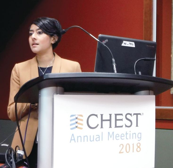
Although health economics weren’t evaluated in this analysis, changes in those quality measures might also impact the bottom line, according to study coauthor Priscilla Chow, DO, a resident at Suburban Community Hospital in East Norriton, Pa.
“Obviously, if the patient is spending less time in the hospital and fewer days on the ventilator and in the ICU, then we can potentially also be more cost effective in our care,” Dr. Chow said in a podium presentation at the meeting.
The use of checklists to standardize processes and reduce errors is a “relatively simple approach” that was adopted from the airline industry and now has been evaluated in a variety of medical care settings, according to Dr. Chow.
Previous studies have demonstrated that checklist-driven care may reduce the incidence of postoperative complications, central line–associated bloodstream infection, ventilator-associated pneumonia, and catheter associated–urinary tract infection.
The present retrospective data analysis by Dr. Chow and colleagues included 200 consecutive patients admitted to the surgical ICU at an urban level 1 trauma center, including 100 patients managed according to the checklist and 100 managed according to standard processes.
Though survival to discharge was comparable between the groups, use of the checklist was associated with a significantly shorter hospital length of stay versus standard care (23.9 vs. 9.5 days). Likewise, the ICU length of stay was shorter in the checklist group (13.0 vs. 6.5 days), and the checklist group had fewer ventilator days (7.7 to 2.8).
Injury Severity Score did not differ between groups, though overall, use of the checklist resulted in more of the underlying topics being addressed in clinical documentation (5.0 vs. 8.7 items).
Dr. Chow and colleagues disclosed that they had no relationships relevant to their study.
SOURCE: Akella K et al. CHEST 2019. Abstract, doi: 10.1016/j.chest.2019.08.201.
NEW ORLEANS – A standardized checklist may help reduce errors and improve common quality measures in critically ill patients, results of a recent retrospective analysis of 200 consecutive patients suggest.
Use of the checklist was linked to significantly shorter hospital length of stay and ICU length of stay as well as fewer days on a ventilator in the analysis, which was presented here at the annual meeting of the American College of Chest Physicians. The 10-item standardized checklist covered a variety topics ranging from comfort, prophylaxis, and sedation to infection control and prevention, nutrition, and medication management review.
Although health economics weren’t evaluated in this analysis, changes in those quality measures might also impact the bottom line, according to study coauthor Priscilla Chow, DO, a resident at Suburban Community Hospital in East Norriton, Pa.
“Obviously, if the patient is spending less time in the hospital and fewer days on the ventilator and in the ICU, then we can potentially also be more cost effective in our care,” Dr. Chow said in a podium presentation at the meeting.
The use of checklists to standardize processes and reduce errors is a “relatively simple approach” that was adopted from the airline industry and now has been evaluated in a variety of medical care settings, according to Dr. Chow.
Previous studies have demonstrated that checklist-driven care may reduce the incidence of postoperative complications, central line–associated bloodstream infection, ventilator-associated pneumonia, and catheter associated–urinary tract infection.
The present retrospective data analysis by Dr. Chow and colleagues included 200 consecutive patients admitted to the surgical ICU at an urban level 1 trauma center, including 100 patients managed according to the checklist and 100 managed according to standard processes.
Though survival to discharge was comparable between the groups, use of the checklist was associated with a significantly shorter hospital length of stay versus standard care (23.9 vs. 9.5 days). Likewise, the ICU length of stay was shorter in the checklist group (13.0 vs. 6.5 days), and the checklist group had fewer ventilator days (7.7 to 2.8).
Injury Severity Score did not differ between groups, though overall, use of the checklist resulted in more of the underlying topics being addressed in clinical documentation (5.0 vs. 8.7 items).
Dr. Chow and colleagues disclosed that they had no relationships relevant to their study.
SOURCE: Akella K et al. CHEST 2019. Abstract, doi: 10.1016/j.chest.2019.08.201.
REPORTING FROM CHEST 2019
Playing harmonica improves COPD
SAN ANTONIO – Playing while also boosting their quality of life, suggest the findings from a small pilot study.
Three months of playing the harmonica about a half hour a day most days of the week led to several improved pulmonary outcome measures in participants, Mary Hart, RRT, MS, of Baylor Scott & White Health in Dallas, reported at the annual meeting of the American College of Chest Physicians.
Ms. Hart played a bit of harmonica during her presentation to demonstrate how playing can help with breathing.
“The harder I push with my diaphragm, the louder I was blowing,” she told attendees. “There’s actually a different amount of effort that you have to use to create sounds with using the harmonica notes.”
Hart said her team found a news article from 1999 about the benefits of playing harmonica, and they became interested in exploring whether it might be a helpful adjunct to respiratory therapy.
Though some previous research has explored potential benefits of harmonica playing in patients with lung disease, one study was too short to demonstrate significant improvement and the other looked at multiple different pulmonary conditions, Ms. Hart said.
The cohort study began with 14 former smokers, average age 72 years, who had completed pulmonary rehabilitation at least 6 months prior to joining the “Harmaniacs,” as the group eventually called themselves.
All participants received a harmonica, an instruction booklet with audio and video supplements, and sheet music for a harmonica in the key of C.
They attended a 2-hour group session once a week with a respiratory therapist and music therapist. The classes focused initially on breathing and relaxation techniques, pacing, and basic harmonica instruction, but the amount of actual playing time increased as the 12-week course went on. Participants were expected to practice their playing for at least a half hour 5 days a week at home.
The group began with the songs “Taps” and “Happy Birthday” because these songs were easy to play. Then they added a song each week, such as “America the Beautiful” and “You Are My Sunshine,” then seasonal favorites such as “We Wish You a Merry Christmas” and “Silent Night,” and easy pop tunes.
The researchers measured both respiratory and quality of life outcomes. Assessments included spirometry, the Six Minute Walk Test, maximal inspiratory pressure (MIP) and maximal expiratory pressure (MEP), the COPD Assessment Test, the modified Medical Research Council Dyspnea Scale, the Patient Health Questionnaire for depression, the St. George’s Respiratory Questionnaire for quality of life, perceived exertion using the Borg scale and assessments by the respiratory therapist and music therapist.
The music therapist listened to and documented participants’ “stories about how they felt about life living with COPD,” and Ms. Hart and her colleagues conducted a respiratory assessment that included data on medication management, adherence to medication, previous hospitalizations and length of stay, perceived shortness of breath, and daily living activities.
In addition to those assessments, the researchers collected data on the length of practice sessions, Borg scores before and after playing, the percentage of time taken for participation in class, the participants’ ability to make a sound, their challenges and triumphs, their tiredness and/or soreness after playing, and the number of people who continued playing after training.
Among the 11 participants who completed the training and all evaluations, the MIP increased by an average 15.36 cmH20 (P = .0017), and their MEP increased by an average 14.36 cmH20 (P = .0061).
Participants increased their distance in the Six Minute Walk Test by an average 60.55 meters (P = .0280), and Ms. Hart reported an improvement in quality of life scores.
In addition to home practice, participants were expected to keep a daily log of how it felt to play and what their biggest challenges and rewards were. The comments they wrote revealed benefits that sometimes surprised even the researchers:
“I can do laundry now.”
“I am more confident.”
“It is relaxing.”
“I want to keep playing forever.”
“It helps me cough up phlegm.”
“I lose track of time and enjoy my playing.”
“I played Happy Birthday at a party for my friend.”
Others express their difficulties as well, such as one person who wrote of being “really frustrated” and another who claimed to “have a hard time playing just one note.”
But the players learned to play as a group as well, even ordering T-shirts for themselves to give concerts. The group now has about 30 songs in its repertoire, Ms. Hart said, and they recently gave a 2-hour concert during which they played all 30 songs twice.
One consistent theme that emerged, Ms. Hart said, was improved control of breathing since playing the harmonica required participants to purse their lips (similar to the way needed for expiratory maneuvers), breathe from their diaphragms, and pace themselves. Playing exercised “the muscles that help pull air in and push air out of the lungs,” Ms. Hart said, and strengthened participants’ abdominal muscles, allowing more effective coughing.
Playing harmonica also increased self-confidence. It provided stress relief for some, and others simply found it fun or enjoyed the socializing opportunities.
The study’s small size and lack of a control group limit the generalizability of its findings.
Baylor Scott & White Central Texas Foundation funded the research. Ms. Hart reported no conflicts of interest.
SOURCE: Hart M et al. CHEST 2018. doi: 10.1016/j.chest.2018.08.669.
REPORTING FROM CHEST 2018
Key clinical point: Playing harmonica improved pulmonary and quality of life outcomes in patients with COPD.
Major finding: Maximal inspiratory pressure increased by an average 15.36 cmH20, maximal expiratory pressure increased by an average 14.36 cmH20, and Six Minute Walk Test distance increased by an average 60.55 meters.
Data source: Cohort study completed by 11 participants with COPD, at least 45 years old, who completed a 12-week harmonica training course.
Disclosures: Baylor Scott & White Central Texas Foundation funded the research. Ms. Hart reported no conflicts of interest.
Source: Hart M et al. CHEST 2018. 10.1016/j.chest.2018.08.669.
Pediatric OSA linked to abnormal metabolic values
SAN ANTONIO – Obstructive sleep apnea (OSA) in children is associated with an abnormal metabolic profile, but not with body mass index (BMI), according to new research.
“Screening for metabolic dysfunction in obese children with obstructive sleep apnea can help identify those at risk for cardiovascular complications,” Kanika Mathur, MD, of the Albert Einstein College of Medicine and the Children’s Hospital at Montefiore, both in New York, told attendees at the annual meeting of the American College of Chest Physicians. Dr. Mathur explained that no consensus currently exists regarding routine cardiac evaluation of children with OSA.
“The American Academy of Pediatrics does not mention any sort of cardiac evaluation in children with OSA while the most recent guidelines from the American Heart Association and the American Thoracic Society recommend echocardiographic evaluation in children with severe obstructive sleep apnea, specifically to evaluate for pulmonary hypertension and right ventricular dysfunction,” Dr. Mathur told attendees.
OSA’s association with obesity, diabetes, and hypertension is well established in adults. It is an independent risk factor for coronary artery disease, heart failure, stroke and atrial fibrillation, and research has suggested OSA treatment can reduce cardiovascular risk in adults, Dr. Mathur explained, but little data on children exist. She and her colleagues set out to understand the relationship of OSA in children with various measures of cardiovascular and metabolic health.
“Despite similar degrees of obesity and systemic blood pressure, pediatric patients with OSA had significantly higher diastolic blood pressure, heart rate, and abnormal metabolic profile, including elevated alanine transaminase, aspartate transaminase, triglycerides and hemoglobin A1c,” they found.
Their study included patients aged 3-21 years with a BMI of at least the 95th percentile who had undergone sleep study and an echocardiogram at the Children’s Hospital at Montefiore between November 2016 and November 2017.
They excluded those with comorbidities related to cardiovascular morbidity: heart disease, neuromuscular disease, sickle cell disease, rheumatologic diseases, significant cranial facial abnormalities, tracheostomy, and any lung disease. However, 7% of the patients had trisomy 21.
Among the 81 children who met their criteria, 37 were male and 44 were female, with an average age of 14 years old and a mean BMI of 39.4 kg/m2 (mean BMI z score of 2.22). Most of the patients (53.1%) had severe OSA (apnea-hypopnea index of at least 10), 21% had moderate OSA (AHI 5-9.9), 12.3% had mild OSA (AHI 2-4.9), and 13.6% did not have OSA. The median AHI of the children was 10.3.
Among all the children, “about half had elevated systolic blood pressure, which is already a risk factor for cardiovascular morbidity,” Mathur reported.
BMI, BMI z score, systolic blood pressure z score, oxygen saturation and cholesterol (overall and both HDL and LDL cholesterol levels) did not significantly differ between children who had OSA and those who did not, but diastolic blood pressure and heart rate did. Those with OSA had a diastolic blood pressure of 65 mm Hg, compared with 58 mm Hg without OSA (P = .008). Heart rate was 89 bpm in the children with OSA, compared with 78 bpm in those without (P = .004).
The children with OSA also showed higher mean levels of several other metabolic biomarkers:
- Alanine transaminase: 26 U/L with OSA vs. 18 U/L without (P = .01).
- Aspartate transaminase: 23 U/L with OSA vs. 18 U/L without (P = .03).
- Triglycerides: 138 mg/dL with OSA vs 84 mg/dL without (P = .004).
- Hemoglobin A1c: 6.2% with OSA vs. 5.4% without (P = .002).
Children with and without OSA did not have any significant differences in left atrial indexed volume, left ventricular volume, left ventricular ejection fraction, or left ventricular mass (measured by M-mode or 5/6 area length formula). Though research has shown these measures to differ in adults with and without OSA, evidence on echocardiographic changes in children has been conflicting, Dr. Mathur noted.
The researchers also conducted subanalyses according to OSA severity, but BMI, BMI Z-score, systolic or diastolic blood pressure Z-score, heart rate and oxygen saturation did not differ between those with mild OSA vs those with moderate or severe OSA. No differences in echocardiographic measurements existed between these subgroups, either.
However, children with moderate to severe OSA did have higher alanine transaminase (27 U/L with moderate to severe vs. 17 U/L with mild OSA; P = .005) and higher triglycerides (148 vs 74; P = .001).
“Certainly we need further evaluation to see the efficacy of obstructive sleep apnea therapies on metabolic dysfunction and whether weight loss needs to be an adjunct therapy for these patients,” Dr. Mathur told attendees. She also noted the need to define the role of echocardiography in managing children with OSA.
The study had several limitations, including its retrospective cross-sectional nature at a single center and its small sample size.
“Additionally, we have a wide variety of ages, which could represent different pathophysiology of the associated metabolic dysfunction in these patients,” Mathur said. “There is an inherent difficulty to performing echocardiograms in a very obese population as well.”
Both the moderators of the pediatrics section, Christopher Carroll, MD, FCCP, of Connecticut Children’s Medical Center in Hartford, and Shahid Sheikh, MD, FCCP, of Nationwide Children’s Hospital in Columbus, Ohio, were impressed with the research. Dr. Carroll called it a “very elegant” study, and Dr. Sheikh noted the need for these studies in pediatrics “so that we don’t have to rely on grown-up data,” which may or may not generalize to children.
SOURCE: CHEST 2018. https://journal.chestnet.org/article/S0012-3692(18)31935-4/fulltext
REPORTING FROM CHEST 2018
Key clinical point: Children with obesity and obstructive sleep apnea have an abnormal metabolic profile.
Major finding: Diastolic blood pressure (65 vs. 58 mm Hg), heart rate (89 vs. 78 bpm), triglycerides (138 vs. 84 mg/dL), alanine transaminase (26 vs. 18 U/L), aspartate transaminase (23 vs. 18 U/L) and hemoglobin A1c (6.2% vs. 5.4%) were all elevated in obese children with OSA, compared with obese children without OSA.
Study details: The findings are based on a retrospective analysis of 81 patients aged 3-21 years, from the Children’s Hospital at Montefiore between November 2016-November 2017.
Disclosures: No external funding was noted. The authors reported having no disclosures.
Breaking the glass ceiling: Women in pulmonary medicine face both barriers and opportunities
SAN ANTONIO – Women in medicine have made great strides in cracking the glass ceiling, but it’s not shattered yet, said Stephanie M. Levine, MD, FCCP, the incoming president of CHEST. 
At a session on women in medicine at the annual meeting of the American College of Chest Physicians, Dr. Levine discussed the challenges of breaking through the metaphorical invisible barrier. The “glass ceiling” refers to multiple ways in which women lack equality with men in medicine: leadership roles, positions and titles, progress in academic medicine, gaps in salaries and compensation, and overall gender parity in specialties.
For example, according to data from the American Association of Medical Colleges for 2017-2018, women comprise 50% of medical school graduates but only 34% of the physician workforce and 22% of leadership roles. Women are 13% less likely to be promoted to professor. They receive salaries an average 21% lower than those of their male peers, said Dr. Levine, professor of medicine and director of the pulmonary/critical care fellowship program at the University of Texas, San Antonio.
Disparities exist particularly within specialties and subspecialties, she said. Women make 85% of what men earn in primary care but, in the specialties, only 75% of what men earn. Among active fellow trainees in the areas of medicine most represented by CHEST, one-third (32%) of critical care physicians and less than a third (29%) of pulmonary physicians are female.
Why the lag in specialty parity?
The reasons for these disparities are complex, Dr. Levine argued, but the problem is not insurmountable. They begin, in a sense, with the problem itself: When there are fewer mentors, role models, sponsors, and leaders, and less overall representation of women in the first place, it is harder for women to advance.
One male audience member, for example, asked how his department could recruit more women, because most turned down interviews despite the fact that more women than men were being invited. “How many women are in your leadership?” Dr. Levine asked. He acknowledged that there were none – and therein lies the likely problem. Applicants are looking for female representation in leadership.
Gender bias and discrimination certainly play a role among peers, leadership, and even patients. Patients referring to female physicians by their first names and asking questions such as “Are you my nurse?” are subtle but cutting examples of the ways in which they reveal implicit bias and reinforce gender stereotypes, Dr. Levine said to weary nods of agreement among the attendees.
Implicit, unconscious bias is also built into the culture of a place and the way things have always been done. Lack of equity in salary, space to work, support, and promotion all compound one another. Work-life integration challenges often do not favor women. Studies have shown that in the hiring process, CVs with female names do not receive as much attention as do CVs with male names, Dr. Levine noted.
Some of the challenges lie with the way women themselves do or do not advocate for themselves. Research has long shown that women do not negotiate as well – or at all – compared with men. Women tend to be less aggressive in seeking higher compensation and leadership roles, possibly because of existing implicit bias against female assertiveness in general.
The catch-22 is that being more assertive or direct can lead others to interpret a woman as being rude or brusk, as one audience member noted when she described how colleagues perceive her simple, direct tone as seeming “upset.”
Conscious bias remains alive and well: The stereotypes that women are caretakers and men are take-charge dominators persist and can reinforce gender disparities in leadership roles.
Women also must make calculations and trade-offs between their academic promotion clocks and their biologic clocks, Dr. Levine explained.
“The 30s are great for both academic and biologic productivity,” she told attendees. The typical age for a person’s first National Institutes of Health Research Project grant (R01) is in the early 40s.
How to improve gender equality
Women bring diverse skills and perspectives to the table, Dr. Levine explained. Women tend to have stronger collaborative skills and greater compassion and empathy, for example. They tend to be less hierarchal and better at mentoring and empowerment, she said.
There are many ways to poke more cracks in the ceiling, starting with diversity and inclusion officers who make it a priority to focus on parity. Formal programs can educate staff and colleagues about implicit bias so that they might more easily recognize it when it kicks in, and training for gatekeepers can lead to more proportional hiring of women at every level.
Institutions should review their policies – salary inequities, diversity in promotion, processes for selecting leaders – and set formal interventional goals that are then evaluated in honest, documented annual reviews.
Some of these policies should address work-life balance as well: Offering part-time and flexible work options during early child-rearing years helps not only mothers, but also fathers who are now taking a more active role in parenting. Slowing or prorating the promotion clock can help those building families, and shifting meetings away from times such as 7:00 a.m. and 6:00 p.m. allow mothers and fathers alike to get their kids to and from school and attend children’s events.
Sponsorship of women is an important strategy in breaking the glass ceiling, Dr. Levine said. Sponsors can support women with untapped leadership potential and do the necessary networking and introductions that help make that advance happen. And it must be done by sponsors with power and influence, including men, Dr. Levine said.
Men can play important roles in promoting gender parity by suggesting women for key roles, leadership positions, and committees and also notifying women of upcoming opportunities, such as editorial board spots and other hot jobs. For women who aspire to be leaders, men can seek to convey leadership skills that may be needed to chair committees and other groups. Search committees need to expand beyond looking for “token women,” she said.
Dr. Levine illustrated her address with her own story. She described how many of these strategies had helped her career and how many male supervisors, mentors, and colleagues helped her, including introducing her to other male leaders who then offered her opportunities to contribute to the American College of Chest Physicians. She ran for CHEST president twice before being elected on her third run in September. She is the fifth woman to lead CHEST.
“Don’t give up,” she encouraged women in the audience, telling them to advocate for themselves and to encourage, mentor, and sponsor their female fellows and junior faculty.
“This will result in closing the gaps and will help women achieve leadership roles and competitive salaries as well as work-life integration,” Dr. Levine said.
SAN ANTONIO – Women in medicine have made great strides in cracking the glass ceiling, but it’s not shattered yet, said Stephanie M. Levine, MD, FCCP, the incoming president of CHEST.
At a session on women in medicine at the annual meeting of the American College of Chest Physicians, Dr. Levine discussed the challenges of breaking through the metaphorical invisible barrier. The “glass ceiling” refers to multiple ways in which women lack equality with men in medicine: leadership roles, positions and titles, progress in academic medicine, gaps in salaries and compensation, and overall gender parity in specialties.
For example, according to data from the American Association of Medical Colleges for 2017-2018, women comprise 50% of medical school graduates but only 34% of the physician workforce and 22% of leadership roles. Women are 13% less likely to be promoted to professor. They receive salaries an average 21% lower than those of their male peers, said Dr. Levine, professor of medicine and director of the pulmonary/critical care fellowship program at the University of Texas, San Antonio.
Disparities exist particularly within specialties and subspecialties, she said. Women make 85% of what men earn in primary care but, in the specialties, only 75% of what men earn. Among active fellow trainees in the areas of medicine most represented by CHEST, one-third (32%) of critical care physicians and less than a third (29%) of pulmonary physicians are female.
Why the lag in specialty parity?
The reasons for these disparities are complex, Dr. Levine argued, but the problem is not insurmountable. They begin, in a sense, with the problem itself: When there are fewer mentors, role models, sponsors, and leaders, and less overall representation of women in the first place, it is harder for women to advance.
One male audience member, for example, asked how his department could recruit more women, because most turned down interviews despite the fact that more women than men were being invited. “How many women are in your leadership?” Dr. Levine asked. He acknowledged that there were none – and therein lies the likely problem. Applicants are looking for female representation in leadership.
Gender bias and discrimination certainly play a role among peers, leadership, and even patients. Patients referring to female physicians by their first names and asking questions such as “Are you my nurse?” are subtle but cutting examples of the ways in which they reveal implicit bias and reinforce gender stereotypes, Dr. Levine said to weary nods of agreement among the attendees.
Implicit, unconscious bias is also built into the culture of a place and the way things have always been done. Lack of equity in salary, space to work, support, and promotion all compound one another. Work-life integration challenges often do not favor women. Studies have shown that in the hiring process, CVs with female names do not receive as much attention as do CVs with male names, Dr. Levine noted.
Some of the challenges lie with the way women themselves do or do not advocate for themselves. Research has long shown that women do not negotiate as well – or at all – compared with men. Women tend to be less aggressive in seeking higher compensation and leadership roles, possibly because of existing implicit bias against female assertiveness in general.
The catch-22 is that being more assertive or direct can lead others to interpret a woman as being rude or brusk, as one audience member noted when she described how colleagues perceive her simple, direct tone as seeming “upset.”
Conscious bias remains alive and well: The stereotypes that women are caretakers and men are take-charge dominators persist and can reinforce gender disparities in leadership roles.
Women also must make calculations and trade-offs between their academic promotion clocks and their biologic clocks, Dr. Levine explained.
“The 30s are great for both academic and biologic productivity,” she told attendees. The typical age for a person’s first National Institutes of Health Research Project grant (R01) is in the early 40s.
How to improve gender equality
Women bring diverse skills and perspectives to the table, Dr. Levine explained. Women tend to have stronger collaborative skills and greater compassion and empathy, for example. They tend to be less hierarchal and better at mentoring and empowerment, she said.
There are many ways to poke more cracks in the ceiling, starting with diversity and inclusion officers who make it a priority to focus on parity. Formal programs can educate staff and colleagues about implicit bias so that they might more easily recognize it when it kicks in, and training for gatekeepers can lead to more proportional hiring of women at every level.
Institutions should review their policies – salary inequities, diversity in promotion, processes for selecting leaders – and set formal interventional goals that are then evaluated in honest, documented annual reviews.
Some of these policies should address work-life balance as well: Offering part-time and flexible work options during early child-rearing years helps not only mothers, but also fathers who are now taking a more active role in parenting. Slowing or prorating the promotion clock can help those building families, and shifting meetings away from times such as 7:00 a.m. and 6:00 p.m. allow mothers and fathers alike to get their kids to and from school and attend children’s events.
Sponsorship of women is an important strategy in breaking the glass ceiling, Dr. Levine said. Sponsors can support women with untapped leadership potential and do the necessary networking and introductions that help make that advance happen. And it must be done by sponsors with power and influence, including men, Dr. Levine said.
Men can play important roles in promoting gender parity by suggesting women for key roles, leadership positions, and committees and also notifying women of upcoming opportunities, such as editorial board spots and other hot jobs. For women who aspire to be leaders, men can seek to convey leadership skills that may be needed to chair committees and other groups. Search committees need to expand beyond looking for “token women,” she said.
Dr. Levine illustrated her address with her own story. She described how many of these strategies had helped her career and how many male supervisors, mentors, and colleagues helped her, including introducing her to other male leaders who then offered her opportunities to contribute to the American College of Chest Physicians. She ran for CHEST president twice before being elected on her third run in September. She is the fifth woman to lead CHEST.
“Don’t give up,” she encouraged women in the audience, telling them to advocate for themselves and to encourage, mentor, and sponsor their female fellows and junior faculty.
“This will result in closing the gaps and will help women achieve leadership roles and competitive salaries as well as work-life integration,” Dr. Levine said.
REPORTING FROM CHEST 2018
Two-thirds of COPD patients not using inhalers correctly
SAN ANTONIO – Two-thirds of U.S. adults with (MDIs), according to new research. About half of patients failed to inhale slowly and deeply to ensure they received the appropriate dose, and about 40% of patients failed to hold their breath for 5-10 seconds afterward so that the medication made its way to their lungs, the findings show.
“There’s a need to educate patients on proper inhalation technique to optimize the appropriate delivery of medication,” Maryam Navaie, DrPH, of Advance Health Solutions in New York told attendees at the annual meeting of the American College of Chest Physicians. She also urged practitioners to think more carefully about what devices to prescribe to patients based on their own personal attributes.
“Nebulizer devices may be a better consideration for patients who have difficulty performing the necessary steps required by handheld inhalers,” Dr. Navaie said.
She and fellow researchers conducted a systematic review to gain more insights into the errors and difficulties experienced by U.S. adults using MDIs for COPD or asthma. They combed through PubMed, EMBASE, PsycINFO, Cochrane, and Google Scholar databases for English language studies about MDI-related errors in U.S. adult COPD or asthma patients published between January 2003 and February 2017.
The researchers included only randomized controlled trials and cross-sectional and observational studies, and they excluded studies with combined error rates across multiple devices so they could better parse out the data. They also used baseline rates only in studies that involved an intervention to reduce errors.
The researchers defined the proportion of overall MDI errors as “the percentage of patients who made errors in equal to or greater than 20% of inhalation steps.” They computed pooled estimates and created forest plots for both overall errors and for errors according to each step in using an MDI.
The eight studies they identified involved 1,221 patients, with ages ranging from a mean 48 to 82 years, 53% of whom were female. Nearly two-thirds of the patients had COPD (63.6%) while 36.4% had asthma. Most of the devices studied were MDIs alone (68.8%), while 31.2% included a spacer.
The pooled weighted average revealed a 66.5% error rate, that is, two-thirds of all the patients were making at least two errors during the 10 steps involved in using their device. The researchers then used individual error rates data in five studies to calculate the overall error rate for each step in using MDIs. The most common error, made by 73.8% of people in those five studies, was failing to attach the inhaler to the spacer. In addition, 68.7% of patients were failing to exhale fully and away from the inhaler before inhaling, and 47.8% were inhaling too fast instead of inhaling deeply.
“So these [findings] actually give you [some specific] ideas of how we could help improve patients’ ability to use the device properly,” Dr. Navaie told attendees, adding that these data can inform patient education needs and interventions.
Based on the data from those five studies, the error rates for all 10 steps to using an MDI were as follows:
- Failed to shake inhaler before use (37.9%).
- Failed to attach inhaler to spacer (73.8%).
- Failed to exhale fully and away from inhaler before inhalation (68.7%).
- Failed to place mouthpiece between teeth and sealed lips (7.4%).
- Failed to actuate once during inhalation (24.4%).
- Inhalation too fast, not deep (47.8%).
- Failed to hold breath for 5-10 seconds (40.1%).
- Failed to remove the inhaler/spacer from mouth (11.3%).
- Failed to exhale after inhalation (33.2%).
- Failed to repeat steps for second puff (36.7%).
Dr. Navaie also noted the investigators were surprised to learn that physicians themselves sometimes make several of these errors in explaining to patients how to use their devices.
“I think for the reps and other people who go out and visit doctors, it’s important to think about making sure the clinicians are using the devices properly,” Dr. Navaie said. She pointed out the potential for patients to forget steps between visits.
“One of the things a lot of our clinicians and key opinion leaders told us during the course of this study is that you shouldn’t just educate the patient at the time you are scripting the device but repeatedly because patients forget,” she said. She recommended having patients demonstrate their use of the device at each visit. If patients continue to struggle, it may be worth considering other therapies, such as a nebulizer, for patients unable to regularly use their devices correctly.
The meta-analysis was limited by the sparse research available in general on MDI errors in the U.S. adult population, so the data on error rates for each individual step may not be broadly generalizable. The studies also did not distinguish between rates among users with asthma vs. users with COPD. Further, too few data exist on associations between MDI errors and health outcomes to have a clear picture of the clinical implications of regularly making multiple errors in MDI use.
Dr. Navaie is employed by Advance Health Solutions, which received Sunovion Pharmaceuticals funding for the study.
SOURCE: Navaie M et al. CHEST 2018. doi: 10.1016/j.chest.2018.08.705.
REPORTING FROM CHEST 2018
Key clinical point: 67% of US adult patients with COPD or asthma report making errors in using metered-dose inhalers.
Major finding: 69% of patients do not exhale fully and away from the inhaler before inhalation; 50% do not inhale slowly and deeply.
Study details: Meta-analysis of eight studies involving 1,221 U.S. adult patients with COPD or asthma who use metered-dose inhalers.
Disclosures: Dr. Navaie is employed by Advance Health Solutions, which received Sunovion Pharmaceuticals funding for the study.
Source: Navaie M et al. CHEST 2018. doi: 10.1016/j.chest.2018.08.705.

Bias in the clinical setting can impact patient care
SAN ANTONIO – Physicians and other health care providers may harbor implicit, or unconscious, biases that contribute to health care disparities, patient communication researcher Stacey Passalacqua, PhD, said here at the annual meeting of the American College of Chest Physicians.
Implicit biases are beliefs or attitudes, for example, about certain social groups, that exist outside of a health care provider’s conscious awareness, said Dr. Passalacqua of the department of communication at the University of Texas, San Antonio. If bias is implicit, it can be difficult self-assess.
among other social, ethnic, and racial groups, Dr. Passalacqua told attendees in workshops at the meeting.
“If a health care provider has negative biases toward a particular patient – maybe they think that these patients doesn’t care that much about their health or that they really have no interest in participating – then obviously that health care provider is far less likely to engage that patient in shared decision making,” she said in a video interview.
Diagnosis and treatment are subject to influence by the bias that physicians have toward certain patient groups, according to Dr. Passalacqua. For example, she said women with heart disease are less likely to be accurately diagnosed.
The bias in the medical setting might be mitigated by the presence of more individuals from the at-risk groups in the health care workforce, she added. In one recent retrospective study, investigators found that after an MI, a woman treated by a male physician was associated with higher mortality, while women and men had similar outcomes when treated by female physicians.
“That is one of the reasons why it is so important to have a diverse workforce, to have health care providers of different ethnicities, of different genders, or different backgrounds, because they are less subject to some of these implicit biases that we know are highly problematic in health care,” she said in the interview.
Dr. Passalacqua had no disclosures related to her presentation.
REPORTING FROM CHEST 2018

Dupilumab offers extra benefits to asthmatic teens
SAN ANTONIO – For adolescents with asthma, treatment with the biologic agent dupilumab provided benefits that were at least comparable with what was seen in adults, results from a retrospective analysis of a randomized, phase 3 study suggest.

Adolescents had reduced asthma exacerbations in line with what was seen in adults and had improvements in lung function that were at a greater magnitude than adults, according to study coauthor Neil M.H. Graham, MD, of Regeneron Pharmaceuticals, Tarrytown, N.Y.
“We think, overall, it’s a very good treatment response from this drug in this high-risk population, and it is generally well tolerated, as we’ve seen in other studies,” Dr. Graham said in a podium presentation at the annual meeting of the American College of Chest Physicians.
Dr. Graham presented results of an analysis of the 1,902-patient, phase 3 Liberty Asthma QUEST trial, published in May 2018 in the New England Journal of Medicine.
Top-line results of QUEST showed that treatment with dupilumab, a fully human anti–IL-4Ra monoclonal antibody, resulted in significantly lower rates of severe asthma exacerbation, along with improved lung function, in patients aged 12 years and older with moderate to severe asthma.
Now, this retrospective analysis shows that, in adolescents, improvements from baseline to week 12 in forced expiratory volume in 1 second (FEV1) were significant and at a greater magnitude than in adults, according to Dr. Graham and his coinvestigators.
The improvement over 12 weeks in FEV1 for adolescents was 0.36 L and 0.27 L, respectively, for the 200- and 300-mg doses of dupilumab (P less than .05 vs. placebo for both), Dr. Graham and his coinvestigators reported. In adults, the improvement was 0.12 L.
The annualized exacerbation rate dropped by 46.4% for those adolescents who received 200 mg dupilumab, though there was no treatment effect versus placebo for dupilumab 300 mg; the investigators said the lack of effect in this retrospective analysis could have been caused by imbalances in prior event rates or the small sample size.
A total of 107 out of 1,902 patients in QUEST were adolescents, and of those, 68 were randomly assigned to dupilumab, according to the report. Injection site reaction was the most common adverse event in adolescents in both dosing groups.
Dr. Graham reported disclosures related to his employment with Regeneron. Study coauthors reported disclosures related to Regeneron, AstraZeneca, Sanofi, Teva Pharmaceutical, GlaxoSmithKline, Boehringer Ingelheim, Merck, Genentech, and others.
SOURCE: Graham NMH et al. CHEST. 2018 Oct. doi: 10.1016/j.chest.2018.08.022.
SAN ANTONIO – For adolescents with asthma, treatment with the biologic agent dupilumab provided benefits that were at least comparable with what was seen in adults, results from a retrospective analysis of a randomized, phase 3 study suggest.
Adolescents had reduced asthma exacerbations in line with what was seen in adults and had improvements in lung function that were at a greater magnitude than adults, according to study coauthor Neil M.H. Graham, MD, of Regeneron Pharmaceuticals, Tarrytown, N.Y.
“We think, overall, it’s a very good treatment response from this drug in this high-risk population, and it is generally well tolerated, as we’ve seen in other studies,” Dr. Graham said in a podium presentation at the annual meeting of the American College of Chest Physicians.
Dr. Graham presented results of an analysis of the 1,902-patient, phase 3 Liberty Asthma QUEST trial, published in May 2018 in the New England Journal of Medicine.
Top-line results of QUEST showed that treatment with dupilumab, a fully human anti–IL-4Ra monoclonal antibody, resulted in significantly lower rates of severe asthma exacerbation, along with improved lung function, in patients aged 12 years and older with moderate to severe asthma.
Now, this retrospective analysis shows that, in adolescents, improvements from baseline to week 12 in forced expiratory volume in 1 second (FEV1) were significant and at a greater magnitude than in adults, according to Dr. Graham and his coinvestigators.
The improvement over 12 weeks in FEV1 for adolescents was 0.36 L and 0.27 L, respectively, for the 200- and 300-mg doses of dupilumab (P less than .05 vs. placebo for both), Dr. Graham and his coinvestigators reported. In adults, the improvement was 0.12 L.
The annualized exacerbation rate dropped by 46.4% for those adolescents who received 200 mg dupilumab, though there was no treatment effect versus placebo for dupilumab 300 mg; the investigators said the lack of effect in this retrospective analysis could have been caused by imbalances in prior event rates or the small sample size.
A total of 107 out of 1,902 patients in QUEST were adolescents, and of those, 68 were randomly assigned to dupilumab, according to the report. Injection site reaction was the most common adverse event in adolescents in both dosing groups.
Dr. Graham reported disclosures related to his employment with Regeneron. Study coauthors reported disclosures related to Regeneron, AstraZeneca, Sanofi, Teva Pharmaceutical, GlaxoSmithKline, Boehringer Ingelheim, Merck, Genentech, and others.
SOURCE: Graham NMH et al. CHEST. 2018 Oct. doi: 10.1016/j.chest.2018.08.022.
REPORTING FROM CHEST 2018
Key clinical point: Dupilumab’s benefits in adolescents with moderate to severe asthma are at least comparable with what has been reported in adults.
Major finding: The improvement over 12 weeks in forced expiratory volume in 1 second for adolescents was 0.36 L and 0.27 L, respectively, for the 200- and 300-mg doses of dupilumab versus 0.12 L for adults.
Study details: A retrospective analysis of 1,902 patients (including 107 adolescents) in Liberty Asthma QUEST, a recently reported randomized, phase 3 trial.
Disclosures: Several study coauthors reported employment with Regeneron. Dr. Graham reported disclosures related to his employment with Regeneron. Other reported disclosures were related to AstraZeneca, Sanofi, Teva Pharmaceutical, GlaxoSmithKline, Boehringer Ingelheim, Merck, and Genentech, among other entities.
Source: Graham NMH et al. CHEST. 2018 Oct. doi: 10.1016/j/chest.2018.08.002.
CT for evaluating pulmonary embolism overused
SAN ANTONIO – The recommended approach to evaluating suspected pulmonary embolism is “greatly underutilized” in the Veterans Health Administration system, Nancy Hsu, MD, said at the annual meeting of the American College of Chest Physicians.
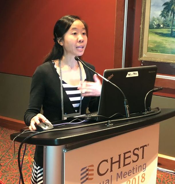
Most Veterans Affairs sites did not require incorporation of a clinical decision rule (CDR) and highly sensitive D-dimer prior to ordering CT pulmonary angiography (CTPA) for suspected pulmonary embolism (PE), according to results of a survey by Dr. Hsu and her coinvestigator, Guy Soo Hoo, MD.
While CTPA has become the imaging modality of choice for evaluating suspected PE, it is overused and potentially avoidable in one-third of cases, said Dr. Hsu, who is with the VA Greater Los Angeles Healthcare System.
“In the 10 years following the advent of CTPA use, there was a 14-fold increase in usage, but there was no change in mortality,” Dr. Hsu said. “This is consistent with overdiagnosis.”
Dr. Hsu said.
Dr. Hsu and Dr. Soo Hoo surveyed 606 individuals at 18 Veterans Integrated Service Networks (VISNs) and 143 medical centers. A total of 120 fully completed questionnaires were analyzed.
Most respondents (63%) were chiefs, and 80% had 11+ years of experience, Dr. Hsu reported.
Almost all respondents (85%) said CDR with or without D-dimer was not required before ordering a CTPA, survey results show, while only about 7% required both.
“A very small minority of [Veterans Integrated Service Networks], or geographic regions, contained even one hospital that adhered to the guidelines,” Dr. Hsu added.
Though further analysis was limited by sample size, the average CTPA yield for PE appeared to be higher when both components were used in the evaluation, according to Dr. Hsu, who noted an 11.9% yield for CDR plus D-dimer.
Use of CTPA appeared lower at sites with CDR and D-dimer testing, Dr. Hsu added.
These results suggest a need for further research to compare CTPA use and yield in sites that have the algorithm in place, Dr. Hsu told attendees at the meeting.
Adherence to the CDR plus D-dimer diagnostic strategy is “modest at best” despite being a Top 5 Choosing Wisely recommendation in pulmonary medicine, Dr. Hsu told attendees.
The biggest barrier to optimal practice may be the fear of having a patient who “falls through the cracks” based on false-negative CDR and D-dimer data, according to Dr. Hsu.
On the other hand, judicious use of CTPA likely avoids negative sequelae related to radiation, contrast exposure, and treatment-related bleeding, Dr. Hsu said.
“It’s all about balancing risks and benefits,” she said from the podium in a discussion of the study results.
Dr. Hsu and Dr. Soo Hoo disclosed that they had no relationships relevant to their research.
SOURCE: Hsu N et al. CHEST. 2018 Oct. doi: 10.1016/j.chest.2018.08.937
SAN ANTONIO – The recommended approach to evaluating suspected pulmonary embolism is “greatly underutilized” in the Veterans Health Administration system, Nancy Hsu, MD, said at the annual meeting of the American College of Chest Physicians.
Most Veterans Affairs sites did not require incorporation of a clinical decision rule (CDR) and highly sensitive D-dimer prior to ordering CT pulmonary angiography (CTPA) for suspected pulmonary embolism (PE), according to results of a survey by Dr. Hsu and her coinvestigator, Guy Soo Hoo, MD.
While CTPA has become the imaging modality of choice for evaluating suspected PE, it is overused and potentially avoidable in one-third of cases, said Dr. Hsu, who is with the VA Greater Los Angeles Healthcare System.
“In the 10 years following the advent of CTPA use, there was a 14-fold increase in usage, but there was no change in mortality,” Dr. Hsu said. “This is consistent with overdiagnosis.”
Dr. Hsu said.
Dr. Hsu and Dr. Soo Hoo surveyed 606 individuals at 18 Veterans Integrated Service Networks (VISNs) and 143 medical centers. A total of 120 fully completed questionnaires were analyzed.
Most respondents (63%) were chiefs, and 80% had 11+ years of experience, Dr. Hsu reported.
Almost all respondents (85%) said CDR with or without D-dimer was not required before ordering a CTPA, survey results show, while only about 7% required both.
“A very small minority of [Veterans Integrated Service Networks], or geographic regions, contained even one hospital that adhered to the guidelines,” Dr. Hsu added.
Though further analysis was limited by sample size, the average CTPA yield for PE appeared to be higher when both components were used in the evaluation, according to Dr. Hsu, who noted an 11.9% yield for CDR plus D-dimer.
Use of CTPA appeared lower at sites with CDR and D-dimer testing, Dr. Hsu added.
These results suggest a need for further research to compare CTPA use and yield in sites that have the algorithm in place, Dr. Hsu told attendees at the meeting.
Adherence to the CDR plus D-dimer diagnostic strategy is “modest at best” despite being a Top 5 Choosing Wisely recommendation in pulmonary medicine, Dr. Hsu told attendees.
The biggest barrier to optimal practice may be the fear of having a patient who “falls through the cracks” based on false-negative CDR and D-dimer data, according to Dr. Hsu.
On the other hand, judicious use of CTPA likely avoids negative sequelae related to radiation, contrast exposure, and treatment-related bleeding, Dr. Hsu said.
“It’s all about balancing risks and benefits,” she said from the podium in a discussion of the study results.
Dr. Hsu and Dr. Soo Hoo disclosed that they had no relationships relevant to their research.
SOURCE: Hsu N et al. CHEST. 2018 Oct. doi: 10.1016/j.chest.2018.08.937
REPORTING FROM CHEST 2018
Key clinical point: The recommended approach to evaluating suspected pulmonary embolism was underutilized in VA facilities.
Major finding: 85% of respondents said incorporation of a clinical decision rule plus highly sensitive D-dimer was not required prior to CTPA.
Study details: Analysis of 120 survey questionnaires completed by individuals working in Veterans Integrated Service Networks and medical centers.
Disclosures: Study authors reported no conflicts of interest.
Source: Hsu N et al. CHEST 2018 Oct. doi: 10/1016/j.chest.2018.08.937.
COPD: Triple trumps dual therapy regardless of baseline reversibility
SAN ANTONIO – Regardless of COPD patients’ bronchodilator reversibility at baseline, versus dual therapies, according to a recent retrospective analysis of a randomized, double-blind study.
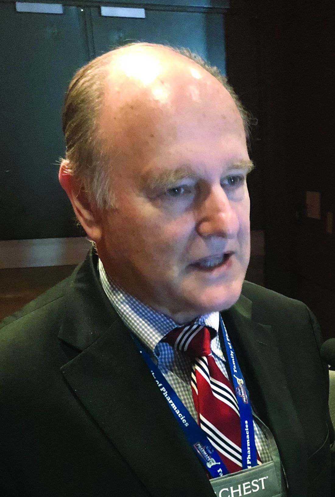
FF/UMEC/VI, a triple-therapy combination of an inhaled corticosteroid, long-acting muscarinic antagonist, and long-acting beta2 agonist (ICS/LAMA/LABA), was superior to both LAMA/LABA and ICS/LABA combinations in reducing the rate of moderate to severe exacerbation and lung function, the analysis showed.
The ICS/LAMA/LABA combination, compared with LAMA/LABA, also significantly reduced the rate of severe exacerbations and time to first moderate to severe exacerbations in both reversible and nonreversible patients, Robert Wise, MD, FCCP, of Johns Hopkins University, Baltimore, said at the annual meeting of the American College of Chest Physicians.
The analysis was based on data from IMPACT, an international, randomized, 52-week study that included more than 10,000 patients with symptomatic COPD, of whom 18% demonstrated reversibility at screening.
“The results across both reversibility subgroups are consistent with those observed in the intention-to-treat or overall study population and show a similar benefit-to-risk profile of the triple therapy across different subtypes based on bronchodilator reversibility,” Dr. Wise told attendees in a podium presentation.
Reversibility was defined as a difference between pre- and postalbuterol assessment of FEV1 of equal to or greater than 12% and equal to or greater than 200 mL at screening, Dr. Wise said.
Reversible patients had a 40% reduction in the rate of moderate to severe exacerbations for FF/UMEC/VI versus UMEC/VI, while nonreversible patients had a 21% reduction, according to data reported in the meeting abstract.
Severe exacerbation rates dropped by 44% and 31%, respectively, in the reversible and nonreversible patients for triple versus dual therapy, he added.
Triple therapy reduced time to first moderate to severe exacerbation versus dual therapy by 25.6% in reversible and 13.6% in nonreversible COPD patients, the data showed.
The FF/UMEC/VI combination also demonstrated improvements versus UMEC/VI in time to first severe exacerbation for both the reversible and nonreversible groups, as well as improved quality of life in both groups as measured by the St. George Respiratory Questionnaire (SGRQ) in both groups.
Results were somewhat different when comparing the FF/UMEC/VI combination with the FF/VI – the ICS/LABA combination – in this post hoc analysis.
Triple therapy did reduce moderate to severe exacerbations and improved lung function regardless of baseline reversibility. However, for the reversible patients, ICS/LAMA/LABA versus ICS/LABA did not significantly reduce risk specifically of severe exacerbations, time to first moderate to severe exacerbation, or increase odds of being an SGRQ responder, Dr. Wise said.
Nonetheless, these findings taken together imply that this ICS/LAMA/LABA combination provides clinically relevant improvements versus dual therapy across a range of important outcomes regardless of baseline reversibility, according to Dr. Wise and colleagues.
Dr. Wise and coinvestigators provided disclosures related to Boehringer Ingelheim, BTG, Chiesi, GlaxoSmithKline, Mereo, Novartis, PneumRx, Prometic, and Pulmonx.
SOURCE: Wise R et al. Chest. 2018 Oct. doi: 10.1016/j.chest.2018.08.662
SAN ANTONIO – Regardless of COPD patients’ bronchodilator reversibility at baseline, versus dual therapies, according to a recent retrospective analysis of a randomized, double-blind study.
FF/UMEC/VI, a triple-therapy combination of an inhaled corticosteroid, long-acting muscarinic antagonist, and long-acting beta2 agonist (ICS/LAMA/LABA), was superior to both LAMA/LABA and ICS/LABA combinations in reducing the rate of moderate to severe exacerbation and lung function, the analysis showed.
The ICS/LAMA/LABA combination, compared with LAMA/LABA, also significantly reduced the rate of severe exacerbations and time to first moderate to severe exacerbations in both reversible and nonreversible patients, Robert Wise, MD, FCCP, of Johns Hopkins University, Baltimore, said at the annual meeting of the American College of Chest Physicians.
The analysis was based on data from IMPACT, an international, randomized, 52-week study that included more than 10,000 patients with symptomatic COPD, of whom 18% demonstrated reversibility at screening.
“The results across both reversibility subgroups are consistent with those observed in the intention-to-treat or overall study population and show a similar benefit-to-risk profile of the triple therapy across different subtypes based on bronchodilator reversibility,” Dr. Wise told attendees in a podium presentation.
Reversibility was defined as a difference between pre- and postalbuterol assessment of FEV1 of equal to or greater than 12% and equal to or greater than 200 mL at screening, Dr. Wise said.
Reversible patients had a 40% reduction in the rate of moderate to severe exacerbations for FF/UMEC/VI versus UMEC/VI, while nonreversible patients had a 21% reduction, according to data reported in the meeting abstract.
Severe exacerbation rates dropped by 44% and 31%, respectively, in the reversible and nonreversible patients for triple versus dual therapy, he added.
Triple therapy reduced time to first moderate to severe exacerbation versus dual therapy by 25.6% in reversible and 13.6% in nonreversible COPD patients, the data showed.
The FF/UMEC/VI combination also demonstrated improvements versus UMEC/VI in time to first severe exacerbation for both the reversible and nonreversible groups, as well as improved quality of life in both groups as measured by the St. George Respiratory Questionnaire (SGRQ) in both groups.
Results were somewhat different when comparing the FF/UMEC/VI combination with the FF/VI – the ICS/LABA combination – in this post hoc analysis.
Triple therapy did reduce moderate to severe exacerbations and improved lung function regardless of baseline reversibility. However, for the reversible patients, ICS/LAMA/LABA versus ICS/LABA did not significantly reduce risk specifically of severe exacerbations, time to first moderate to severe exacerbation, or increase odds of being an SGRQ responder, Dr. Wise said.
Nonetheless, these findings taken together imply that this ICS/LAMA/LABA combination provides clinically relevant improvements versus dual therapy across a range of important outcomes regardless of baseline reversibility, according to Dr. Wise and colleagues.
Dr. Wise and coinvestigators provided disclosures related to Boehringer Ingelheim, BTG, Chiesi, GlaxoSmithKline, Mereo, Novartis, PneumRx, Prometic, and Pulmonx.
SOURCE: Wise R et al. Chest. 2018 Oct. doi: 10.1016/j.chest.2018.08.662
REPORTING FROM CHEST 2018
Key clinical point: Triple therapy with fluticasone furoate, umeclidinium, and vilanterol (FF/UMEC/VI) is superior to UMEC/VI in COPD patients regardless of baseline bronchodilator reversibility.
Major finding: Reversible patients had a 40% reduction in the rate of moderate to severe exacerbations for FF/UMEC/VI versus UMEC/VI, while nonreversible patients had a 21% reduction.
Study details: Retrospective analysis of IMPACT, an international, randomized, 52-week study that included more than 10,000 patients with symptomatic COPD, of whom 18% demonstrated reversibility at screening.
Disclosures: Study authors reported disclosures related to Boehringer Ingelheim, BTG, Chiesi, GlaxoSmithKline, Mereo, Novartis, PneumRx, Prometic, and Pulmonx.
Source: Wise R et al. Chest. 2018 Oct. doi: 10.1016/j/chest.2018.08.662.
Managing asthma in children: Pets don’t always have to go
SAN ANTONIO – It may not always be necessary to tell parents of children with asthma to get rid of the household pet, a recent study suggests.
Children with had significant improvements in a variety of asthma measures, regardless of whether parents reported pets or smoking at home, according to results of the 4-year, 471-patient prospective study.
Those results suggest that clinicians should be working to make sure the guidelines are being closely followed before, for example, telling parents they need to consider getting rid of the family pet, said Shahid Sheikh, MD, FCCP, of Nationwide Children’s Hospital, Columbus, Ohio.
“As the guidelines still work, we need to focus and develop the connections with the family to make sure the patients are on the right treatment, and that they’re getting the medications,” Dr. Sheikh said in an interview at the annual meeting of the American College of Chest Physicians.
The prospective cohort study by Dr. Sheikh and his colleagues, presented in a poster session, included children referred to a pediatric asthma center with the diagnosis of uncontrolled asthma. All patients received asthma care according to National Asthma Education and Prevention Program Expert Panel Report 3 guidelines.
Medications were changed as needed, and the asthma action plan was revised accordingly and reviewed with the family at each visit, Dr. Sheikh reported. After a baseline evaluation, clinic visits for the study occurred at 3 months, 6 months, and then at 1, 2, 3, and 4 years.
Out of 471 patients, 258 had pets, and 125 were in homes where smoking took place, according to parent reports.
Asthma control test scores were 15.1 at baseline for children in no-pet households, and 16.5 for those with pets; by the 3-month visit, scores increased to 20.1 and 20.3 for the no-pet and pet groups, and at 4 years, those scores had edged up to 22.2 and 22.7 (P = .371), Dr. Sheikh reported.
Similarly, after care was started, there was no significant difference between the no-pet and pet groups in mean percent of predicted forced expiratory volume in 1 second (FEV1), wheezing, nighttime cough, albuterol use, and other factors over the 4 years of follow-up, he said.
Likewise, looking at the data by nonsmoking vs. smoking households, asthma control test scores at baseline were 16.1 and 15.1, respectively, and at 4 years they were 22.2 and 22.3 (P = .078), with a similar lack of difference in predicted FEV1, wheezing, and all other factors evaluated.
Getting rid of the family pet may need to be a consideration for some families, but based on these data, that might not be necessary for the majority of families, Dr. Sheikh said in the interview.
“On the other hand, we are not saying that if you are smoking, you should continue to smoke,” he added.
“What we are saying is that smoking is bad, but if your child is not getting better, I don’t want to blame your smoking for it. There may be something else which may be more important than smoking which we are missing – the child may not be getting the medicine, or may not be on the right medicine, or may have other comorbidities.”
Dr. Sheikh and his coinvestigators disclosed that they had no relationships relevant to the study.
SOURCE: Sheikh S et al. CHEST 2018. doi: 10.1016/j.chest.2018.08.666.
REPORTING FROM CHEST 2018
Key clinical point: Exposure to pets and tobacco smoke may have very little effect on the improvement of asthma in children who are being managed according to guidelines.
Major finding: ACT scores were 15.1 and 16.5 at baseline for children in no-pet and pet households, respectively, and were 22.2 and 22.7 at the 4-year evaluation.
Study details: A 4-year prospective cohort study of 471 children with uncontrolled asthma seen in a pediatric asthma center.
Disclosures: The study authors had no disclosures.
Source: Sheikh S et al. CHEST 2018. doi: 10.1016/j.chest.2018.08.666.