User login
Weekly tocilizumab provides durable benefits for giant cell arteritis patients
ATLANTA –
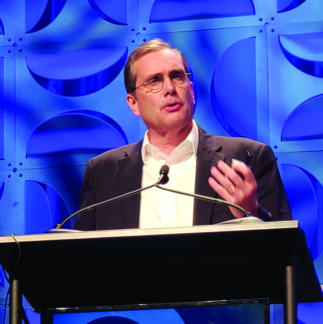
Of 250 patients treated in the double-blind portion of the trial, 215 entered the extension period (part 2 of the trial), and 197 (92%) completed all 3 years of the trial. A total of 38 of 81 (47%) extension participants in clinical remission (CR) at week 52 after receiving tocilizumab (Actemra) weekly maintained it throughout the extension, and 13 of 36 (36%) treated every other week maintained it, John Stone, MD, reported at the annual meeting of the American College of Rheumatology.
Notably, more of those 51 patients treated with tocilizumab who maintained CR throughout the extension were treatment free at the end of the extension, compared with those originally assigned to the placebo groups (65% vs. 45%), and their median time to first flare after stopping treatment was longer, said Dr. Stone, director of clinical rheumatology at Massachusetts General Hospital and associate professor of medicine in the division of rheumatology at Harvard Medical School, both in Boston.
For example, times to first flare were 575 and 428 days for the weekly and biweekly tocilizumab dosing groups – which each had 26-week prednisone tapering – but they were only 162 days in a placebo group with 26-week prednisone tapering and 295 days for patients in another placebo group with 52-week prednisone tapering, Dr. Stone explained. Retreatment with tocilizumab restored CR in patients who experienced flares after discontinuing treatment, he added.
Importantly, cumulative glucocorticoid doses over the 3 years were also lower in the tocilizumab groups (2,647 and 3,782 mg/day with weekly and biweekly tocilizumab and 5,248, and 5,323 mg/day in the placebo groups, respectively), Dr. Stone said.
No new safety signals were observed during the extension. Rates of serious adverse events (SAEs) and serious infections per 100 patient-years during the entire 3 years of the study were comparable among those who never received tocilizumab and those who received at least 1 dose (23.2 and 25.4 for SAEs and 4.6 and 3.5 for serious infections).
Detailed results of the extension period
GiACTA enrolled patients with GCA and randomized them to receive weekly or biweekly treatment with the interleukin-6 receptor–alpha inhibitor tocilizumab at doses of 162 mg delivered subcutaneously with a 26-week prednisone taper, or to the placebo groups with a 26- or 52-week prednisone taper. The results, reported in 2017, demonstrated the superiority of tocilizumab over placebo for providing sustained glucocorticoid-free remission and ultimately led to approval from Food and Drug Administration of the biologic agent for the treatment of GCA.
The current analysis was carried out to determine long-term safety, to explore maintenance of efficacy after discontinuation, and to “gain insight into the long-term glucocorticoid-sparing effects of tocilizumab,” Dr. Stone said.
He noted that the extension period was not randomized because of the way that patients in the “original four randomized groups had sorted themselves out into very different groups by virtue of their response in part 1 [of the trial],” which made randomization impossible because of the varying categories of patients.
Patients in the extension were also treated at investigators’ discretion to prevent “catastrophic GCA flares, vision loss, and other problems” that could potentially occur with discontinuation of the blinded injections in part 1, he said.
Nevertheless, the analysis “provides some incredibly important information about the management of GCA now,” he said, adding that the dramatic effect of the original randomization was still profound at 3 years “despite the fact that investigators could treat the patients as they wished.”
“Even at 3 years there was still a profound difference in the cumulative glucocorticoid [use], compared to those patients who were randomized to one of the placebo versus prednisone groups,” he explained.
However, the most important question from this study relates to what happened when weekly tocilizumab was stopped: Of the 81 patients in part 2 who were in CR on tocilizumab after part 1 of the study, 59 (73%) were not started on any treatment by their investigator, and of those, 25 (42%) maintained treatment-free remission for 2 full years following discontinuation of tocilizumab, he said.
What happened when prednisone was stopped?
In the absence of a direct comparison group (since the extension wasn’t a randomized, controlled part of the trial), the best comparison to that is what happens when prednisone is stopped, he noted.
“And we did that experiment in part 1” of the trial, he said. Overall, 68% of patients in the prednisone/placebo groups flared in 1-year of follow-up, and most of those did so even before they stopped prednisone. In the group that received placebo plus a 52-week prednisone taper, 49% of patients flared and “every single one of those patients flared before they got off prednisone,” he said.
“So in that context, 2 years of treatment-free remission following weekly tocilizumab discontinuation seems very good,” he added.
Of the patients who flared in part 2 of the trial, 11 whose investigator elected to treat them with only tocilizumab reentered remission in a median of 15.8 days, 13 treated with tocilizumab and glucocorticoids reentered remission in a median of 8.5 days (1 didn’t reenter remission at all), and 15 treated with only glucocorticoids reentered remission in a median of 38 days.
“There are a number of biases in these data, but they do point out the fact that some patients can be treated with tocilizumab alone,” Dr. Stone said. “I would be very careful doing that.”
The trial was not powered for the comparison of weekly versus biweekly dosing, he said. However, “multiple lines of investigation and analysis support the idea that weekly tocilizumab is more effective at controlling disease,” he added. But “if you want to be assured that you are doing everything you can to control the disease, weekly tocilizumab would be better than every-other-week tocilizumab,” he said.
GiACTA was funded by Roche. Dr. Stone reported research grants from Roche/Genentech and consulting fees from Chugal, Roche/Genentech, and Xencor.
SOURCE: Stone J et al. Arthritis Rheumatol. 2019;71(suppl 10), Abstract 808.
ATLANTA –
Of 250 patients treated in the double-blind portion of the trial, 215 entered the extension period (part 2 of the trial), and 197 (92%) completed all 3 years of the trial. A total of 38 of 81 (47%) extension participants in clinical remission (CR) at week 52 after receiving tocilizumab (Actemra) weekly maintained it throughout the extension, and 13 of 36 (36%) treated every other week maintained it, John Stone, MD, reported at the annual meeting of the American College of Rheumatology.
Notably, more of those 51 patients treated with tocilizumab who maintained CR throughout the extension were treatment free at the end of the extension, compared with those originally assigned to the placebo groups (65% vs. 45%), and their median time to first flare after stopping treatment was longer, said Dr. Stone, director of clinical rheumatology at Massachusetts General Hospital and associate professor of medicine in the division of rheumatology at Harvard Medical School, both in Boston.
For example, times to first flare were 575 and 428 days for the weekly and biweekly tocilizumab dosing groups – which each had 26-week prednisone tapering – but they were only 162 days in a placebo group with 26-week prednisone tapering and 295 days for patients in another placebo group with 52-week prednisone tapering, Dr. Stone explained. Retreatment with tocilizumab restored CR in patients who experienced flares after discontinuing treatment, he added.
Importantly, cumulative glucocorticoid doses over the 3 years were also lower in the tocilizumab groups (2,647 and 3,782 mg/day with weekly and biweekly tocilizumab and 5,248, and 5,323 mg/day in the placebo groups, respectively), Dr. Stone said.
No new safety signals were observed during the extension. Rates of serious adverse events (SAEs) and serious infections per 100 patient-years during the entire 3 years of the study were comparable among those who never received tocilizumab and those who received at least 1 dose (23.2 and 25.4 for SAEs and 4.6 and 3.5 for serious infections).
Detailed results of the extension period
GiACTA enrolled patients with GCA and randomized them to receive weekly or biweekly treatment with the interleukin-6 receptor–alpha inhibitor tocilizumab at doses of 162 mg delivered subcutaneously with a 26-week prednisone taper, or to the placebo groups with a 26- or 52-week prednisone taper. The results, reported in 2017, demonstrated the superiority of tocilizumab over placebo for providing sustained glucocorticoid-free remission and ultimately led to approval from Food and Drug Administration of the biologic agent for the treatment of GCA.
The current analysis was carried out to determine long-term safety, to explore maintenance of efficacy after discontinuation, and to “gain insight into the long-term glucocorticoid-sparing effects of tocilizumab,” Dr. Stone said.
He noted that the extension period was not randomized because of the way that patients in the “original four randomized groups had sorted themselves out into very different groups by virtue of their response in part 1 [of the trial],” which made randomization impossible because of the varying categories of patients.
Patients in the extension were also treated at investigators’ discretion to prevent “catastrophic GCA flares, vision loss, and other problems” that could potentially occur with discontinuation of the blinded injections in part 1, he said.
Nevertheless, the analysis “provides some incredibly important information about the management of GCA now,” he said, adding that the dramatic effect of the original randomization was still profound at 3 years “despite the fact that investigators could treat the patients as they wished.”
“Even at 3 years there was still a profound difference in the cumulative glucocorticoid [use], compared to those patients who were randomized to one of the placebo versus prednisone groups,” he explained.
However, the most important question from this study relates to what happened when weekly tocilizumab was stopped: Of the 81 patients in part 2 who were in CR on tocilizumab after part 1 of the study, 59 (73%) were not started on any treatment by their investigator, and of those, 25 (42%) maintained treatment-free remission for 2 full years following discontinuation of tocilizumab, he said.
What happened when prednisone was stopped?
In the absence of a direct comparison group (since the extension wasn’t a randomized, controlled part of the trial), the best comparison to that is what happens when prednisone is stopped, he noted.
“And we did that experiment in part 1” of the trial, he said. Overall, 68% of patients in the prednisone/placebo groups flared in 1-year of follow-up, and most of those did so even before they stopped prednisone. In the group that received placebo plus a 52-week prednisone taper, 49% of patients flared and “every single one of those patients flared before they got off prednisone,” he said.
“So in that context, 2 years of treatment-free remission following weekly tocilizumab discontinuation seems very good,” he added.
Of the patients who flared in part 2 of the trial, 11 whose investigator elected to treat them with only tocilizumab reentered remission in a median of 15.8 days, 13 treated with tocilizumab and glucocorticoids reentered remission in a median of 8.5 days (1 didn’t reenter remission at all), and 15 treated with only glucocorticoids reentered remission in a median of 38 days.
“There are a number of biases in these data, but they do point out the fact that some patients can be treated with tocilizumab alone,” Dr. Stone said. “I would be very careful doing that.”
The trial was not powered for the comparison of weekly versus biweekly dosing, he said. However, “multiple lines of investigation and analysis support the idea that weekly tocilizumab is more effective at controlling disease,” he added. But “if you want to be assured that you are doing everything you can to control the disease, weekly tocilizumab would be better than every-other-week tocilizumab,” he said.
GiACTA was funded by Roche. Dr. Stone reported research grants from Roche/Genentech and consulting fees from Chugal, Roche/Genentech, and Xencor.
SOURCE: Stone J et al. Arthritis Rheumatol. 2019;71(suppl 10), Abstract 808.
REPORTING FROM ACR 2019
Marked increase in psoriasis seen with TNFi use in pediatric inflammatory diseases
ATLANTA – in a review of 4,111 patients at the Children’s Hospital of Philadelphia.

The finding confirms a clinical suspicion that biologics can cause psoriasis in children, just as has been shown in adults, said lead investigator Lisa Buckley, MD, a rheumatology fellow at the hospital when she conducted the study, but now a pediatric rheumatologist at Vanderbilt University, Nashville, Tenn. The study was recently published in Arthritis Care & Research.
“I don’t think this will change my prescribing habits” because tumor necrosis factor inhibitors (TNFi) are so useful, but “what this will change is how much information I give to families about the risk of psoriasis, especially in kids with a family history,” which also predisposed children in the study to psoriasis, Dr. Buckley said . “Anecdotally, psoriasis has not been part of the traditional risk-benefit conversation with families. This has added that to my” discussion, he added.
TNFi “psoriasis can be a really big deal for these children, especially in adolescence. They don’t want to go to school and things like that. Children and parents often prioritize [it] over the underlying disease,” she said.
For now, how to best manage TNFi psoriasis is uncertain. Children often are in remission when it starts, and a decision has to be made whether to discontinue treatment, reduce the dose, or add something for the psoriasis. There are no clear answers at the moment. “This is the beginning of the beginning of studies looking at this. It just proves that there actually is a problem,” Dr. Buckley said at the annual meeting of the American College of Rheumatology.
About three-quarters of the children had inflammatory bowel disease, and most of the rest had juvenile idiopathic arthritis. Just 2% had chronic nonbacterial osteomyelitis. Billing codes were used to confirm diagnosis, new-onset psoriasis, and incident TNFi exposure, defined as at least one prescription for adalimumab, etanercept, or infliximab.
Overall, 1,614 children (39%) were treated with a TNFi and 2,497 (61%) were not. There were 58 cases of psoriasis in the TNFi group for an incidence ratio of 12.3 cases per 1,000 person-years, and a standard IR – observed psoriasis cases over expected cases in the general pediatrics population – of 30.
There were 25 cases among children not treated with a TNFi, for an IR of 3.8 per 1,000 person-years and SIR of 9.3.
In the end, TNFi exposure was associated with a marked increase in psoriasis risk (hazard ratio, 3.84; 95% confidence interval, 2.28-6.47; P less than .001). Family history was positive in 8% of subjects and was a known risk factor; it was the only other independent predictor (HR, 3.11; 95% CI, 1.79-5.41; P less than .001).
Obesity, which was linked in previous studies to psoriasis and was higher in the non-TNFI group, did not influence risk, nor did methotrexate, which was also used more commonly in the TNFi group. “We thought that including patients on methotrexate, which is a treatment for psoriasis, might have altered the outcomes, but it didn’t seem to make any difference in developing psoriasis,” Dr. Buckley said.
It’s possible that children on a TNFi had higher disease activity, and that in itself increased the risk of psoriasis, but there isn’t an association in the literature between high disease activity and psoriasis, she said. In past reports, TNFi-induced psoriasis was most likely to occur in adults who were in disease remission.
Subjects were aged about 11 years on average and about equally split between boys and girls; three-quarters were white. Children previously diagnosed with psoriasis were excluded. Average follow up was about 2 years.
The National Institutes of Health funded the work. The investigators didn’t report any relevant disclosures.
SOURCE: Buckley L et al. Arthritis Rheumatol. 2019;71(suppl 10), Abstract 1816.
ATLANTA – in a review of 4,111 patients at the Children’s Hospital of Philadelphia.
The finding confirms a clinical suspicion that biologics can cause psoriasis in children, just as has been shown in adults, said lead investigator Lisa Buckley, MD, a rheumatology fellow at the hospital when she conducted the study, but now a pediatric rheumatologist at Vanderbilt University, Nashville, Tenn. The study was recently published in Arthritis Care & Research.
“I don’t think this will change my prescribing habits” because tumor necrosis factor inhibitors (TNFi) are so useful, but “what this will change is how much information I give to families about the risk of psoriasis, especially in kids with a family history,” which also predisposed children in the study to psoriasis, Dr. Buckley said . “Anecdotally, psoriasis has not been part of the traditional risk-benefit conversation with families. This has added that to my” discussion, he added.
TNFi “psoriasis can be a really big deal for these children, especially in adolescence. They don’t want to go to school and things like that. Children and parents often prioritize [it] over the underlying disease,” she said.
For now, how to best manage TNFi psoriasis is uncertain. Children often are in remission when it starts, and a decision has to be made whether to discontinue treatment, reduce the dose, or add something for the psoriasis. There are no clear answers at the moment. “This is the beginning of the beginning of studies looking at this. It just proves that there actually is a problem,” Dr. Buckley said at the annual meeting of the American College of Rheumatology.
About three-quarters of the children had inflammatory bowel disease, and most of the rest had juvenile idiopathic arthritis. Just 2% had chronic nonbacterial osteomyelitis. Billing codes were used to confirm diagnosis, new-onset psoriasis, and incident TNFi exposure, defined as at least one prescription for adalimumab, etanercept, or infliximab.
Overall, 1,614 children (39%) were treated with a TNFi and 2,497 (61%) were not. There were 58 cases of psoriasis in the TNFi group for an incidence ratio of 12.3 cases per 1,000 person-years, and a standard IR – observed psoriasis cases over expected cases in the general pediatrics population – of 30.
There were 25 cases among children not treated with a TNFi, for an IR of 3.8 per 1,000 person-years and SIR of 9.3.
In the end, TNFi exposure was associated with a marked increase in psoriasis risk (hazard ratio, 3.84; 95% confidence interval, 2.28-6.47; P less than .001). Family history was positive in 8% of subjects and was a known risk factor; it was the only other independent predictor (HR, 3.11; 95% CI, 1.79-5.41; P less than .001).
Obesity, which was linked in previous studies to psoriasis and was higher in the non-TNFI group, did not influence risk, nor did methotrexate, which was also used more commonly in the TNFi group. “We thought that including patients on methotrexate, which is a treatment for psoriasis, might have altered the outcomes, but it didn’t seem to make any difference in developing psoriasis,” Dr. Buckley said.
It’s possible that children on a TNFi had higher disease activity, and that in itself increased the risk of psoriasis, but there isn’t an association in the literature between high disease activity and psoriasis, she said. In past reports, TNFi-induced psoriasis was most likely to occur in adults who were in disease remission.
Subjects were aged about 11 years on average and about equally split between boys and girls; three-quarters were white. Children previously diagnosed with psoriasis were excluded. Average follow up was about 2 years.
The National Institutes of Health funded the work. The investigators didn’t report any relevant disclosures.
SOURCE: Buckley L et al. Arthritis Rheumatol. 2019;71(suppl 10), Abstract 1816.
REPORTING FROM ACR 2019
EHR treat-to-target prompts spur RA medication management decisions
ATLANTA – Opportunities to escalate or deescalate medications for patients with rheumatoid arthritis via an electronic health record at the point of care led rheumatologists at the Geisinger Medical Center in Danville, Pa., to increase the number of such decisions in their practice.
“Opportunities for escalation and de-escalation of therapy are common, even in a well-managed RA population,” Eric D. Newman, MD, director of the department of rheumatology at Geisinger, said in a presentation at the annual meeting of the American College of Rheumatology. “In our hands, over a third of the time, there was an opportunity to change therapy.”
Dr. Newman and colleagues developed a new treat-to-target tab for their (Patient Centric Electronic Redesign) PACER project, an EHR-adjacent system that captures patient and provider data and presents the information in different views, based on desired action items. The target in the study was low disease activity or remission, which was assessed using Clinical Disease Activity Index (CDAI) scores.
The treat-to-target tab offered three options to rheumatologists in real time when meeting with a patient: an escalation opportunity, which was defined as the patient’s two most recent CDAI scores showing moderate to high disease activity; and a deescalation opportunity, defined as a look-back up to 1 year during which at least two CDAI measures were within low to-moderate disease range. There was also a third “leave-alone” option to neither escalate nor deescalate therapy, but the rheumatologist was prompted to explain why if that option is selected, Dr. Newman noted.
In the first phase of releasing the treat-to-target tab, there was low adoption among the 17 rheumatologists at Geisinger: 82% of rheumatologists did not use the tab for escalation therapy, and 64% did not use the tab for deescalation therapy.
That prompted Dr. Newman and colleagues to develop a new version of the treat-to-target tab for phase 2 of the program. “Once they complete their CDAI, if there’s an opportunity, there would be a bright orange button that would glow right in front of them,” Dr. Newman said. “It was really hard to miss, and all they would have to do is click it and it would bring them right to the treat-to-target tab.”
To increase rheumatologists’ use of the new treat-to-target tab, the amount of time spent using the tab and making decisions is presented to them, he said. “It’s actually now part of our quality measure bundle, so every month, they get a leaderboard to see how they compare with their partners,” he noted. “It’s a great way to drive down variability and get everybody approaching the same sort of mean.”
Overall, between July 2018 and May 2019, there were 1,428 treat-to-target opportunities, consisting of 34.2% of RA office visits. Of these, 11.3% were escalation opportunities and 22.9% were deescalation opportunities, Dr. Newman said.
Between phase 1 and phase 2, the rheumatologists’ nonuse of the treat-to-target tab decreased from 82% to 36% for escalation opportunities, and decisions to escalate therapy increased from 10% to 46%. Similarly, nonuse of the treat-to-target tab for deescalation therapy decreased from 64% to 34%, and decisions to deescalate therapy increased from 5% to 17%.
In 49% of escalation opportunities and 80% of deescalation opportunities in phase 2 of the program, rheumatologists made the decision to leave the patient alone. The reasons for not escalating therapy for a patient included an inaccurate CDAI (34%), patient decision (15%), risks outweighing the benefits (15%), and other (37%). “This is interesting, because if you take 49% times 34%, that means that only 17% of the time they felt it didn’t represent what was going on in our department, which is not what we heard from them verbally before this project,” he said. For deescalation opportunities in which the rheumatologist left the patient alone, the most common reasons were hard-to-control disease (46%), patient preference (29%), and poor prognostic factors (25%).
“Keep in mind, some of these patients may have actually already been deescalated prior to this,” Dr. Newman noted. “We’ve actually done some previous work 3 years ago – we provided a visual signal to our physicians that there was a deescalation opportunity, so we may have already accounted for that portion of the population to some extent.”
Dr. Newman said a future goal of the treat-to-target system is to developed more specific treat-to-target strategies to improve the “signal-to-noise” ratio in the system. “Now [that] it’s fully embedded into our routine RA care delivery across our system, our next steps are going to be to use this tool to proactively drive value-concordant decision making and monitor the effect this has on both disease control as well as cost of care,” he said.
Dr. Newman reported no relevant financial disclosures.
SOURCE: Newman ED et al. Arthritis Rheumatol. 2019;71(suppl 10). Abstract 1862.
REPORTING FROM ACR 2019
B-cell-poor RA responds better to tocilizumab than to rituximab
ATLANTA – Tocilizumab proved more effective than rituximab in B-cell-poor but not in B-cell-rich patients with RA who have had an inadequate response to disease-modifying antirheumatic drugs (DMARDs) and tumor necrosis factor inhibition in the randomized, open-label, 48-week, phase 4 R4-RA trial.

If validated, the findings of the trial – the first randomized, controlled, biopsy-driven trial in RA – could have “massive implications” for treatment selection and improved outcomes, Constantino Pitzalis, MD, reported during a press conference at the annual meeting of the American College of Rheumatology.
Of 164 RA patients who were failing or intolerant to conventional synthetic (cs) DMARD therapy and at least one tumor necrosis factor inhibitor (TNFi), 83 were randomized to receive rituximab and 81 received tocilizumab. Of those patients, 49.1% were considered B-cell poor (BCP) based on synovial tissue biopsies obtained at trial entry.
The BCP patients treated with tocilizumab were numerically more likely than those treated with rituximab to achieve the coprimary endpoint of Clinical Disease Activity Index (CDAI) improvement of at least 50% from baseline at 16 weeks (56.1% vs. 44.7%), and they were significantly more likely – twice as likely, in fact – to achieve the coprimary endpoint of CDAI improvement of at least 50% from baseline at 16 weeks as well as CDAI score less than 10, indicating a major treatment response (46.3% vs. 23.7%), said Dr. Pitzalis, head of the Centre for Experimental Medicine & Rheumatology at Queen Mary University of London.
BCP patients receiving tocilizumab also were significantly more likely to achieve a number of secondary endpoints, he noted.
In the B-cell-rich (BCR) population, no significant differences were seen in the majority of endpoints with tocilizumab versus rituximab, he said.
Study participants had a mean age of 55-56 years and were intolerant of or refractory to csDMARDs and at least one TNFi. They were recruited from 19 centers in Europe, were randomized and treated with standard doses of rituximab or tocilizumab, and were stratified based on histologic classification (BCP vs. BCR). Baseline characteristics were comparable among the treatment groups, Dr. Pitzalis said.
Adverse events occurred in 62 and 68 patients in the rituximab and tocilizumab groups, respectively, and serious adverse events occurred in 8 and 12 patients, respectively, thus 40% of all serious adverse events occurred in the rituximab group and 60% in the tocilizumab group. Infections and serious infections each occurred in three patients in each group, and two patients in each group discontinued treatment because of adverse events.
B cells are pivotal to RA pathogenesis, as demonstrated by the efficacy of the B-cell-depleting agent, rituximab. However, rituximab, which is licensed for use following failure of csDMARDs and TNFi therapy, is only effective for achieving a 50% improvement in ACR response criteria at 6 months in about 30% of such patients, Dr. Pitzalis noted. In a recent early RA cohort, he and his colleagues found synovial heterogeneity, with more than half of patients showing low or no synovial B-cell infiltration.
“So why would you give them rituximab?” he asked, explaining the rationale for the R4-RA trial: The hypothesis was that alternative biologic agents targeting alternative pathways may be more effective in BCP patients.
“The results showed quite clearly that rituximab was inferior to tocilizumab in this patient group,” he said. “We demonstrated that tocilizumab is more effective than rituximab in achieving low disease activity in patients who had, on synovial biopsy, low levels of B-cell infiltration.
“The study really highlights the importance of integrating molecular pathology into the clinical algorithms, because making the diagnosis is not sufficient. We really need to know what the pathology of the patient is so we can give the right drug to the right patients.”
Donald Thomas, MD, a rheumatologist in private practice in Silver Spring, Md., called the study “fascinating,” and noted during a question-and-answer period during the press conference that, during his lifetime, he “has probably wasted tens of thousands to hundreds of thousands of dollars,” using the trial-and-error approach to treatment.
“We can only treat by trial and error ... so being able to pinpoint therapy is just phenomenal,” he added, further noting in an interview after the press conference that finding the right treatment can be “such a struggle.”
“I literally have patients who have gone through 10 medicines and wasted thousands of dollars,” he said.
In response to a question from Dr. Thomas about the potential for rheumatologists to do their own synovial biopsies to help guide treatment in the event the findings are validated, Dr. Pitzalis said that is both feasible and an important goal.
“All the biopsies [in the study] were carried out by rheumatologists,” he noted. “We have trained over 150 rheumatologists worldwide, including at 15 centers in the United States. ... We want to empower the rheumatologists to do it.”
He added that “this is early data ... and will require validation in larger trials,” but said the point is that “we can’t continue to just give these drugs and see if the patient responds.”
R4-RA is funded by the National Institute of Health Research Efficacy and Mechanism Evaluation program. Dr. Pitzalis and some of the other study authors reported financial relationships with Roche/Genentech, which markets tocilizumab and rituximab, and other pharmaceutical companies.
SOURCE: Pitzalis C et al. Arthritis Rheumatol. 2019;71(suppl 10), Abstract 2911.
ATLANTA – Tocilizumab proved more effective than rituximab in B-cell-poor but not in B-cell-rich patients with RA who have had an inadequate response to disease-modifying antirheumatic drugs (DMARDs) and tumor necrosis factor inhibition in the randomized, open-label, 48-week, phase 4 R4-RA trial.
If validated, the findings of the trial – the first randomized, controlled, biopsy-driven trial in RA – could have “massive implications” for treatment selection and improved outcomes, Constantino Pitzalis, MD, reported during a press conference at the annual meeting of the American College of Rheumatology.
Of 164 RA patients who were failing or intolerant to conventional synthetic (cs) DMARD therapy and at least one tumor necrosis factor inhibitor (TNFi), 83 were randomized to receive rituximab and 81 received tocilizumab. Of those patients, 49.1% were considered B-cell poor (BCP) based on synovial tissue biopsies obtained at trial entry.
The BCP patients treated with tocilizumab were numerically more likely than those treated with rituximab to achieve the coprimary endpoint of Clinical Disease Activity Index (CDAI) improvement of at least 50% from baseline at 16 weeks (56.1% vs. 44.7%), and they were significantly more likely – twice as likely, in fact – to achieve the coprimary endpoint of CDAI improvement of at least 50% from baseline at 16 weeks as well as CDAI score less than 10, indicating a major treatment response (46.3% vs. 23.7%), said Dr. Pitzalis, head of the Centre for Experimental Medicine & Rheumatology at Queen Mary University of London.
BCP patients receiving tocilizumab also were significantly more likely to achieve a number of secondary endpoints, he noted.
In the B-cell-rich (BCR) population, no significant differences were seen in the majority of endpoints with tocilizumab versus rituximab, he said.
Study participants had a mean age of 55-56 years and were intolerant of or refractory to csDMARDs and at least one TNFi. They were recruited from 19 centers in Europe, were randomized and treated with standard doses of rituximab or tocilizumab, and were stratified based on histologic classification (BCP vs. BCR). Baseline characteristics were comparable among the treatment groups, Dr. Pitzalis said.
Adverse events occurred in 62 and 68 patients in the rituximab and tocilizumab groups, respectively, and serious adverse events occurred in 8 and 12 patients, respectively, thus 40% of all serious adverse events occurred in the rituximab group and 60% in the tocilizumab group. Infections and serious infections each occurred in three patients in each group, and two patients in each group discontinued treatment because of adverse events.
B cells are pivotal to RA pathogenesis, as demonstrated by the efficacy of the B-cell-depleting agent, rituximab. However, rituximab, which is licensed for use following failure of csDMARDs and TNFi therapy, is only effective for achieving a 50% improvement in ACR response criteria at 6 months in about 30% of such patients, Dr. Pitzalis noted. In a recent early RA cohort, he and his colleagues found synovial heterogeneity, with more than half of patients showing low or no synovial B-cell infiltration.
“So why would you give them rituximab?” he asked, explaining the rationale for the R4-RA trial: The hypothesis was that alternative biologic agents targeting alternative pathways may be more effective in BCP patients.
“The results showed quite clearly that rituximab was inferior to tocilizumab in this patient group,” he said. “We demonstrated that tocilizumab is more effective than rituximab in achieving low disease activity in patients who had, on synovial biopsy, low levels of B-cell infiltration.
“The study really highlights the importance of integrating molecular pathology into the clinical algorithms, because making the diagnosis is not sufficient. We really need to know what the pathology of the patient is so we can give the right drug to the right patients.”
Donald Thomas, MD, a rheumatologist in private practice in Silver Spring, Md., called the study “fascinating,” and noted during a question-and-answer period during the press conference that, during his lifetime, he “has probably wasted tens of thousands to hundreds of thousands of dollars,” using the trial-and-error approach to treatment.
“We can only treat by trial and error ... so being able to pinpoint therapy is just phenomenal,” he added, further noting in an interview after the press conference that finding the right treatment can be “such a struggle.”
“I literally have patients who have gone through 10 medicines and wasted thousands of dollars,” he said.
In response to a question from Dr. Thomas about the potential for rheumatologists to do their own synovial biopsies to help guide treatment in the event the findings are validated, Dr. Pitzalis said that is both feasible and an important goal.
“All the biopsies [in the study] were carried out by rheumatologists,” he noted. “We have trained over 150 rheumatologists worldwide, including at 15 centers in the United States. ... We want to empower the rheumatologists to do it.”
He added that “this is early data ... and will require validation in larger trials,” but said the point is that “we can’t continue to just give these drugs and see if the patient responds.”
R4-RA is funded by the National Institute of Health Research Efficacy and Mechanism Evaluation program. Dr. Pitzalis and some of the other study authors reported financial relationships with Roche/Genentech, which markets tocilizumab and rituximab, and other pharmaceutical companies.
SOURCE: Pitzalis C et al. Arthritis Rheumatol. 2019;71(suppl 10), Abstract 2911.
REPORTING FROM ACR 2019
Even low-dose steroids increase DMARD infection risk
ATLANTA – Concomitant use of even low-dose steroids increases the risk of serious infections with antirheumatic drugs, according to a review of 170,357 Medicare patients by investigators at the University of Pennsylvania, Philadelphia.

Infections are a well-known side effect of high-dose glucocorticoids, but there’s been debate about prednisone doses in the 5-10 mg/day range. Guidelines generally advise tapering RA patients off steroids after they start a biologic or methotrexate, but that doesn’t always happen because there’s a common perception that low-dose steroids are safe, said lead investigator Michael George, MD, assistant professor of medicine and epidemiology at the university.
“Many people continue low-dose steroids over the long term, but even low dose seems to be associated with infection. It’s a small risk, but it should be something you are aware of; for some patients, it might be quite important,” he said in an interview at the annual meeting of the American College of Rheumatology.
The team wanted to mimic real-world practice, so they compared infection incidence between the 53% of patients who were not on low-dose steroids with the 47% who were after at least 6 months of disease-modifying antirheumatic drug (DMARD) therapy. About 56% of patients were on methotrexate, with the rest on biologics or a targeted synthetic DMARD (tsDMARD). Average follow up was an additional 6 months, but some people were followed for several years; prednisone 5 mg/day or less was the most common dose.
There were 20,630 serious infections requiring hospitalization, most often urinary tract infection, pneumonia, bacteremia/septicemia, and skin or soft-tissue infections. The crude incidence was 11 per 100 person-years.
After propensity-score weighting to balance out about 50 potential confounders, the predicted 1-year incidence of infection was 9.3% among patients not on steroids. Among those on up to 5 mg/day of prednisone, it was 12.5%; among those on 5-10 mg/day, 17.2%; and among those on more than 10 mg/day, 23.9%.
Glucocorticoids were associated with a 37% increased rate of serious infections, even with doses at or below 5mg/day. The effect “was really similar” whether people were on a biologic, tsDMARD, or methotrexate, which was “surprising,” Dr. George said.
“When I see a patient now who is on long-term, low-dose prednisone, I don’t just say ‘okay, that’s probably safe.’ I think really hard about how much benefit they’re getting. For some people, that means I try to get them off it,” he said. For those who flare otherwise, “I might continue them on it, but recognize there is likely some risk.”
The magnitude of the infection risk was similar to that reported with tumor necrosis factors inhibitors, which might reassure patients who are reluctant to switch to a tumor necrosis factor inhibitor.
“Now I can say you’ve been taking 10 mg prednisone a day, and that’s probably at least as risky,” Dr. George said.
Frequency of office visits, hospitalizations, and ED visits, as well as prior infections, comorbidities, nursing-home admissions, and use of durable medical equipment were among the potential confounders controlled for in the analysis. They stood in for direct markers of RA severity, which weren’t available in the data. “We spent a lot of time trying to make sure our groups were as similar as possible in every way except prednisone use,” he said.
Patients were in their late 60s on average, 71% white, and 81% were women. People with other autoimmune rheumatic diseases, cancer, or HIV were excluded. Dr. George said the next step is to run the same analysis in a younger cohort.
The work was funded by the National Institutes of Health. Dr. George disclosed relationships with AbbVie and Bristol-Myers Squibb.
SOURCE: George M et al. ACR 2019, Abstract 848
ATLANTA – Concomitant use of even low-dose steroids increases the risk of serious infections with antirheumatic drugs, according to a review of 170,357 Medicare patients by investigators at the University of Pennsylvania, Philadelphia.
Infections are a well-known side effect of high-dose glucocorticoids, but there’s been debate about prednisone doses in the 5-10 mg/day range. Guidelines generally advise tapering RA patients off steroids after they start a biologic or methotrexate, but that doesn’t always happen because there’s a common perception that low-dose steroids are safe, said lead investigator Michael George, MD, assistant professor of medicine and epidemiology at the university.
“Many people continue low-dose steroids over the long term, but even low dose seems to be associated with infection. It’s a small risk, but it should be something you are aware of; for some patients, it might be quite important,” he said in an interview at the annual meeting of the American College of Rheumatology.
The team wanted to mimic real-world practice, so they compared infection incidence between the 53% of patients who were not on low-dose steroids with the 47% who were after at least 6 months of disease-modifying antirheumatic drug (DMARD) therapy. About 56% of patients were on methotrexate, with the rest on biologics or a targeted synthetic DMARD (tsDMARD). Average follow up was an additional 6 months, but some people were followed for several years; prednisone 5 mg/day or less was the most common dose.
There were 20,630 serious infections requiring hospitalization, most often urinary tract infection, pneumonia, bacteremia/septicemia, and skin or soft-tissue infections. The crude incidence was 11 per 100 person-years.
After propensity-score weighting to balance out about 50 potential confounders, the predicted 1-year incidence of infection was 9.3% among patients not on steroids. Among those on up to 5 mg/day of prednisone, it was 12.5%; among those on 5-10 mg/day, 17.2%; and among those on more than 10 mg/day, 23.9%.
Glucocorticoids were associated with a 37% increased rate of serious infections, even with doses at or below 5mg/day. The effect “was really similar” whether people were on a biologic, tsDMARD, or methotrexate, which was “surprising,” Dr. George said.
“When I see a patient now who is on long-term, low-dose prednisone, I don’t just say ‘okay, that’s probably safe.’ I think really hard about how much benefit they’re getting. For some people, that means I try to get them off it,” he said. For those who flare otherwise, “I might continue them on it, but recognize there is likely some risk.”
The magnitude of the infection risk was similar to that reported with tumor necrosis factors inhibitors, which might reassure patients who are reluctant to switch to a tumor necrosis factor inhibitor.
“Now I can say you’ve been taking 10 mg prednisone a day, and that’s probably at least as risky,” Dr. George said.
Frequency of office visits, hospitalizations, and ED visits, as well as prior infections, comorbidities, nursing-home admissions, and use of durable medical equipment were among the potential confounders controlled for in the analysis. They stood in for direct markers of RA severity, which weren’t available in the data. “We spent a lot of time trying to make sure our groups were as similar as possible in every way except prednisone use,” he said.
Patients were in their late 60s on average, 71% white, and 81% were women. People with other autoimmune rheumatic diseases, cancer, or HIV were excluded. Dr. George said the next step is to run the same analysis in a younger cohort.
The work was funded by the National Institutes of Health. Dr. George disclosed relationships with AbbVie and Bristol-Myers Squibb.
SOURCE: George M et al. ACR 2019, Abstract 848
REPORTING FROM ACR 2019
Methotrexate may affect joint erosions but not pain in patients with erosive hand OA
ATLANTA – , according to results from the small, prospective, double-blind, randomized, placebo-controlled ADEM trial.

“Our study failed to show the superiority of methotrexate over placebo on pain evolution, but our results on structural evolution and the presence of inflammatory parameters as predictors of erosive evolution in nonerosive diseases may lead us to discuss the place of methotrexate in early steps of the disease evolution, and underlines the importance of the part played by the interaction between synovitis and subchondral bone in erosive progression,” Christian Roux, MD, PhD, of the department of rheumatology at Côte d’Azur University, Nice, France, said in his presentation at the annual meeting of the American College of Rheumatology.
Dr. Roux and colleagues enrolled 64 patients in the ADEM trial, where patients with symptomatic erosive hand osteoarthritis (EHOA) were randomized to receive 10 mg of methotrexate (MTX) per week or placebo. At 3 months, researchers assessed patients for pain using the Visual Analog Scale (VAS) score for hand pain, and secondary outcome measures at 12 months included VAS score for hand pain, radiographic progression using Verbruggen-Veys Anatomical Phase Score and Gent University Scoring System, and MRI.
Patients were included in the study if they were between 45 and 85 years old with a VAS pain score greater than 40, had failed classic therapeutics (acetaminophen, topical NSAIDs, and symptomatic slow-acting drugs), and had at least one erosive lesion. At baseline, the MTX and placebo groups were not significantly different with regard to gender (91% vs. 97% female), mean body mass index (24.6 kg/m2 vs. 24.2 kg/m2) and mean age (67.5 years vs. 64.9 years). Radiologic data showed joint loss, erosive, and erosive plus remodeling measurements were also similar between groups at baseline.
The mean VAS score for patients in the MTX group decreased from 65.7 at baseline to 48.2 at 3 months (–17.5; P = .07), compared with a decrease from 63.9 to 55.5 (–8.4; P = .002). At 12 months, VAS scores for patients in the MTX group decreased to 47.5, compared with a decrease in the placebo group to 48.2. However, the between-group differences for VAS scores were not significant at 3 months (P = .2) and at 12 months (P = .6).
“We have different hypotheses on the failure of our study on our main outcome, which was pain,” he said. “The first is a low-dose of methotrexate, and the second may be ... a placebo effect, which is very, very important in osteoarthritis.”
Dr. Roux noted the results from the ADEM trial were similar to a recent study in which 90 patients with hand OA were randomized to receive etanercept or placebo. At 24 weeks, there was no statistically significant difference between VAS pain in the etanercept group (between group difference, −5.7; 95% confidence interval, −15.9 to 4.5; P = .27) and the placebo groups, and at 1 year (between-group difference, –8.5; 95% CI, −18.6 to 1.6; P = .10), although the results favored patients receiving anti-tumor necrosis factor therapy (Ann Rheum Dis. 2018;77:1757-64. doi: 10.1136/annrheumdis-2018-213202).
With regard to the Verbruggen-Veys score, joint degradation was not significantly higher in the placebo group (29.4%), compared with the MTX group (7.7%), but there was a significantly higher number of erosive joints progressing to a remodeling phase in the MTX group (27.2%), compared with the placebo group (15.2%) at 12 months.
Dr. Roux said two factors are likely predictors of erosive disease based on data in ADEM: the level of interleukin-6 at baseline (odds ratio, 1.04; 95% CI, 1.03-1.06; P less than .0001), and joints with synovitis at baseline (OR, 4.7; 95% CI, 1.25-17.90; P = .02).
“Our study has several limitations, but we like to see our study as a pilot study,” he added, noting that a study analyzing bone turnover in patients with different doses of methotrexate and a longer disease duration is needed.
The authors reported no conflicts of interest.
SOURCE: Ferraro S et al. Arthritis Rheumatol. 2019;71(suppl 10), Abstract 1759.
ATLANTA – , according to results from the small, prospective, double-blind, randomized, placebo-controlled ADEM trial.
“Our study failed to show the superiority of methotrexate over placebo on pain evolution, but our results on structural evolution and the presence of inflammatory parameters as predictors of erosive evolution in nonerosive diseases may lead us to discuss the place of methotrexate in early steps of the disease evolution, and underlines the importance of the part played by the interaction between synovitis and subchondral bone in erosive progression,” Christian Roux, MD, PhD, of the department of rheumatology at Côte d’Azur University, Nice, France, said in his presentation at the annual meeting of the American College of Rheumatology.
Dr. Roux and colleagues enrolled 64 patients in the ADEM trial, where patients with symptomatic erosive hand osteoarthritis (EHOA) were randomized to receive 10 mg of methotrexate (MTX) per week or placebo. At 3 months, researchers assessed patients for pain using the Visual Analog Scale (VAS) score for hand pain, and secondary outcome measures at 12 months included VAS score for hand pain, radiographic progression using Verbruggen-Veys Anatomical Phase Score and Gent University Scoring System, and MRI.
Patients were included in the study if they were between 45 and 85 years old with a VAS pain score greater than 40, had failed classic therapeutics (acetaminophen, topical NSAIDs, and symptomatic slow-acting drugs), and had at least one erosive lesion. At baseline, the MTX and placebo groups were not significantly different with regard to gender (91% vs. 97% female), mean body mass index (24.6 kg/m2 vs. 24.2 kg/m2) and mean age (67.5 years vs. 64.9 years). Radiologic data showed joint loss, erosive, and erosive plus remodeling measurements were also similar between groups at baseline.
The mean VAS score for patients in the MTX group decreased from 65.7 at baseline to 48.2 at 3 months (–17.5; P = .07), compared with a decrease from 63.9 to 55.5 (–8.4; P = .002). At 12 months, VAS scores for patients in the MTX group decreased to 47.5, compared with a decrease in the placebo group to 48.2. However, the between-group differences for VAS scores were not significant at 3 months (P = .2) and at 12 months (P = .6).
“We have different hypotheses on the failure of our study on our main outcome, which was pain,” he said. “The first is a low-dose of methotrexate, and the second may be ... a placebo effect, which is very, very important in osteoarthritis.”
Dr. Roux noted the results from the ADEM trial were similar to a recent study in which 90 patients with hand OA were randomized to receive etanercept or placebo. At 24 weeks, there was no statistically significant difference between VAS pain in the etanercept group (between group difference, −5.7; 95% confidence interval, −15.9 to 4.5; P = .27) and the placebo groups, and at 1 year (between-group difference, –8.5; 95% CI, −18.6 to 1.6; P = .10), although the results favored patients receiving anti-tumor necrosis factor therapy (Ann Rheum Dis. 2018;77:1757-64. doi: 10.1136/annrheumdis-2018-213202).
With regard to the Verbruggen-Veys score, joint degradation was not significantly higher in the placebo group (29.4%), compared with the MTX group (7.7%), but there was a significantly higher number of erosive joints progressing to a remodeling phase in the MTX group (27.2%), compared with the placebo group (15.2%) at 12 months.
Dr. Roux said two factors are likely predictors of erosive disease based on data in ADEM: the level of interleukin-6 at baseline (odds ratio, 1.04; 95% CI, 1.03-1.06; P less than .0001), and joints with synovitis at baseline (OR, 4.7; 95% CI, 1.25-17.90; P = .02).
“Our study has several limitations, but we like to see our study as a pilot study,” he added, noting that a study analyzing bone turnover in patients with different doses of methotrexate and a longer disease duration is needed.
The authors reported no conflicts of interest.
SOURCE: Ferraro S et al. Arthritis Rheumatol. 2019;71(suppl 10), Abstract 1759.
REPORTING FROM ACR 2019
CVD risk in black SLE patients 18 times higher than in whites
ATLANTA – Black race was the single greatest predictor of cardiovascular disease (CVD) events in systemic lupus erythematosus, with black patients having an 18-fold higher risk than white patients from 2 years before to 8 years after diagnosis, according to a review of 336 patients in the Georgia Lupus Registry that was presented at the annual meeting of the American College of Rheumatology.
The greatest risk was in the first 2 years after diagnosis, which has been reported before in white patients, but not before in a mostly (75%) black cohort.
Lupus is known to strike earlier and be more aggressive in black patients, so “we were expecting racial disparities in incident CVD, but” the magnitude of the increased risk “was very surprising. This study [identifies] a population that needs more attention, more targeted CVD prevention. We have to intervene early and be on top of everything,” especially for black patients, said lead investigator Shivani Garg, MD, an assistant professor of rheumatology at the University of Wisconsin–Madison.
Lipids, blood pressure, and the other usual CVD risk factors, as well as lupus itself, have to be optimally controlled; glucocorticoid use limited as much as possible; and there needs to be improved adherence to hydroxychloroquine, which has been shown to reduce CVD events in lupus patients, she said in an interview.
The 336 patients, mostly women (87%) from the Atlanta area, were diagnosed during 2002-2004 at a mean age of 40 years. Dr. Garg and associates reviewed CVD events – ischemic heart disease, stroke, transient ischemic attack, and peripheral vascular disease – and death over 16 years, beginning 2 years before diagnosis.
About 22% of subjects had a CVD event, most commonly within 2 years after diagnosis. The risk was 500% higher in black patients overall (adjusted hazard ratio, 6.4; 95% confidence interval, 2.4-17.5; P = .0003), and markedly higher in the first 10 years (aHR, 18; 95% CI, 2.2-141; P less than .0001). The findings were not adjusted for socioeconomic factors.
In the first 12 years of the study, the mean age at lupus diagnosis was 46 years and the first CVD event occurred at an average of 48 years. From 12 to 16 years follow-up, the mean age of diagnosis was 38 years, and the first CVD event occurred at 52 years.
Age older than 65 years (aHR, 7.9; 95% CI, 2.2-29) and the presence of disease-associated antibodies (aHR, 2.1; 95% CI, 1.01-4.4) increased CVD risk, which wasn’t surprising, but another predictor – discoid lupus – was unexpected (aHR, 3.2; 95% CI, 1.5-6.8). “A lot of times, we’ve considered discoid rash to be a milder form, but these patients have some kind of chronic, smoldering inflammation that is leading to atherosclerosis,” Dr. Garg said.
At diagnosis, 84% of the subjects had lupus hematologic disorders, 69% immunologic disorders, and 14% a discoid rash. CVD risk factor data were not collected.
There was no external funding, and the investigators reported no disclosures.
SOURCE: Garg S et al. Arthritis Rheumatol. 2019;71(suppl 10), Abstract 805.
REPORTING FROM ACR 2019
Guselkumab improves psoriatic arthritis regardless of prior TNFi use
ATLANTA – Guselkumab improved outcomes in psoriatic arthritis patients regardless of past treatment with tumor necrosis factor inhibitors in the phase 3 DISCOVER-1 trial.
The anti-interleukin-23p19 monoclonal antibody is approved in the United States for the treatment of moderate to severe plaque psoriasis (PsO).
Benefits in psoriatic arthritis (PsA) were seen in both biologic-naive and tumor necrosis factor inhibitor (TNFi)–treated patients and occurred with both 4- and 8-week dosing regimens, Atul Deodhar, MD, reported during a plenary session at the annual meeting of the American College of Rheumatology.
For example, the primary endpoint of ACR 20 response at 24 weeks was achieved in 58.6% and 52.8% of patients randomized to receive 100 mg of guselkumab delivered subcutaneously either at baseline and every 4 weeks or at baseline, week 4, and then every 8 weeks, respectively, compared with 22.2% of those randomized to receive placebo, said Dr. Deodhar, professor of medicine at Oregon Health & Science University, Portland.
Greater proportions of patients in the guselkumab groups achieved ACR 20 response at week 16; ACR 50 response at weeks 16 and 24; ACR 70 response at week 24; Psoriasis Area and Severity Index 75, 90, and 100 responses at week 24; and minimal disease activity response at week 24, he said, adding that improvements were also seen with guselkumab versus placebo for the controlled major secondary endpoints of change from baseline in Health Assessment Questionnaire–Disability Index score, Short Form 36 Health Survey score, and investigator global assessment (IGA) of PsO response.
The response rates with guselkumab versus placebo were seen regardless of prior TNFi use, he said.
The study included 381 patients with active PsA, defined as three or more swollen joints, three or more tender joints, and C-reactive protein of 0.3 mg/dL or greater despite standard therapies. About 30% were exposed to up to two TNFi therapies and 10% were nonresponders or inadequate responders to those therapies.
Concomitant use of select nonbiologic disease-modifying antirheumatic drugs, oral corticosteroids, and NSAIDs was allowed, and patients with less than 5% improvement in tender plus swollen joints at week 16 could initiate or increase the dose of the permitted medications while continuing study treatment, Dr. Deodhar said.
The mean body surface area with PsO involvement was 13.4%; 42.5% of patients had an IGA of 3-4 for skin involvement. Mean swollen and tender joint counts were 9.8 and 19.3, respectively, indicating a population with moderate to severe disease, he added.
Serious adverse events, serious infections, and death occurred in 2.4%, 0.5%, and 0.3% of patients, respectively.
“Both guselkumab regimens were safe and well tolerated through week 24,” Dr. Deodhar said, noting that the safety profile was consistent with that established in the treatment of PsO and described in the label.
DISCOVER-1 was funded by Janssen Research & Development. Dr. Deodhar reported relationships (advisory board activity, consulting, and/or research grant funding) with several pharmaceutical companies including Janssen. Several coauthors are employees of Janssen.
SOURCE: Deodhar A et al. Arthritis Rheumatol. 2019;71(suppl 10), Abstract 807.
Patients taking TNF inhibitors can safely receive Zostavax
ATLANTA – A group of patients using a tumor necrosis factor inhibitor safely received the live-attenuated varicella vaccine Zostavax without any cases of herpes zoster in the first 6 weeks after vaccination in the blinded, randomized, placebo-controlled Varicella Zoster Vaccine (VERVE) trial .
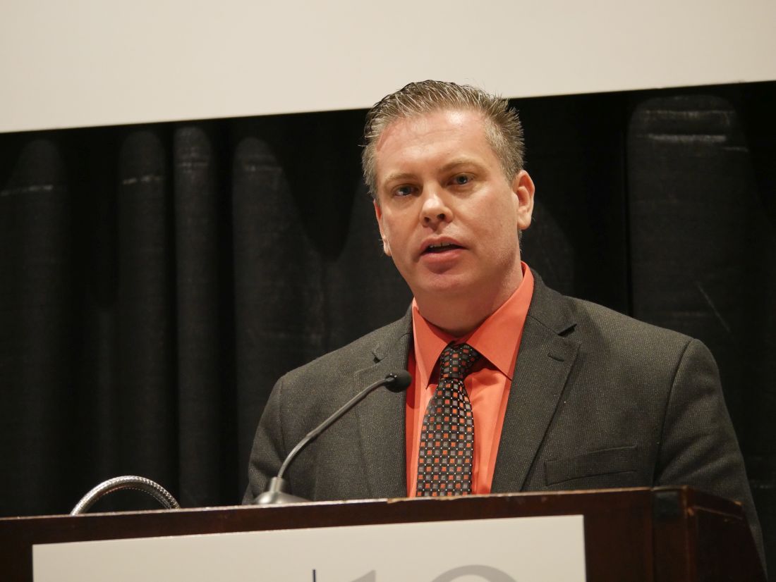
According to guidelines from the Centers for Disease Control and Prevention’s Advisory Committee on Immunization Practices, there is a theoretical concern that patients using a tumor necrosis factor inhibitor (TNFi) and other biologic therapies who receive a live-attenuated version of the varicella vaccine (Zostavax) could become infected with varicella from the vaccine. Patients with RA and psoriatic arthritis as well as other autoimmune and inflammatory conditions who are likely to receive TNFi therapy are also at risk for herpes zoster reactivation, Jeffrey Curtis, MD, professor of medicine in the division of clinical immunology and rheumatology of the University of Alabama at Birmingham, said in his presentation at the annual meeting of the American College of Rheumatology. There also exists a risk for patients receiving low-dose glucocorticoids.
“The challenge, of course, is there’s not a great definition and there certainly is not a well-standardized assay for how immunocompromised someone is, and so that led to the uncertainty in this patient population for this and other live-virus vaccines,” Dr. Curtis said.
Dr. Curtis and colleagues enrolled 627 participants from 33 centers into the VERVE trial. Participants were aged at least 50 years, were taking a TNFi, and had not previously received Zostavax.
Patients in both groups had a mean age of about 63 years and about two-thirds were women. The most common indications for TNFi use in the Zostavax group and the placebo group were RA (59.2% vs. 56.0%, respectively), psoriatic arthritis (24.3% vs. 23.9%), and ankylosing spondylitis (7.2% vs. 8.5%), while the anti-TNF agents used were adalimumab (38.1% vs. 27.4%), infliximab (28.4% vs. 34.2%), etanercept (19.0% vs. 23.5%), golimumab (10.0% vs. 8.1%), and certolizumab pegol (4.5% vs. 6.8%). In addition, some patients in the Zostavax and placebo groups were also taking concomitant therapies with TNFi, such as oral glucocorticoids (9.7% vs. 11.4%).
The researchers randomized participants to receive Zostavax or placebo (saline) and then followed them for 6 weeks, and looked for signs of wild-type or vaccine-strain varicella infection. If participants were suspected to have varicella, they were assessed clinically, underwent polymerase chain reaction testing, and rashes were photographed. At baseline and at 6 weeks, the researchers collected serum and peripheral blood mononuclear cells to determine patient immunity to varicella. After 6 months, participants were unmasked to the treatment arm of the study.
Dr. Curtis and colleagues found no confirmed varicella infection cases at 6 weeks. “To the extent that 0 cases out of 317 vaccinated people is reassuring, there were no cases, so that was exceedingly heartening as a result,” he said.
Out of 20 serious adverse events total in the groups, 15 events occurred before 6 months, including 8 suspected varicella cases in the Zostavax group and 7 in the placebo group. However, there were no positive cases of varicella – either wild type or vaccine type – after polymerase chain reaction tests. Overall, there were 268 adverse events in 195 participants, with 73 events (27.2%) consisting of injection-site reactions. The researchers also found no difference in the rate of disease flares, and found no differences in adverse reactions between groups, apart from a higher rate of injection-site reactions in the varicella group (19.4% vs. 4.2%).
With regard to immunogenicity, the humoral immune response was measured through IgG, which showed an immune response in the varicella group at 6 weeks (geometric mean fold ratio, 1.33; 95% confidence interval, 1.18-1.51), compared with the placebo group (GMFR, 1.02; 95% CI, 0.91-1.14); cell-mediated immune response was measured by interferon-gamma, which also showed an immune response in the live-vaccine group (GMFR, 1.49; 95% CI, 1.14-1.94), compared with participants who received placebo (GMFR, 1.14; 95% CI, 0.87-1.48). In preliminary 1-year data, IgG immune response was elevated in the varicella group (GMFR, 1.46; 95% CI, 1.08-1.99), but there was no elevated immune response for interferon-gamma (GMFR, 0.78; 95% CI, 0.49-1.25).
“I think the trial is encouraging not only for its result with the live zoster vaccine and TNF-treated patients, but also challenge the notion that, if you need to, a live-virus vaccine may in fact be able to be safely given to people with autoimmune and inflammatory diseases, even those treated with biologics like tumor necrosis factor inhibitors,” Dr. Curtis said.
As patients in VERVE consented to long-term follow-up in health plan claims and EHR data, it will be possible to follow these patients in the future to assess herpes zoster reactivation. Dr. Curtis also noted that a new trial involving the recombinant, adjuvanted zoster vaccine (Shingrix) is currently in development and should begin next year.
The VERVE trial was supported by the National Institute of Arthritis and Musculoskeletal and Skin Diseases. Dr. Curtis reported serving as a current member of the Center for Disease Control and Prevention’s Advisory Committee on Immunization Practices Herpes Zoster Work Group. He and some of the other authors reported financial relationships with many pharmaceutical companies.
SOURCE: Curtis J et al. Arthritis Rheumatol. 2019;71(suppl 10), Abstract 824.
ATLANTA – A group of patients using a tumor necrosis factor inhibitor safely received the live-attenuated varicella vaccine Zostavax without any cases of herpes zoster in the first 6 weeks after vaccination in the blinded, randomized, placebo-controlled Varicella Zoster Vaccine (VERVE) trial .
According to guidelines from the Centers for Disease Control and Prevention’s Advisory Committee on Immunization Practices, there is a theoretical concern that patients using a tumor necrosis factor inhibitor (TNFi) and other biologic therapies who receive a live-attenuated version of the varicella vaccine (Zostavax) could become infected with varicella from the vaccine. Patients with RA and psoriatic arthritis as well as other autoimmune and inflammatory conditions who are likely to receive TNFi therapy are also at risk for herpes zoster reactivation, Jeffrey Curtis, MD, professor of medicine in the division of clinical immunology and rheumatology of the University of Alabama at Birmingham, said in his presentation at the annual meeting of the American College of Rheumatology. There also exists a risk for patients receiving low-dose glucocorticoids.
“The challenge, of course, is there’s not a great definition and there certainly is not a well-standardized assay for how immunocompromised someone is, and so that led to the uncertainty in this patient population for this and other live-virus vaccines,” Dr. Curtis said.
Dr. Curtis and colleagues enrolled 627 participants from 33 centers into the VERVE trial. Participants were aged at least 50 years, were taking a TNFi, and had not previously received Zostavax.
Patients in both groups had a mean age of about 63 years and about two-thirds were women. The most common indications for TNFi use in the Zostavax group and the placebo group were RA (59.2% vs. 56.0%, respectively), psoriatic arthritis (24.3% vs. 23.9%), and ankylosing spondylitis (7.2% vs. 8.5%), while the anti-TNF agents used were adalimumab (38.1% vs. 27.4%), infliximab (28.4% vs. 34.2%), etanercept (19.0% vs. 23.5%), golimumab (10.0% vs. 8.1%), and certolizumab pegol (4.5% vs. 6.8%). In addition, some patients in the Zostavax and placebo groups were also taking concomitant therapies with TNFi, such as oral glucocorticoids (9.7% vs. 11.4%).
The researchers randomized participants to receive Zostavax or placebo (saline) and then followed them for 6 weeks, and looked for signs of wild-type or vaccine-strain varicella infection. If participants were suspected to have varicella, they were assessed clinically, underwent polymerase chain reaction testing, and rashes were photographed. At baseline and at 6 weeks, the researchers collected serum and peripheral blood mononuclear cells to determine patient immunity to varicella. After 6 months, participants were unmasked to the treatment arm of the study.
Dr. Curtis and colleagues found no confirmed varicella infection cases at 6 weeks. “To the extent that 0 cases out of 317 vaccinated people is reassuring, there were no cases, so that was exceedingly heartening as a result,” he said.
Out of 20 serious adverse events total in the groups, 15 events occurred before 6 months, including 8 suspected varicella cases in the Zostavax group and 7 in the placebo group. However, there were no positive cases of varicella – either wild type or vaccine type – after polymerase chain reaction tests. Overall, there were 268 adverse events in 195 participants, with 73 events (27.2%) consisting of injection-site reactions. The researchers also found no difference in the rate of disease flares, and found no differences in adverse reactions between groups, apart from a higher rate of injection-site reactions in the varicella group (19.4% vs. 4.2%).
With regard to immunogenicity, the humoral immune response was measured through IgG, which showed an immune response in the varicella group at 6 weeks (geometric mean fold ratio, 1.33; 95% confidence interval, 1.18-1.51), compared with the placebo group (GMFR, 1.02; 95% CI, 0.91-1.14); cell-mediated immune response was measured by interferon-gamma, which also showed an immune response in the live-vaccine group (GMFR, 1.49; 95% CI, 1.14-1.94), compared with participants who received placebo (GMFR, 1.14; 95% CI, 0.87-1.48). In preliminary 1-year data, IgG immune response was elevated in the varicella group (GMFR, 1.46; 95% CI, 1.08-1.99), but there was no elevated immune response for interferon-gamma (GMFR, 0.78; 95% CI, 0.49-1.25).
“I think the trial is encouraging not only for its result with the live zoster vaccine and TNF-treated patients, but also challenge the notion that, if you need to, a live-virus vaccine may in fact be able to be safely given to people with autoimmune and inflammatory diseases, even those treated with biologics like tumor necrosis factor inhibitors,” Dr. Curtis said.
As patients in VERVE consented to long-term follow-up in health plan claims and EHR data, it will be possible to follow these patients in the future to assess herpes zoster reactivation. Dr. Curtis also noted that a new trial involving the recombinant, adjuvanted zoster vaccine (Shingrix) is currently in development and should begin next year.
The VERVE trial was supported by the National Institute of Arthritis and Musculoskeletal and Skin Diseases. Dr. Curtis reported serving as a current member of the Center for Disease Control and Prevention’s Advisory Committee on Immunization Practices Herpes Zoster Work Group. He and some of the other authors reported financial relationships with many pharmaceutical companies.
SOURCE: Curtis J et al. Arthritis Rheumatol. 2019;71(suppl 10), Abstract 824.
REPORTING FROM ACR 2019