User login
Study finds differences for HCC in women
SAN DIEGO – Hepatocellular carcinoma is the third leading cause of cancer-related death in the United States and its incidence is increasing worldwide. While it affects men much more frequently than women, approximately 4 to 1, the differences in risk factors between men and women have never been studied.
At the annual Digestive Disease Week, Meaghan Phipps, MD, of New York–Presbyterian Hospital, described in a video interview how she and her colleagues set up a retrospective study of these differences in 5,327 patients at five large academic centers around the country. She and her colleagues found that women tended to present later, and with less severe disease, which was more likely to be treated with resection than transplantation. Women had better overall survival. Women were significantly more likely to present without cirrhosis and with nonalcoholic fatty liver disease than were men. Dr. Phipps noted that they did not characterize the women in their study by menopausal status, and suggested that this would be an important thing to look at in a future prospective study because it has long been thought that estrogen confers some protection against hepatocellular carcinoma.
REPORTING FROM DDW 2019

Adding drugs to gastric balloons increases weight loss
SAN DIEGO – In a multicenter study involving four academic medical centers, the addition of weight loss drugs to intragastric balloons resulted in better weight loss 12 months after balloon placement.
In a video interview at the annual Digestive Disease Week, study investigator Reem Sharaiha, MD, explained that one of the drawbacks of intragastric balloons is that, although they produce weight loss for the 6 or 12 months that they are in place, patients tend to regain that weight after they are removed. The study, involving 111 patients, was designed to determine whether the addition of weight loss drugs could mitigate this effect and improve weight loss, said Dr. Sharaiha of Weill Cornell Medical Center, New York.
Adding drugs such as metformin or weight loss drugs tailored to patients’ particular weight issues (cravings, anxiety, or fast gastric emptying) at the 3- or 6-month mark while the intragastric balloon was in place helped patients continue losing weight after balloon removal. At 12 months, the percentage of total body weight lost was significantly greater in the intragastric balloon group with concurrent pharmacotherapy (21.4% vs. 13.1%).
SOURCE: Shah SL et al. DDW 2019, Abstract 1105.
REPORTING FROM DDW 2019
Treatment-resistant GERD reported by more than half of patients
SAN DIEGO – Gastroesophageal reflux disease refractory to proton pump inhibitors may affect nearly half of those treated, according to the findings of a population-based sample of more than 70,000 Americans.
As part of the National Institutes of Health GI Patient Reported Outcomes Measurement Information System (NIH GI-PROMIS) questionnaire, respondents could download a free app called “My GI Health,” which led them through a series of questions about GI diseases. Sean Delshad, MD, MBA, of Cedars-Sinai Medical Center Los Angeles, and his colleagues examined data on symptom responses about GERD and heartburn.
Their somewhat surprising findings were that 44% of respondents had ever had GERD and that 70% of those respondents had symptoms in the past week. GERD seemed to be more common in women than in men, and in non-Hispanic whites more than other demographic groups. The rate of proton pump inhibitor–refractory GERD was reported at 54%.
Dr. Delshad discussed the implications of the study results for treatment and research in a video interview at the annual Digestive Disease Week.
REPORTING FROM DDW 2019

Physical activity linked to less cirrhosis-related mortality
SAN DIEGO – People who were more physically active, including those who did strength training, had significantly reduced risks of cirrhosis-related and liver cancer–related mortality, based on 26 years of prospective data from 113,000 participants in the Nurses Health Study and the Health Professionals Follow-Up Study.
Adults in the highest quintile of physical activity in the study had a 73% lower risk for cirrhosis-related death than did those in the lowest quintile, according to researchers at Massachusetts General Hospital and Harvard Medical School, Boston, who presented the study findings at Digestive Disease Week 2019.
One of the researchers, Tracey Simon, MD, MPH, of Massachusetts General Hospital, Boston, broke down the major take-home messages from the study in this video interview.
For example, vigorous activity was not necessary to improve hepatic health, she said. Walking for 4 hours per week made a big difference.
Dr. Simon has no relevant financial disclosures.
REPORTING FROM DDW 2019

Button batteries that pass to the stomach may warrant rapid endoscopic removal
SAN DIEGO – A button battery lodged in a child’s esophagus is an acknowledged emergency, but there is less evidence about retrieval of button batteries that have passed to the stomach. Observation alone has been recommended when an x-ray determines that the button battery has passed to the stomach within 2 hours of ingestion, when the battery is less than 20 mm, and the child is aged at least 5 years.
At the annual Digestive Disease Week, Racha Khalaf, MD, and Thomas Walker, MD, both of Children’s Hospital Colorado, Aurora, presented data that call this approach into question. Their retrospective cohort study of 4 years’ worth of records from four pediatric centers in the United States identified 68 cases in which a pediatric gastroenterologist had endoscopically removed the button battery. In 60% of those cases, the battery had already caused mucosal damage varying from minor to deep necrosis and perforation.
Further, the degree of injury was not correlated with symptoms, strengthening the recommendation for retrieving the button battery from the stomach.
In our exclusive video interview, Dr. Khalaf and Dr. Walker discussed the impact of their findings for guidelines for pediatric gastroenterologists and Poison Control Center advice to parents about ingestion of button batteries.
Their study was partly supported by a Cystic Fibrosis Foundational Grant Award and by National Institutes of Health Training Grants.
REPORTING FROM DDW 2019
Looking back at 10 years of the AGA Center for GI Innovation and Technology

SAN FRANCISCO – Jay Pasricha, MD, director of the Johns Hopkins Center for Neurogastroenterology, in Baltimore, reminisced about the early days of the AGA Center for GI Innovation and Technology in an interview at the AGA Tech Summit. “I was a founder,” he said, “along with Joel Brill and others.”
He goes back to when the idea was first pitched to the AGA Institute Council in 2009 as a technology center. He recalls that the first summit was held in Palo Alto, Calif., and that it was a “terrific success” because it filled a void. Dr. Pasricha said that the CGIT has fulfilled most if not all of its early expectations and – in some cases – went beyond expectations. Importantly, it transformed how people thought about GI as a specialty – GI was considered a risk-averse specialty previously. CGIT helped to develop relationships with many stakeholders, including the Food and Drug Administration. Dr. Pasricha predicts that CGIT will continue to do well because of its leadership and because AGA is completely invested in its success.
REPORTING FROM 2019 AGA TECH SUMMIT

Leveraging consumer technology in gastroenterology practice
SAN FRANCISCO – Dr. Michael Docktor, a pediatric gastroenterologist at Boston Hospital, described myriad digital tools that physicians – especially gastroenterologists – as well as patients are now using. Some tools may be implemented to track stool output or diet for diseases like irritable bowel syndrome or Crohn’s disease, he said in an interview at the AGA Tech Summit, sponsored by the AGA Center for GI Innovation and Technology.
There are patient-facing applications that provide data that can be used by both patients and their physicians to better understand the disease. These data can help in diagnosis and management and give the GI doctor a “window into the 99% of the time that they aren’t with the patient.” Other apps can build a timeline of the disease that can help the patient get a better understanding of their disease and learn to distinguish a flare from a bad day with poor food choices. Dr. Docktor described the AGA Tech Summit as a place to try out new ideas and work with like-minded doctors.
REPORTING FROM 2019 AGA TECH SUMMIT
Interventional endoscopic ultrasonography is hitting its stride

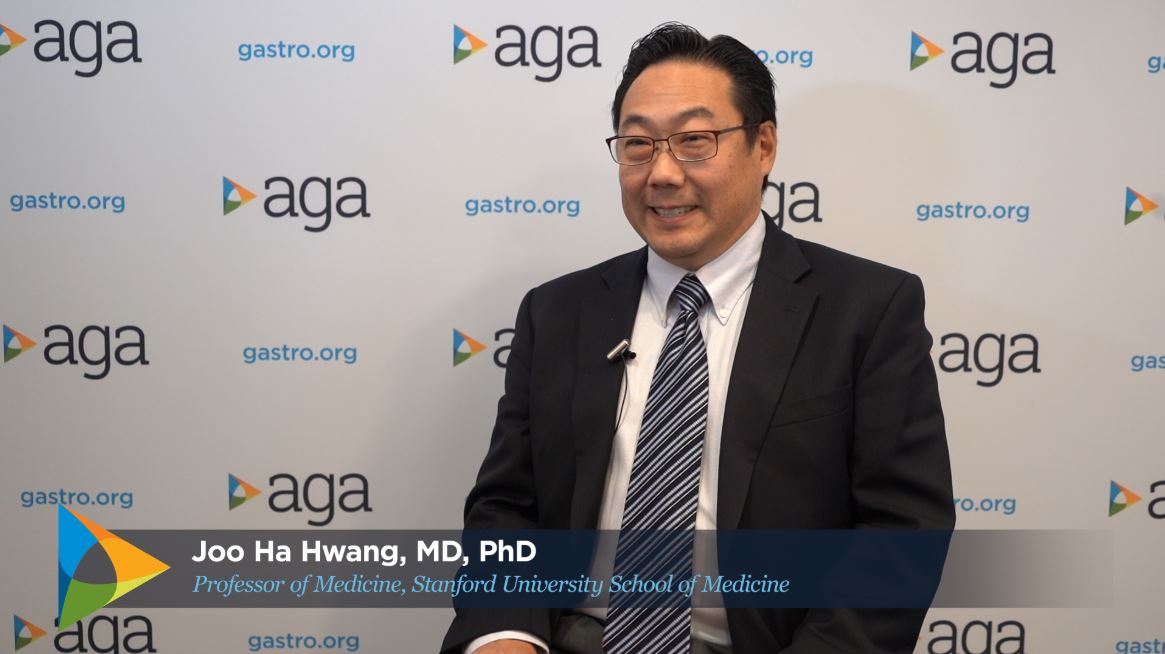
SAN FRANCISCO – Interventional endoscopic ultrasonography (EUS) recently has brought transluminal stent placement to problems like walled off pancreatic necroses, Joo Ha Hwang, MD, PhD, professor of medicine at Stanford (Calif.) University, said in an interview at the AGA Tech Summit, sponsored by the AGA Center for GI Innovation and Technology. He sees EUS taking over surgical territory beyond the usual biopsies in the gastrointestinal and biliary tracts and soon moving into tumor treatment in these areas. The field is “rapidly expanding,” he said, and the value of these minimally invasive procedures means that the field will soon need to train more interventional endoscopists.
SAN FRANCISCO – Interventional endoscopic ultrasonography (EUS) recently has brought transluminal stent placement to problems like walled off pancreatic necroses, Joo Ha Hwang, MD, PhD, professor of medicine at Stanford (Calif.) University, said in an interview at the AGA Tech Summit, sponsored by the AGA Center for GI Innovation and Technology. He sees EUS taking over surgical territory beyond the usual biopsies in the gastrointestinal and biliary tracts and soon moving into tumor treatment in these areas. The field is “rapidly expanding,” he said, and the value of these minimally invasive procedures means that the field will soon need to train more interventional endoscopists.
SAN FRANCISCO – Interventional endoscopic ultrasonography (EUS) recently has brought transluminal stent placement to problems like walled off pancreatic necroses, Joo Ha Hwang, MD, PhD, professor of medicine at Stanford (Calif.) University, said in an interview at the AGA Tech Summit, sponsored by the AGA Center for GI Innovation and Technology. He sees EUS taking over surgical territory beyond the usual biopsies in the gastrointestinal and biliary tracts and soon moving into tumor treatment in these areas. The field is “rapidly expanding,” he said, and the value of these minimally invasive procedures means that the field will soon need to train more interventional endoscopists.
REPORTING FROM 2019 AGA TECH SUMMIT
Artificial intelligence and machine learning in gastroenterology
SAN FRANCISCO – Artificial intelligence (AI) is using computer technology to solve particular kinds of medical problems, Sushovan Guha, MD, PhD, AGAF, chair, division of gastroenterology, University of Arizona, Phoenix, said in an interview at the AGA Tech Summit, sponsored by the AGA Center for GI Innovation and Technology.

Computers are good at doing many processes in a short period of time and executing repetitive tasks with no errors, whereas humans tend to introduce errors after many repetitions. Using algorithms by which physicians assess and diagnose colonic lesions, computer software can learn the criteria that diagnose adenomas and assist in the process of diagnosis, Dr. Guha said.
Computers are also ideal for managing and analyzing large amounts of data – this ability has so far been used to personalize cancer treatment – and is now being used to suggest the best treatment and predict remission in patients with inflammatory bowel disease. AI can use anatomical data combined with endoscopy to predict GI bleeding so that physicians can target therapy. Dr. Guha predicts that there will be an “explosion” of applications of AI in gastroenterology in the next 5-10 years.
SAN FRANCISCO – Artificial intelligence (AI) is using computer technology to solve particular kinds of medical problems, Sushovan Guha, MD, PhD, AGAF, chair, division of gastroenterology, University of Arizona, Phoenix, said in an interview at the AGA Tech Summit, sponsored by the AGA Center for GI Innovation and Technology.
Computers are good at doing many processes in a short period of time and executing repetitive tasks with no errors, whereas humans tend to introduce errors after many repetitions. Using algorithms by which physicians assess and diagnose colonic lesions, computer software can learn the criteria that diagnose adenomas and assist in the process of diagnosis, Dr. Guha said.
Computers are also ideal for managing and analyzing large amounts of data – this ability has so far been used to personalize cancer treatment – and is now being used to suggest the best treatment and predict remission in patients with inflammatory bowel disease. AI can use anatomical data combined with endoscopy to predict GI bleeding so that physicians can target therapy. Dr. Guha predicts that there will be an “explosion” of applications of AI in gastroenterology in the next 5-10 years.
REPORTING FROM 2019 AGA TECH SUMMIT

Female authorship trends in academic gastroenterology over 4 decades
WASHINGTON – Gastroenterology is still a majority male specialty, although women are entering the field at higher and higher rates. Female first authorship tripled from 1995 to 2010 (from 11% to 32%) and female senior authorship tripled from 2000 to 2010 (from 7% to 24%), but gains have not been equal in all areas and have not continued in all areas.
Eileen J. Benz, MD, of Beaumont Hospital, Dublin, described in a video interview at the annual Digestive Disease Week® a study she and her colleagues conducted to analyze published research in the journal Gastroenterology for the changing prevalence of female authorship over 4 decades.
The researchers reviewed all research published in the January and July issues of Gastroenterology during 1971-2010 (865 abstracts); animal trials were excluded. The sex of the first author and the last author (considered the senior author) of each paper was recorded, as was the type of study (basic science, clinical trials, or epidemiologic research). The increase in female senior authorship lagged behind the increase in first authorship, which likely reflects the promotion of female gastroenterologists over time into senior academic positions.
Also noted was that basic science and epidemiology research have the highest number of female authors overall, and these areas seem to continue to add female authors, whereas the number of female authors in clinical trials research seems to have stagnated since 1996. Dr. Benz hypothesizes that both bench science and epidemiology have research time built in, but that for a physician who may have other demands on her time, clinical trials research is an add-on for which there may not be protected time.
Reporting from DDW 2018