User login
Economic Forecast: Rough Road Ahead for Dermatologists
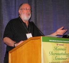
WAIKOLOA, HAWAII – Dr. Brett M. Coldiron is a sort of latter-day Paul Revere, travelling far and wide to spread the alarm to his fellow dermatologists – not of Redcoats a’coming, but of the need to prepare for looming economic hard times.
"I wasn’t invited to speak at your meeting, so I invited myself," he declared by way of introduction at the Hawaii Dermatology Seminar sponsored by Skin Disease Education Foundation (SDEF), as he launched into an analysis of dermatology’s near-term financial future.

"I’m here to present your 5-year economic plan – what you can expect. I think you can plan your next 5 years based on these predictions. It’s not pretty; and it’s not kind," cautioned Dr. Coldiron, whose expertise regarding health care policy and reform has been forged through long-term involvement representing dermatology on the American Medical Association’s Relative Value Scale Update, Health Care Finance, and Government Health Care Policy committees.
His core message to his colleagues boiled down to this: "Don’t build palaces. It’s time to hunker down."
Dermatology is a heavily procedurally oriented, small specialty – less then 1% of all physicians – which has experienced dramatic growth in procedure volume over the past couple decades. As such, it is a high-priority target for congressional cost-cutting efforts. In the first 4 months of next year, Dr. Coldiron said he expects reimbursement for codes pertaining to actinic keratosis treatment to be cut by 25%-50%, along with a roughly 20% reduction in payment for Mohs surgery. And that’s just the beginning.
Dermatology and other small specialties will bear the brunt of any cost-savings attempts by Congress. Dermatology has powerful enemies in Congress, the Centers for Medicare and Medicaid Services, and the American Medical Association, who view dermatologists as overpaid, wasteful abusers of the system, explained Dr. Coldiron, who is president of the American College of Mohs Surgery and a 2013 member of the American Academy of Dermatology’s board of directors.
Among his predictions for the next 5 years:
• Hospitals and pharmacies, if squeezed too hard, will simply close. Insurers will move into other lines of business coverage. Pharmaceutical companies will reduce their research and development budgets. Thus, reducing physician income will be one of the few politically acceptable health care cost-cutting avenues available.
• There will be more bundling of minor procedures into evaluation and management fees.
• The government will attempt to force all physicians to accept Medicaid. "They’ll probably try to tie it to your acceptance of Medicare. Or maybe they’ll say, ‘We paid for 4 years of postgraduate education; now you owe us 4 years of taking Medicaid,’ " he said.
• The use of physician assistants and nurse practitioners will grow in dermatology. This will result in increased utilization and more intense billing audits along with reimbursement cuts aimed at cancelling out the economic impact of greater utilization.
• Cosmetic procedures and reconstructive surgery will remain safe havens. "They may try to pass a cosmetic procedure tax, but I think the fact that you have another source of income is very important," noted Dr. Coldiron, a dermatologist at the University of Cincinnati.
He recommended that dermatologists temper their income projections for the coming half-decade: "Don’t promise big salaries to new associates, only a percentage of income collected."
Also, read the fine print before jumping on board one of the accountable care organizations that are springing up. "This is government-driven managed care with capitation. What they’re going to do is extract from the specialists and give back to primary care. It’s kind of a loser’s game," he said.
Beyond the next 5 years, however, the outlook for dermatology is bright, Dr. Coldiron stressed.
"Be strong. We are not greedy specialists; we are a frontline specialty fighting an epidemic of skin cancer. We are needed by our patients and by the health care system. The pendulum will eventually swing back our way," he concluded.
Dr. Coldiron reported having no relevant financial conflicts. SDEF and this news organization are owned by Elsevier.
WAIKOLOA, HAWAII – Dr. Brett M. Coldiron is a sort of latter-day Paul Revere, travelling far and wide to spread the alarm to his fellow dermatologists – not of Redcoats a’coming, but of the need to prepare for looming economic hard times.
"I wasn’t invited to speak at your meeting, so I invited myself," he declared by way of introduction at the Hawaii Dermatology Seminar sponsored by Skin Disease Education Foundation (SDEF), as he launched into an analysis of dermatology’s near-term financial future.
"I’m here to present your 5-year economic plan – what you can expect. I think you can plan your next 5 years based on these predictions. It’s not pretty; and it’s not kind," cautioned Dr. Coldiron, whose expertise regarding health care policy and reform has been forged through long-term involvement representing dermatology on the American Medical Association’s Relative Value Scale Update, Health Care Finance, and Government Health Care Policy committees.
His core message to his colleagues boiled down to this: "Don’t build palaces. It’s time to hunker down."
Dermatology is a heavily procedurally oriented, small specialty – less then 1% of all physicians – which has experienced dramatic growth in procedure volume over the past couple decades. As such, it is a high-priority target for congressional cost-cutting efforts. In the first 4 months of next year, Dr. Coldiron said he expects reimbursement for codes pertaining to actinic keratosis treatment to be cut by 25%-50%, along with a roughly 20% reduction in payment for Mohs surgery. And that’s just the beginning.
Dermatology and other small specialties will bear the brunt of any cost-savings attempts by Congress. Dermatology has powerful enemies in Congress, the Centers for Medicare and Medicaid Services, and the American Medical Association, who view dermatologists as overpaid, wasteful abusers of the system, explained Dr. Coldiron, who is president of the American College of Mohs Surgery and a 2013 member of the American Academy of Dermatology’s board of directors.
Among his predictions for the next 5 years:
• Hospitals and pharmacies, if squeezed too hard, will simply close. Insurers will move into other lines of business coverage. Pharmaceutical companies will reduce their research and development budgets. Thus, reducing physician income will be one of the few politically acceptable health care cost-cutting avenues available.
• There will be more bundling of minor procedures into evaluation and management fees.
• The government will attempt to force all physicians to accept Medicaid. "They’ll probably try to tie it to your acceptance of Medicare. Or maybe they’ll say, ‘We paid for 4 years of postgraduate education; now you owe us 4 years of taking Medicaid,’ " he said.
• The use of physician assistants and nurse practitioners will grow in dermatology. This will result in increased utilization and more intense billing audits along with reimbursement cuts aimed at cancelling out the economic impact of greater utilization.
• Cosmetic procedures and reconstructive surgery will remain safe havens. "They may try to pass a cosmetic procedure tax, but I think the fact that you have another source of income is very important," noted Dr. Coldiron, a dermatologist at the University of Cincinnati.
He recommended that dermatologists temper their income projections for the coming half-decade: "Don’t promise big salaries to new associates, only a percentage of income collected."
Also, read the fine print before jumping on board one of the accountable care organizations that are springing up. "This is government-driven managed care with capitation. What they’re going to do is extract from the specialists and give back to primary care. It’s kind of a loser’s game," he said.
Beyond the next 5 years, however, the outlook for dermatology is bright, Dr. Coldiron stressed.
"Be strong. We are not greedy specialists; we are a frontline specialty fighting an epidemic of skin cancer. We are needed by our patients and by the health care system. The pendulum will eventually swing back our way," he concluded.
Dr. Coldiron reported having no relevant financial conflicts. SDEF and this news organization are owned by Elsevier.
EXPERT ANALYSIS FROM THE HAWAII DERMATOLOGY SEMINAR SPONSORED BY SKIN DISEASE EDUCATION FOUNDATION
Term 'Hemangioma' Used Incorrectly in 71% of Cases
WAIKOLOA, HAWAII – The term "hemangioma" is used erroneously in 70% of cases of vascular anomalies, a study has shown.
"We’re not doing so well. That’s a high rate of misses," Dr. Moise L. Levy said in highlighting the findings.
The study, conducted by investigators at Harvard Medical School, Boston, involved an analysis of all 320 English language articles in PubMed that were published in 2009 with the word "hemangioma" in the title or abstract.

The investigators sought to learn whether the approved binary classification system for vascular anomalies introduced by the International Society for the Study of Vascular Anomalies (ISSVA) 15 years ago was being utilized by physicians and the consequences, in terms of treatment appropriateness, when it wasn’t.
Overall, the term "hemangioma" was used incorrectly in 71% of cases. Improper use of the term diagnostically was associated with a 21% rate of erroneous treatment. In contrast, patients whose lesions were described using ISSVA terminology had a 0% rate of improper treatment.
Physicians in some specialties did better than others. Radiologists used the term inappropriately in 84% of cases. Pediatricians, internists, general surgeons, ob.gyns., and pathologists who authored papers misused the term "hemangioma" in 60%-70% of cases. And, dermatologists, plastic surgeons, pediatric surgeons, and otolaryngologists used the term inappropriately in 32% of cases.
The investigators noted that the 71% incidence of incorrect nomenclature and 21% rate of incorrect treatment probably underestimate the true rates in clinical practice (Plast. Reconstr. Surg. 2011;127:347-51).
Dr. Levy said the study results indicate most physicians still use the antiquated method of classifying vascular anomalies learned during training. They’re using the term "hemangioma" to describe a wide variety of vascular birthmarks with differing etiologies and behavior patterns, ranging from strawberry hemangiomas to port wine stains, capillary hemangiomas, infantile hemangiomas, and lower-segment hemangiomas, he said.
The ISSVA system of classification divides vascular anomalies into two categories: tumors and malformations. Tumors show rapid growth in the neonatal period, followed in most cases by slow involution. These lesions are comprised of biologically active tissues that procreate in culture. Examples include infantile hemangioma, tufted angioma, and pyogenic granuloma.
Malformations are by definition present at birth. Their growth rate is commensurate with that of the child. Examples include capillary, venous, and lymphatic malformations, explained Dr. Levy, a pediatric dermatologist at Dell Children’s Medical Center of Central Texas in Austin.
In the Harvard study, misdiagnosis of malformations occurred significantly more often than for vascular tumors. The high misdiagnosis rates underscore a key point: Nonspecialists should consider referral of any patient with a complicated lesion, or one raising aesthetic concern, or when the diagnosis is unclear, Dr. Levy said at the Hawaii Dermatology Seminar sponsored by Skin Disease Education Foundation (SDEF).
"The bottom line is we need to encourage our primary care providers to get cases to us early rather than after a problem is discovered," he said.
Infantile hemangiomas are the most common type of vascular tumors in infancy. They vary tremendously in their threat level. The first weeks to months of life are a critical time in the growth of these lesions. Useful guidance as to when to refer children with infantile hemangiomas and when reassurance can be offered is provided by a multicenter prospective study published by pediatric dermatologists several years ago, Dr. Levy continued.
The investigators concluded that infantile hemangiomas posing the greatest risk for serious morbidity based upon morphology and/or anatomic site include large, segmented facial lesions because of the possibility of PHACES syndrome (posterior fossa malformations, hemangiomas, arterial anomalies, cardiac defects, eye abnormalities, and sternal clefting).
Other red flag infantile hemangiomas identified in the study as warranting referral included perioral, periocular, perineal, axillary, or retrobulbar lesions; segmental lesions on the central neck, or what in later life would be called the beard area; and segmental lesions overlying the lumbosacral spine (Pediatrics 2008;122:360-7).
Dr. Levy reported having no relevant financial disclosures. SDEF and this news organization are owned by Elsevier.
WAIKOLOA, HAWAII – The term "hemangioma" is used erroneously in 70% of cases of vascular anomalies, a study has shown.
"We’re not doing so well. That’s a high rate of misses," Dr. Moise L. Levy said in highlighting the findings.
The study, conducted by investigators at Harvard Medical School, Boston, involved an analysis of all 320 English language articles in PubMed that were published in 2009 with the word "hemangioma" in the title or abstract.
The investigators sought to learn whether the approved binary classification system for vascular anomalies introduced by the International Society for the Study of Vascular Anomalies (ISSVA) 15 years ago was being utilized by physicians and the consequences, in terms of treatment appropriateness, when it wasn’t.
Overall, the term "hemangioma" was used incorrectly in 71% of cases. Improper use of the term diagnostically was associated with a 21% rate of erroneous treatment. In contrast, patients whose lesions were described using ISSVA terminology had a 0% rate of improper treatment.
Physicians in some specialties did better than others. Radiologists used the term inappropriately in 84% of cases. Pediatricians, internists, general surgeons, ob.gyns., and pathologists who authored papers misused the term "hemangioma" in 60%-70% of cases. And, dermatologists, plastic surgeons, pediatric surgeons, and otolaryngologists used the term inappropriately in 32% of cases.
The investigators noted that the 71% incidence of incorrect nomenclature and 21% rate of incorrect treatment probably underestimate the true rates in clinical practice (Plast. Reconstr. Surg. 2011;127:347-51).
Dr. Levy said the study results indicate most physicians still use the antiquated method of classifying vascular anomalies learned during training. They’re using the term "hemangioma" to describe a wide variety of vascular birthmarks with differing etiologies and behavior patterns, ranging from strawberry hemangiomas to port wine stains, capillary hemangiomas, infantile hemangiomas, and lower-segment hemangiomas, he said.
The ISSVA system of classification divides vascular anomalies into two categories: tumors and malformations. Tumors show rapid growth in the neonatal period, followed in most cases by slow involution. These lesions are comprised of biologically active tissues that procreate in culture. Examples include infantile hemangioma, tufted angioma, and pyogenic granuloma.
Malformations are by definition present at birth. Their growth rate is commensurate with that of the child. Examples include capillary, venous, and lymphatic malformations, explained Dr. Levy, a pediatric dermatologist at Dell Children’s Medical Center of Central Texas in Austin.
In the Harvard study, misdiagnosis of malformations occurred significantly more often than for vascular tumors. The high misdiagnosis rates underscore a key point: Nonspecialists should consider referral of any patient with a complicated lesion, or one raising aesthetic concern, or when the diagnosis is unclear, Dr. Levy said at the Hawaii Dermatology Seminar sponsored by Skin Disease Education Foundation (SDEF).
"The bottom line is we need to encourage our primary care providers to get cases to us early rather than after a problem is discovered," he said.
Infantile hemangiomas are the most common type of vascular tumors in infancy. They vary tremendously in their threat level. The first weeks to months of life are a critical time in the growth of these lesions. Useful guidance as to when to refer children with infantile hemangiomas and when reassurance can be offered is provided by a multicenter prospective study published by pediatric dermatologists several years ago, Dr. Levy continued.
The investigators concluded that infantile hemangiomas posing the greatest risk for serious morbidity based upon morphology and/or anatomic site include large, segmented facial lesions because of the possibility of PHACES syndrome (posterior fossa malformations, hemangiomas, arterial anomalies, cardiac defects, eye abnormalities, and sternal clefting).
Other red flag infantile hemangiomas identified in the study as warranting referral included perioral, periocular, perineal, axillary, or retrobulbar lesions; segmental lesions on the central neck, or what in later life would be called the beard area; and segmental lesions overlying the lumbosacral spine (Pediatrics 2008;122:360-7).
Dr. Levy reported having no relevant financial disclosures. SDEF and this news organization are owned by Elsevier.
EXPERT ANALYSIS FROM THE SDEF HAWAII DERMATOLOGY SEMINAR
Oral Spironolactone Hailed for Acne in Women
WAIKOLOA, HAWAII – The androgen receptor–blocker spironolactone is highly effective and safe but underused for the treatment of acne in women, according to Dr. Julie C. Harper.
"The longer I’ve been in practice, the wider the age range where I use spironolactone as my drug of choice," Dr. Harper said at the Hawaii Dermatology Seminar sponsored by Skin Disease Education Foundation (SDEF).

Spironolactone is her first-line therapy for acne in postmenopausal women and in those who have had a hysterectomy. In such patients "it’s a really great drug to use by itself" because the women can’t get pregnant; the drug has a pregnancy category C rating, with an associated risk of feminization of a male fetus exposed to the drug late in the first trimester, explained Dr. Harper, a dermatologist at the University of Alabama, Birmingham.
"I have a tendency in my practice to start with an oral contraceptive in women of child-bearing potential and then maybe add spironolactone if I need to," she said.
However, Dr. Harper said that she will turn to spironolactone in women as young as 16 years of age with a contraindication to OCs. First, however, she carefully explains that the drug is safe as long as they don’t take it when pregnant. Spironolactone also cannot be used by nursing mothers because the drug’s major metabolite, canrenone, has been detected in breast milk.
Acne is a common problem in women. A survey of 1,013 adults in which Dr. Harper was a coinvestigator concluded that the prevalence of acne was significantly greater in women than men across all age groups. For example, the prevalence was 26% among 40- to 49-year-old women, compared with 12% in men of the same age, and 15% among women age 50-plus, compared to 7% in men (J. Am. Acad. Dermatol. 2008;58:56-9).
Acne is not only more common in women than men, it also differs in its characteristic presentation, which in women is typically lower-face acne in a U-shaped distribution.
Most women with adult acne have normal levels of circulating androgens. A leading hypothesis holds that their acne is the result of end-organ hyper-responsiveness to androgens. Androgen receptors are present in the sebaceous glands and the follicular wall, where comedones develop. This would explain why spironolactone is so effective clinically; the drug not only blocks the androgen receptor, it also reduces androgen synthesis in the adrenal gland and gonads, Dr. Harper noted.
She prescribes spironolactone in lower-range doses of 25-100 mg/day. At those doses she doesn’t bother to check serum potassium levels in relatively healthy young women so long as they’re not on other medications that may cause hyperkalemia, such as ACE inhibitors or chronic NSAIDs.
"You get good efficacy with fewer side effects at a maximum dose of 100 mg," according to the dermatologist.
Concomitant use of an OC lessens the menstrual irregularities and breast tenderness that can occur as side effects of spironolactone therapy. All combination OCs are probably effective in the treatment of acne. Four are approved for this indication: Ortho Tri-Cyclen, Estrostep, YAZ, and Beyaz.
Spironolactone should be thought of as long-term therapy. When spironolactone or an OC prescribed for adult acne is stopped, it’s highly likely that the acne will return within several months.
"When I plan to use spironolactone, I tell my patients they’re going to be on this for a long time if it works," Dr. Harper said.
The Food and Drug Administration has a long-standing Black Box Warning regarding spironolactone. Such warnings can send a chill down the spine of physicians and patients alike. However, there are widespread misconceptions about what this particular warning actually says. Here it is: "Spironolactone has been shown to be a tumorigen in chronic toxicity studies in rats. Spironolactone should be used only in those conditions described under Indications and Usage. Unnecessary use of this drug should be avoided."
The dosages used in those rat studies, by the way, were 25-100 times higher than those used in patients therapeutically.
"So we’re talking about rats, we’re talking about benign tumors, and we’re talking about super-duper high doses. That’s what’s in the Black Box Warning for spironolactone," she said.
Spironolactone is off-label therapy for acne, a point that caused another speaker at the seminar to bristle.
"If we used medications only for their approved indications, we couldn’t practice two-thirds of what we do," declared Dr. Theodore Rosen, professor of dermatology at Baylor College of Medicine, Houston.
"The FDA has said they’re not in the business of dictating how you practice medicine," he added. "You can use any approved drug in any fashion if you have some reasonable justification for it. If a plaintiff’s lawyer claims ‘This doctor used spironolactone off-label,’ she could line up 100 experts who’d say this is standard-of-care, and the standard of care determines what’s right and what’s wrong."
Dr. Harper is on the speakers bureaus for Allergan, Coria, Galderma, Intendis, Medicis, and Stiefel.
SDEF and this news organization are owned by Elsevier.
WAIKOLOA, HAWAII – The androgen receptor–blocker spironolactone is highly effective and safe but underused for the treatment of acne in women, according to Dr. Julie C. Harper.
"The longer I’ve been in practice, the wider the age range where I use spironolactone as my drug of choice," Dr. Harper said at the Hawaii Dermatology Seminar sponsored by Skin Disease Education Foundation (SDEF).
Spironolactone is her first-line therapy for acne in postmenopausal women and in those who have had a hysterectomy. In such patients "it’s a really great drug to use by itself" because the women can’t get pregnant; the drug has a pregnancy category C rating, with an associated risk of feminization of a male fetus exposed to the drug late in the first trimester, explained Dr. Harper, a dermatologist at the University of Alabama, Birmingham.
"I have a tendency in my practice to start with an oral contraceptive in women of child-bearing potential and then maybe add spironolactone if I need to," she said.
However, Dr. Harper said that she will turn to spironolactone in women as young as 16 years of age with a contraindication to OCs. First, however, she carefully explains that the drug is safe as long as they don’t take it when pregnant. Spironolactone also cannot be used by nursing mothers because the drug’s major metabolite, canrenone, has been detected in breast milk.
Acne is a common problem in women. A survey of 1,013 adults in which Dr. Harper was a coinvestigator concluded that the prevalence of acne was significantly greater in women than men across all age groups. For example, the prevalence was 26% among 40- to 49-year-old women, compared with 12% in men of the same age, and 15% among women age 50-plus, compared to 7% in men (J. Am. Acad. Dermatol. 2008;58:56-9).
Acne is not only more common in women than men, it also differs in its characteristic presentation, which in women is typically lower-face acne in a U-shaped distribution.
Most women with adult acne have normal levels of circulating androgens. A leading hypothesis holds that their acne is the result of end-organ hyper-responsiveness to androgens. Androgen receptors are present in the sebaceous glands and the follicular wall, where comedones develop. This would explain why spironolactone is so effective clinically; the drug not only blocks the androgen receptor, it also reduces androgen synthesis in the adrenal gland and gonads, Dr. Harper noted.
She prescribes spironolactone in lower-range doses of 25-100 mg/day. At those doses she doesn’t bother to check serum potassium levels in relatively healthy young women so long as they’re not on other medications that may cause hyperkalemia, such as ACE inhibitors or chronic NSAIDs.
"You get good efficacy with fewer side effects at a maximum dose of 100 mg," according to the dermatologist.
Concomitant use of an OC lessens the menstrual irregularities and breast tenderness that can occur as side effects of spironolactone therapy. All combination OCs are probably effective in the treatment of acne. Four are approved for this indication: Ortho Tri-Cyclen, Estrostep, YAZ, and Beyaz.
Spironolactone should be thought of as long-term therapy. When spironolactone or an OC prescribed for adult acne is stopped, it’s highly likely that the acne will return within several months.
"When I plan to use spironolactone, I tell my patients they’re going to be on this for a long time if it works," Dr. Harper said.
The Food and Drug Administration has a long-standing Black Box Warning regarding spironolactone. Such warnings can send a chill down the spine of physicians and patients alike. However, there are widespread misconceptions about what this particular warning actually says. Here it is: "Spironolactone has been shown to be a tumorigen in chronic toxicity studies in rats. Spironolactone should be used only in those conditions described under Indications and Usage. Unnecessary use of this drug should be avoided."
The dosages used in those rat studies, by the way, were 25-100 times higher than those used in patients therapeutically.
"So we’re talking about rats, we’re talking about benign tumors, and we’re talking about super-duper high doses. That’s what’s in the Black Box Warning for spironolactone," she said.
Spironolactone is off-label therapy for acne, a point that caused another speaker at the seminar to bristle.
"If we used medications only for their approved indications, we couldn’t practice two-thirds of what we do," declared Dr. Theodore Rosen, professor of dermatology at Baylor College of Medicine, Houston.
"The FDA has said they’re not in the business of dictating how you practice medicine," he added. "You can use any approved drug in any fashion if you have some reasonable justification for it. If a plaintiff’s lawyer claims ‘This doctor used spironolactone off-label,’ she could line up 100 experts who’d say this is standard-of-care, and the standard of care determines what’s right and what’s wrong."
Dr. Harper is on the speakers bureaus for Allergan, Coria, Galderma, Intendis, Medicis, and Stiefel.
SDEF and this news organization are owned by Elsevier.
EXPERT ANALYSIS FROM THE SDEF HAWAII DERMATOLOGY SEMINAR
Extramammary Paget's Needs More Than Mohs
WAIKOLOA, HAWAII – Extramammary Paget’s disease poses a particular challenge because of its multifocal/multicentric nature.
"What you see with extramammary Paget’s is not necessarily what you get," Dr. Theodore Rosen cautioned at the Hawaii Dermatology Seminar sponsored by Skin Disease Education Foundation (SDEF).
This is an uncommon neoplasia that’s typically described as an erythematous, erosive, itchy patch or plaque having a strawberries-and-cream appearance. Yet there may be other areas of subclinical involvement at a distance from the obvious lesion.
"That’s why Mohs surgery for this condition may be difficult without something being done in advance," said Dr. Rosen, professor of dermatology at Baylor College of Medicine, Houston.
He suggested applying topical 5% imiquimod or 5-fluorouracil to identify all of the active foci by lighting up the affected areas to allow more precise surgery or ablative therapy.
Dr. Rosen does not, however, recommend using imiquimod as primary therapy. He noted that a review of 27 published cases of 5% imiquimod for the treatment of extramammary Paget’s disease reported a 22% failure rate (Arch. Dermatol. 2011;147:704-8).
Mohs surgery shows promise for treatment of extramammary Paget’s disease, with lower recurrence rates than reported for wide surgical excision. However, experience to date with Mohs surgery for extensive disease is limited. For this reason, most authorities still consider wide surgical excision the gold standard therapy for extramammary Paget’s disease, despite published recurrence rates of 42%-54% even with clear surgical margins. Use of preoperative topical 5-fluorouracil or 5% imiquimod should substantially reduce those high recurrence rates, the dermatologist said.
Extramammary Paget’s disease is typically slow-growing for a decade or more before invading the dermis, at which point it quickly becomes widely metastatic.
"Once extramammary Paget’s disease has broken through the dermal/epidermal junction, it becomes a very nasty, bad-acting disease," Dr. Rosen said.
It’s well recognized that extramammary Paget’s is associated with an increased risk of underlying internal malignancy of the lower gastrointestinal or genitourinary tract. This increased risk is typically described as being in the 10%-20% range. But that figure may be too low. A recent report from investigators at Houston’s M.D. Anderson Cancer Center involving 20 consecutive patients with extramammary Paget’s on the penis or scrotum indicated that 8 of them – fully 40% – had an associated underlying internal adenocarcinoma (J. Urol. 2011;186:97-102).
The risk of underlying internal malignancy is known to be considerably greater in white patients than in Asians. It’s very uncommon for black patients with extramammary Paget’s to have an associated internal malignancy.
Because the full workup for an associated occult internal adenocarcinoma is elaborate and costly, a means of determining which patients are at greater or lesser risk would be welcome in order to guide the extent of testing. Cytokeratin staining may be the solution. Spanish dermatologists have reported that cutaneous extramammary Paget’s disease is characteristically positive for cytokeratin 7, negative for cytokeratin 20, and positive for cystic disease fluid protein 15.
In contrast, endodermal extramammary Paget’s, which is more strongly associated with internal malignancy, is positive for cytokeratin 7 and 20 and negative for cystic disease fluid protein 15, according to the investigators (Clin. Exp. Dermatol. 2008;33:595-8). A cautionary note: Dr. Rosen said that to his knowledge these findings haven’t yet been confirmed by other groups.
He reported having no financial conflicts.
SDEF and this news organization are owned by Elsevier.
EXPERT ANALYSIS FROM THE SDEF HAWAII DERMATOLOGY SEMINAR
First-Ever Acne Treatment Guidelines for Children Revealed
WAIKOLOA, HAWAII – New acne management recommendations from the American Acne and Rosacea Society are the first guidelines to specifically address pediatric acne.
"Acne is a common problem, and the presentations and differential diagnosis differ among the various ages of childhood and adolescence. We had a strong desire to increase recognition and improve management of pediatric and adolescent acne across the spectrum of primary and specialty care," explained Dr. Lawrence F. Eichenfield, cochair of the guideline-writing panel comprised of general pediatricians, pediatric dermatologists, and acne experts.
The comprehensive guidelines provide a simple and efficient classification scheme in which acne is categorized as comedonal, inflammatory/mixed, or nodular, and graded as globally mild, moderate, or severe. The guidelines offer detailed algorithms for the treatment of each category.
The treatment algorithms are flexible, with multiple options available based upon considerations including financial cost and treatment history.
Regimen complexity is another major consideration. Treatment adherence in pediatric and adolescent acne patients is notoriously a much bigger problem than in adults. Simple, once-daily combination products addressing multiple acne pathogenic mechanisms are advantageous.
Adolescent Acne
The treatment algorithm for the adolescent with mild comedonal or inflammatory/mixed lesions begins with two broad options for initial therapy. Both are topical regimens. The first consists of monotherapy with benzoyl peroxide or a topical retinoid, which can be an inexpensive option. The second is topical fixed-dose combination therapy, which can cost far more but achieves faster clearance, Dr. Eichenfield said at the seminar sponsored by Skin Disease Education Foundation (SDEF). Recommended combinations include benzoyl peroxide with a topical antibiotic or retinoid.
Alternatively, the panel noted that topical dapsone can be used either as initial monotherapy or in place of a topical antibiotic. The panel was in agreement that a topical antibiotic should only be prescribed in conjunction with benzoyl peroxide in order to help prevent the emergence of bacterial resistance, he said.
Doxycycline and other oral antibiotics are to be reserved for treatment of moderate to severe acne, and their use should be limited to 3-6 months, according to the guidelines.
"An oral antibiotic alone is substandard care now. It needs to be accompanied by a topical retinoid, a retinoid/benzoyl peroxide, or retinoid/topical antibiotic combination to minimize resistance," Dr. Eichenfield said.
The guidelines note that it is appropriate for primary care physicians to immediately refer an adolescent who presents with severe acne to a dermatologist. Many such patients will be best-treated with oral isotretinoin, he noted.
Preadolescent Acne
Preadolescent acne arising in children aged 7-12 is common and considered normal. It typically begins with comedones over the forehead and midface, with truncal lesions being far less common.
Treatment follows the same algorithms as adolescent acne, with the caveat that most preadolescent therapy is off-label, since, until quite recently, nearly all treatment studies were restricted to patients aged 12 years and older. Therefore, in formulating a treatment strategy for preadolescents, the panel had to shift gears and switch from the evidence-based approach emphasized elsewhere in the guidelines to expert consensus, said Dr. Eichenfield, professor of clinical pediatrics and dermatology at the University of California, San Diego.
Infantile Acne
Infantile acne generally doesn’t show up until after the first several months of life. It is comedonal, although papules, pustules, nodules, and cysts may also be present. Infantile acne can do significant lasting damage, and treatment is warranted.
Neonatal Acne
Acne developing within the first 6 weeks after birth is classified as neonatal acne. However, the erythematous papules and pustules located on the face, neck, scalp, and torso are not true acne. The skin lesions, also known as neonatal cephalic pustulosis, are associated with skin colonization by Malassezia globosa and M. sympodialis. It is a self-limited condition, although it may clear faster if treated using topical ketoconazole cream or another anti-yeast medication.
Among the other issues addressed in the report are diet and acne, the appropriate use of the various classes of medications, when and how to use oral contraceptive pills for acne, the important distinction between neonatal and infantile acne, how to prescribe the big gun – isotretinoin – in young patients, and when to refer a child with acne for a endocrinology workup, said Dr. Eichenfield.
He explained that acne arising in mid-childhood – age 1-7 years – is a red flag for an increased risk of an endocrinologic disorder. Referral to a pediatric endocrinologist is warranted if a child displays any abnormalities in height and growth, blood pressure, or displays signs of early sexual maturation. Dr. Eichenfield picks up the phone personally, he said, to talk to the pediatric endocrinologist, and to make sure the child won’t wait long for an endocrinologic evaluation.
Publication of the guidelines is pending, he noted. In the meantime, physicians can obtain an introduction to the guidelines, including full details of the treatment algorithms, while earning 1 hour of CME credit by viewing a 56-minute video featuring Dr. Eichenfield and other guideline panelists at www.acneandrosacea.org.
The acne guidelines project was supported by the American Acne and Rosacea Society.
Dr. Eichenfield reported receiving research support or serving as a consultant to Galderma, Johnson & Johnson, Medicis, Stiefel, Valeant, and Ortho-McNeil (Jansen Pharmaceuticals).
SDEF and this news organization are owned by Elsevier.
EXPERT ANALYSIS FROM THE SDEF HAWAII DERMATOLOGY SEMINAR
Numbing Agents Cause More Pain in PDT
WAIKOLOA, HAWAII – Using EMLA or lidocaine cream in conjunction with topical 5-aminolevulinic acid photodynamic therapy may boost uptake of the photosensitizer, but the clinical outcomes can be unpredictable, according to Dr. E. Victor Ross.
"Our worst scenarios have been when we’ve used EMLA on the skin. We’ve stopped using EMLA or lidocaine cream on the skin in ALA-PDT because of the very pronounced, enhanced photodynamic effects. You get great long-term results, but such a bad short-term result that lots of people didn’t want to go through it," said Dr. Ross of Scripps Clinic Laser and Cosmetic Dermatology Center in Carmel Valley, Calif.

Using a topical numbing agent seemed to be an attractive option because patients often complain that photodynamic therapy (PDT) is painful. Plus, there seemed to be an added benefit: The anesthetic cream increased aminolevulanic acid (ALA) uptake by the skin, such that ALA incubation times before application of the light source could be greatly compressed. But clinical outcomes were too unpredictable, he said at the seminar sponsored by Skin Disease Education Foundation (SDEF). After several patients had unintended florid full photopeels involving 1½ weeks of downtime, it was time to abandon the practice.
When ALA-PDT first appeared about 10 years ago, the indication was for the treatment of actinic keratoses (AKs). Today this approved indication remains the No. 1 reason Dr. Ross utilizes the therapy, he said. But PDT’s role has expanded off label to include cosmetic procedures and the treatment of warts, nonmelanoma skin cancer, nevus sebaceous, as well as acne, which he considers "one of the great opportunities for PDT." In addition, in Asia, dermatologists are now refining the use of PDT with hematoporphyrin derivatives for the treatment of port wine stains and other vascular lesions.
Many U.S. dermatologists have incorporated PDT into their practices, with ALA being more widely used as a photosensitizer than methyl aminolevulinate (MAL). Yet PDT is a therapy that hasn’t been fully optimized; it is still fraught with side effects and suboptimal results, he said. Moreover, some fundamental issues regarding PDT remain unanswered: For example, the optimal duration of photosensitizer incubation time for various indications is still controversial.
Dr. Ross said he opts for what he considers a middle-of-the-road approach, with ALA application times of about 90-120 minutes, which he views as having an optimal balance between side effects and effectiveness. Even so, he noted that among the 8-10 patients per week he treats with ALA-PDT on average, 1 or 2 experience mild side effects.
"I don’t think we’re quite ‘there’ yet with PDT, although we’re getting closer. There are still so many tricks involved in making it work without side effects. We’ve still got some work to do," he said.
In addition to advising his colleagues to stay away from topical anesthetic creams, he offered additional tips for the use of ALA-PDT. Among them:
• Sending acne into long-term remission via PDT remains the Holy Grail, he said. The objective is to enhance the fluorescence of protoporphyrin 9 at the sebaceous gland while sparing the epidermis.
Some investigators are using low-intensity blue light at the skin surface while the photosensitizer is incubating in order to bleach it out of the epidermis, or, alternatively, warming and cooling the skin.
The best results Dr. Ross said he has seen have come through an arduous regimen involving three 3-hour-long ALA applications scheduled a month apart. There’s a delayed effect, with significant improvement coming at 3-6 months.
"It’s a tough, tough therapy to get through. There are lots of pustules and papules, and the acne invariably gets worse before it gets better. This doesn’t play into the hands of the typical teenager, who wants to get better right away," Dr. Ross said.
• A creamy solution of ALA will create more protoporphyrin 9 than an aqueous solution will.
• A red light source should be considered for deeper structures, such as basal cell carcinomas or sebaceous glands, and blue light for treating more superficial skin lesions. Although continuous blue light is 40 times more potent per photon than red light in exciting protoporphyrin 9, it doesn’t penetrate as deeply.
• Performing low-density fractional CO2 ablative laser therapy prior to application of the photosensitizing agent is "an exciting advance" in PDT, he said.
The innovation was developed by an international team led by investigators at the Wellman Center for Photomedicine at Massachusetts General Hospital, Boston, who published a split-face randomized study involving 15 patients with a total of 212 facial AKs. At the 3-month follow-up, the complete response rate of grade II-III lesions treated with MAL-PDT preceded by fractional ablative laser therapy was 87.5%, vs. 58.8% with conventional MAL-PDT. The complete response rate of grade I AKs was 100% with fractional laser/MAL-PDT, vs. 79% for MAL-PDT alone.
The fractional laser–pretreated areas also displayed significantly greater improvement in photoaging and fewer new AKs at follow-up: 3, compared with 11. But these superior outcomes came at a price: higher pain scores during illumination and significantly worse erythema and crusting post treatment (Br. J. Dermatol. 2012 Feb. 20 [doi:10.1111/j.1365-2133.2012.10893.x]).
Fractional CO2 laser pretreatment enhances conversion of the photosensitizing agent to protoporphyrin 9, Dr. Ross explained. He compared the tiny holes in the skin created by the fractional laser to the process of aerating a lawn. The holes create conduits for the photosensitizer to bypass the stratum corneum, which is the major obstacle to uptake of ALA or MAL. Once the photosensitizer skips past the stratum corneum, it quickly spreads laterally throughout the epidermis.
However, Dr. Ross offered a note of caution regarding this novel approach. He said that he performed the therapy recently and found that 30 minutes of ALA incubation was too much.
"If you do these procedures, I would say go very light and just leave the ALA for less than 30 minutes to start, because the response you’re going to get when using a fractional laser beforehand is profound. It’s a huge difference," he said.
Dr. Ross reported that he serves as a consultant to and receives research support from Palomar. He also disclosed receiving research support from Candela, Cutera, Lumenis, Sciton, and Ulthera.
SDEF and this news organization are owned by Elsevier.
WAIKOLOA, HAWAII – Using EMLA or lidocaine cream in conjunction with topical 5-aminolevulinic acid photodynamic therapy may boost uptake of the photosensitizer, but the clinical outcomes can be unpredictable, according to Dr. E. Victor Ross.
"Our worst scenarios have been when we’ve used EMLA on the skin. We’ve stopped using EMLA or lidocaine cream on the skin in ALA-PDT because of the very pronounced, enhanced photodynamic effects. You get great long-term results, but such a bad short-term result that lots of people didn’t want to go through it," said Dr. Ross of Scripps Clinic Laser and Cosmetic Dermatology Center in Carmel Valley, Calif.
Using a topical numbing agent seemed to be an attractive option because patients often complain that photodynamic therapy (PDT) is painful. Plus, there seemed to be an added benefit: The anesthetic cream increased aminolevulanic acid (ALA) uptake by the skin, such that ALA incubation times before application of the light source could be greatly compressed. But clinical outcomes were too unpredictable, he said at the seminar sponsored by Skin Disease Education Foundation (SDEF). After several patients had unintended florid full photopeels involving 1½ weeks of downtime, it was time to abandon the practice.
When ALA-PDT first appeared about 10 years ago, the indication was for the treatment of actinic keratoses (AKs). Today this approved indication remains the No. 1 reason Dr. Ross utilizes the therapy, he said. But PDT’s role has expanded off label to include cosmetic procedures and the treatment of warts, nonmelanoma skin cancer, nevus sebaceous, as well as acne, which he considers "one of the great opportunities for PDT." In addition, in Asia, dermatologists are now refining the use of PDT with hematoporphyrin derivatives for the treatment of port wine stains and other vascular lesions.
Many U.S. dermatologists have incorporated PDT into their practices, with ALA being more widely used as a photosensitizer than methyl aminolevulinate (MAL). Yet PDT is a therapy that hasn’t been fully optimized; it is still fraught with side effects and suboptimal results, he said. Moreover, some fundamental issues regarding PDT remain unanswered: For example, the optimal duration of photosensitizer incubation time for various indications is still controversial.
Dr. Ross said he opts for what he considers a middle-of-the-road approach, with ALA application times of about 90-120 minutes, which he views as having an optimal balance between side effects and effectiveness. Even so, he noted that among the 8-10 patients per week he treats with ALA-PDT on average, 1 or 2 experience mild side effects.
"I don’t think we’re quite ‘there’ yet with PDT, although we’re getting closer. There are still so many tricks involved in making it work without side effects. We’ve still got some work to do," he said.
In addition to advising his colleagues to stay away from topical anesthetic creams, he offered additional tips for the use of ALA-PDT. Among them:
• Sending acne into long-term remission via PDT remains the Holy Grail, he said. The objective is to enhance the fluorescence of protoporphyrin 9 at the sebaceous gland while sparing the epidermis.
Some investigators are using low-intensity blue light at the skin surface while the photosensitizer is incubating in order to bleach it out of the epidermis, or, alternatively, warming and cooling the skin.
The best results Dr. Ross said he has seen have come through an arduous regimen involving three 3-hour-long ALA applications scheduled a month apart. There’s a delayed effect, with significant improvement coming at 3-6 months.
"It’s a tough, tough therapy to get through. There are lots of pustules and papules, and the acne invariably gets worse before it gets better. This doesn’t play into the hands of the typical teenager, who wants to get better right away," Dr. Ross said.
• A creamy solution of ALA will create more protoporphyrin 9 than an aqueous solution will.
• A red light source should be considered for deeper structures, such as basal cell carcinomas or sebaceous glands, and blue light for treating more superficial skin lesions. Although continuous blue light is 40 times more potent per photon than red light in exciting protoporphyrin 9, it doesn’t penetrate as deeply.
• Performing low-density fractional CO2 ablative laser therapy prior to application of the photosensitizing agent is "an exciting advance" in PDT, he said.
The innovation was developed by an international team led by investigators at the Wellman Center for Photomedicine at Massachusetts General Hospital, Boston, who published a split-face randomized study involving 15 patients with a total of 212 facial AKs. At the 3-month follow-up, the complete response rate of grade II-III lesions treated with MAL-PDT preceded by fractional ablative laser therapy was 87.5%, vs. 58.8% with conventional MAL-PDT. The complete response rate of grade I AKs was 100% with fractional laser/MAL-PDT, vs. 79% for MAL-PDT alone.
The fractional laser–pretreated areas also displayed significantly greater improvement in photoaging and fewer new AKs at follow-up: 3, compared with 11. But these superior outcomes came at a price: higher pain scores during illumination and significantly worse erythema and crusting post treatment (Br. J. Dermatol. 2012 Feb. 20 [doi:10.1111/j.1365-2133.2012.10893.x]).
Fractional CO2 laser pretreatment enhances conversion of the photosensitizing agent to protoporphyrin 9, Dr. Ross explained. He compared the tiny holes in the skin created by the fractional laser to the process of aerating a lawn. The holes create conduits for the photosensitizer to bypass the stratum corneum, which is the major obstacle to uptake of ALA or MAL. Once the photosensitizer skips past the stratum corneum, it quickly spreads laterally throughout the epidermis.
However, Dr. Ross offered a note of caution regarding this novel approach. He said that he performed the therapy recently and found that 30 minutes of ALA incubation was too much.
"If you do these procedures, I would say go very light and just leave the ALA for less than 30 minutes to start, because the response you’re going to get when using a fractional laser beforehand is profound. It’s a huge difference," he said.
Dr. Ross reported that he serves as a consultant to and receives research support from Palomar. He also disclosed receiving research support from Candela, Cutera, Lumenis, Sciton, and Ulthera.
SDEF and this news organization are owned by Elsevier.
EXPERT ANALYSIS FROM THE SDEF HAWAII DERMATOLOGY SEMINAR
Mineral Makeup Is a Dermatologist's 'Fire Extinguisher'
WAIKOLOA, HAWAII – Mineral makeup has been game changing for everything from covering acne blemishes to masking postprocedure discoloration, noted Dr. Howard K. Steinman.
"Mineral makeup has done to medical makeup what fillers and Botox have done to the field of facial cosmetic surgery. You now have little excuse for not using makeup in your office," said Steinman, director of dermatologic and skin cancer surgery at Scott and White Medical Center, Temple, Texas.
Mineral makeup is easy for patients to learn how to apply effectively; anyone on the office staff can be trained to teach patients how, he said. The products take up a trivial amount of office space, cost very little, pose negligible risk, and generate considerable patient satisfaction.
"Mineral makeup achieves really spectacular results," said Dr. Steinman at the annual Hawaii Dermatology Seminar sponsored by Skin Disease Education Foundation (SDEF).
Patients go to a dermatologist because they want cure or control of a medical condition, symptomatic relief, and an improved appearance. But while successful treatment of melasma, for example, may take months, the patient’s appearance can be improved immediately in the interim – on day 1 of therapy –through the use of mineral makeup.
Mineral makeup provides complete coverage for macular medical conditions including acne erythema, rosacea, vitiligo, sarcoidosis, port wine stains, and spider vessels. These products are also the answer to postprocedure discoloration following chemical peels, laser resurfacing, and fillers.
"It’s your fire extinguisher for procedural misadventures. The nice thing about having mineral makeups in your office is you don’t have to worry about these things as much because the problem is going to leave the office fixed," Dr. Steinman explained.
Green-tinted concealer is a popular but ineffective solution for red discoloration of skin. The results tend to be muddy looking and unconvincing. Mineral makeup, he said, is far superior.
Dr. Steinman singled out four companies as the major players in the mineral makeup market: Youngblood, Bare Minerals, Color Science, and Jane Iredale. All have been in business a long time, offer products of excellent quality, and provide reasonably good service to physicians, he said.
He prefers Youngblood in his own office because his wife, a paramedical camouflage therapist trained to cover burn scars and disfiguring surgical wounds, prefers the quality.
"My suggestion is to have your esthetician, or whoever will be doing your makeup, try them all and decide which one they prefer," said Dr. Steinman.
He also recommends Dermacolor (Kryolan), a specialized camouflage foundation system made in Germany that is formulated for postoperative skin and contains an opaque sunscreen. Unlike mineral makeup, Dermacolor is waterproof even with swimming. The company offers more than 70 premixed colors, although a selection of about 20 is sufficient to cover virtually any clinical situation, he said.
Dr. Steinman suggested that the makeup artist match the patient’s skin tones before surgery so Dermacolor is ready to be applied at the first postoperative visit.
Dr. Steinman said a relatively new comprehensive line of makeup products he recommends is Cover FX. The product texture and coverage are outstanding, it’s easy for patients to apply, and the company caters to physicians, he said.
Camouflage makeup does have limitations. It won’t effectively cover three-dimensional lesions, such as acne scars or keloids. And it can’t do much for burn scars with absent hair and skin pores, he noted.
That being said, Dr. Steinman considers makeup in the office an indispensable part of his practice. "Makeup is just essential, I think, if you’re going to do cosmetic surgery or you want to treat patients with medical macular problems," he said.
Dr. Steinman reported having no financial relationships with any of the companies he discussed.
SDEF and this news organization are owned by Elsevier.
*This post was updated on 3/7/12.
EXPERT ANALYSIS FROM THE SDEF HAWAII DERMATOLOGY SEMINAR
Off-Label Use of Aprepitant Quells Refractory Pruritus
WAIKOLOA, HAWAII – Oral aprepitant has shown considerable promise as a novel therapy in patients with refractory pruritus, including those with severe itching induced by biologic therapies for cancer.
"I think it’s a tool we should consider using in our patients with pruritus who do not respond to the usual measures," Dr. Mario E. Lacouture said at the annual Hawaii Dermatology Seminar sponsored by Skin Disease Education Foundation (SDEF).
Aprepitant (Emend) is approved as an antiemetic agent for cancer patients undergoing chemotherapy. But a growing body of data, including two single-arm proof-of-concept studies, has shown a high response rate in patients with itching resistant to standard treatment, including corticosteroids, UV therapy, and antihistamines, noted Dr. Lacouture, a dermatologist at Memorial Sloan-Kettering Cancer Center, New York.
Severe itching is increasingly recognized as an important side effect of targeted biologic agents for cancer, including erlotinib (Tarceva) and cetuximab (Erbitux). At last year’s meeting of the American Society of Clinical Oncology, Dr. Daniele Santini and coworkers at the Università "Campus Bio-Medico" di Roma presented a study involving 22 patients with severe pruritus treated with aprepitant using a regimen of 125 mg on day 1 and 80 mg on days 3 and 5.
After the single 3-dose cycle of aprepitant, the patients’ median pruritus intensity plummeted from a baseline of 8 down to 1 on a 0-10 visual analog scale (VAS). Twenty of 22 patients experienced a greater than 50% reduction in pruritus. The median duration of benefit following a 3-dose cycle given over 5 days was 25 days.
In the second proof-of-concept study, dermatologists at the Westfälische Wilhelms-Universität Münster (Germany) reported on 20 patients with refractory chronic pruritus from nonmalignant underlying disorders placed on aprepitant at 80 mg/day for 1 week. Sixteen of the 20 patients experienced a marked reduction in itch intensity. From a baseline mean of 8.4 points on the VAS, the group as a whole averaged a 42% improvement to a post-treatment score of 4.3.
A significant reduction in itching was observed as early as 2 days after initiating aprepitant in the study population (PLoS One. 2010;5:e10968).
Patients with itching related to prurigo nodularis or atopic disease responded better than those without the conditions, and patients under age 60 obtained greater improvement than older patients. However, patients with chronic kidney disease as the underlying cause of their itching achieved only minimal benefit.
Side effects were confined to mild, non-treatment-limiting nausea, dizziness, and drowsiness in three patients.
Between the two proof-of-concept studies and smaller published case series, Dr. Lacouture said that he is aware of 63 reported patients with severe refractory pruritus, including 6 with Sézary syndrome and 2 with mycosis fungoides, who were treated with aprepitant on various dosing regimens. The result was an improvement from a mean baseline VAS of 8.4 to 2.3.
Aprepitant’s mechanism of benefit in cases of severe pruritus is believed to lie in the drug’s high affinity as an antagonist of neurokinin receptor 1, which is expressed in the skin and CNS, he explained. Substance P, an important mediator in the initiation and maintenance of pruritus, binds to this receptor.
Dry skin due to radiation therapy, chemotherapy, or cachexia is the most common cause of pruritus in oncology. But he added that pruritus is intrinsically often a prominent feature of a variety of paraneoplastic diseases and malignancies, and aprepitant is worthy of study in refractory cases.
Pruritic paraneoplastic dermatoses in which the skin findings are an indication of potential concurrent underlying malignancy include dermatomyositis, erythroderma, generalized granuloma annulare, and acrokeratosis paraneoplastica.
Hematologic malignancies where itch often figures prominently include Hodgkin’s disease, polycythemia vera, and Waldenström macroglobulinemia. The solid cancers most frequently accompanied by severe itching include malignancies of the brain, prostate, rectum, and vulva.
Dr. Lacouture reported serving as a consultant to more than a dozen pharmaceutical companies, although Merck, which markets aprepitant, is not among them.
SDEF and this news organization are owned by Elsevier.
EXPERT ANALYSIS FROM THE SDEF HAWAII DERMATOLOGY SEMINAR
Prompt Treatment of Neonatal HSV Saves Lives
WAIKOLOA, HAWAII – Ruling out herpes simplex virus infection is the first order of business in any neonate who displays interrupted skin integrity, whether in the form of erosions, vesicles, pustules, erythroderma, or purpura.
"Always think about [herpes simplex virus (HSV)] in neonates with interrupted skin integrity. HSV is first, second, third, fourth, and fifth on my differential diagnosis list," said Dr. Albert C. Yan, director of pediatric dermatology at Children’s Hospital of Philadelphia.
"This can be a devastating disease. The main issue is that a lot of people don’t recognize it until the later stages. Prompt recognition and initiation of empiric therapy when you’re even remotely thinking about neonatal HSV can significantly reduce morbidity and mortality. Once the work-up is negative, you can stop," he said at the Hawaii Dermatology Seminar sponsored by Skin Disease Education Foundation (SDEF).
Dr. Yan highlighted findings from a recent large retrospective cohort study led by investigators at Cincinnati Children’s Hospital Medical Center that underscored the importance of early treatment. The study included 1,086 neonates with HSV at 41 tertiary-care children’s hospitals. The babies were a median of 10 days old when hospitalized.
Three-quarters of the neonates were started on intravenous acyclovir within 1 day after hospital admission. Their in-hospital mortality was 6.6%, as compared to 9.5% in the remaining one-quarter of children who received delayed acyclovir, defined as treatment initiated on day 2-7 after admission. Of neonates with delayed acyclovir, 86% started on the antiviral agent on day 2 or 3 of hospitalization.
In a multivariate logistic regression analysis adjusted for differences in illness severity between the two groups, delayed commencement of intravenous acyclovir was independently associated with a highly significant 2.6-fold increased risk of in-hospital mortality (Pediatrics 2011;128:1153-60).
Dr. Yan noted that common systemic signs of neonatal HSV in term babies include lethargy, temperature instability, hypotension, and transaminitis.
"HSV is first, second, third, fourth, and fifth on my differential diagnosis list."
In contrast, premature babies with neonatal HSV are much more likely to have respiratory distress as a presenting feature. Their onset is earlier – typically within 2 weeks in the NICU – and the infection’s mortality and neurologic sequelae are greater than in term infants.
Both term infants and preemies can develop neonatal HSV in the absence of maternal genital HSV lesions at delivery or even a maternal history of HSV.
Most cases of neonatal HSV infection involve HSV-1; however, HSV-2 disease is far more severe. "Those are kids you’re going to have to monitor more closely because they’re more likely to have dissemination and CNS disease and more likely to have a fatal outcome," the pediatric dermatologist cautioned at the seminar sponsored by the Skin Disease Education Foundation.
Skin vesicles are often absent in neonates with HSV. To maximize the diagnostic yield of testing by PCR or culture, it’s important to obtain swabs from multiple sites, including oral and nasopharyngeal secretions, blood, cerebrospinal fluid, and the trachea, as well as skin.
Dr. Yan reported having no financial conflicts.
SDEF and this news organization are owned by Elsevier.
EXPERT ANALYSIS FROM THE HAWAII DERMATOLOGY SEMINAR
'Early, Proactive' Management of Chemo Toxicites Improves QOL
Skin conditions in cancer patients can lead to a decreased quality of life because of the psychosocial impact, financial burden, poor health, and disruption of anticancer treatments, said Dr. Mario E. Lacouture at the annual Hawaii Dermatology Seminar sponsored by Skin Disease Education Foundation (SDEF).
Clinical trials are underway to address management, cost, and quality of life, but in the meantime, "an early and proactive approach toward toxicities is advisable," he said.
Dr. Lacouture, a dermatologist at Memorial Sloan-Kettering Cancer Center in New York, discussed common cancer-associated dermatology at the seminar.
• Rashes. A RAF inhibitor–induced rash can occur within 1-2 weeks of starting treatment, Dr. Lacouture noted. This rash is associated with sensations of burning or itching in photo-exposed areas. In most cases, the rash can be managed with topical steroids and oral GABA agonists. Vemurafenib use also has been associated with rashes, as well as with photosensitivity, itching, and alopecia, he said.
In addition, mTOR inhibitor–induced rashes involving erythematous pruritic papules and ulcerlike lesions have been observed in patients on everolimus and temsirolimus.
• Skin cancers. Data from several studies suggest that the incidence of squamous cell carcinomas in patients on RAF inhibitors is 7%, and the incidence of actinic keratoses in these patients is 4%, according to Dr. Lacouture. These conditions usually develop after approximately 6 months of treatment. They can be managed by surgery or destruction; and there have been no reports of metastases.
• Hand-foot syndrome. Hand-Foot Syndrome (HFSR) is a common side effect in cancer patients that usually occurs within 45 days of starting treatment, said Dr. Lacouture. HFSR is often associated with the use of sorafenib, sunitinib, and pazopanib, and the histology of these patients shows epidermal necrosis, he noted. Patients who develop HFSR may need to temporarily reduce their medications. Additional management of HFSR includes avoiding excess pressure on the affected areas and keeping them cool and moist to reduce patient discomfort.
• Hair and nail changes. Changes in hair can occur in patients being treated with epidermal growth factor receptor inhibitors (EGFRIs), including a slower hair growth rate and alopecia, Dr. Lacouture said. Changes in hair texture such as increased brittleness and hair curling also have been observed, he said. Other hair-related adverse events can include hirsutism on the face and eyelash trichomegaly. In addition, paronychia has been observed in patients on EGFRIs for more than 6 months.
• Radiation dermatitis. Approximately 50% of cancer patients develop radiation dermatitis, including 87% of breast cancer patients, reported Dr. Lacouture. However, the treatment of radiation dermatitis remains challenging, he said. In several studies, patients who used a nonsteroidal topical, including aloe vera and trolamine, showed no significant improvement in radiation dermatitis, compared with controls. However, studies are ongoing, and significant improvements in radiation dermatitis in breast cancer patients have been seen with topical corticosteroids including mometasone, beclomethasone, and betamethasone.
Dr. Lacouture disclosed financial relationships with multiple companies including Amgen, Bristol-Myers Squibb, Genentech, GlaxoSmithKline, Merck, and Roche. SDEF and this news organization are owned by Elsevier.
EXPERT ANALYSIS FROM THE SDEF HAWAII DERMATOLOGY SEMINAR