User login
Eflornithine + Laser 99% Effective for Pseudofolliculitis Barbae
WAIKOLOA, HAWAII – The use of eflornithine cream may increase the effectiveness of laser hair removal for treating pseudofolliculitis barbae, according to Dr. Andrew F. Alexis.
Laser hair removal has proved to be a game changer in the treatment of pseudofolliculitis barbae, a common chronic, inflammatory dermatosis that’s often been a difficult therapeutic challenge, Dr. Alexis said at the seminar sponsored by Skin Disease Education Foundation (SDEF). And the use of adjunctive eflornithine cream makes laser therapy even more effective, based on a recent study, which is one of the few rigorous studies conducted in pseudofolliculitis barbae (PFB) patients, noted Dr. Alexis, director of the skin of color center at St. Luke’s–Roosevelt Hospital and a dermatologist at Columbia University, New York.
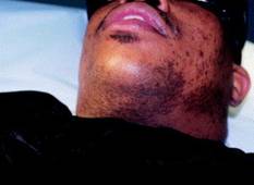
The double-blind placebo controlled study was carried out by U.S. military physicians. PFB, which is predominantly a disorder of black men, has at times been a source of racial tension in the military because the simplest treatment for PFB is to stop shaving and grow a beard, a form of individual expression at odds with regulations.
The study included 27 men with PFB. They received laser therapy once every 4 weeks for 16 weeks. In addition, they applied eflornithine cream to one side of their bearded neck region and placebo to the other side twice daily.
At 16 weeks, the laser plus eflornithine cream side produced a median 99.5% reduction in hair count and inflammatory papules. This was a significantly better result than the median 85% reduction on the laser plus placebo–treated side (J. Am. Acad. Dermatol. 2012 Jan. 8; in press).
The PFB study follows an earlier study by other investigators who demonstrated that eflornithine cream as an adjunct to laser hair removal for facial hirsutism in women was more effective than laser therapy alone (J. Am. Acad. Dermatol. 2007;57:54-9).
Dr. Alexis said performing laser hair removal safely in darker-skinned patients with PFB requires attention to several key principles: longer wavelengths, lower fluences, longer pulse durations, and plenty of epidermal cooling.
"The No. 1 thing is to use longer wavelengths, because the goal is deeper penetration to maximize the ratio of the temperature in the bulb of the follicle to the temperature in the epidermis," he explained.
The long-pulsed 1,064-nm Nd:YAG laser has the lowest rate of associated epidermal burns, hypopigmentation, and other adverse events in darker-skinned patients, as has been shown in a review of a wide assortment of lasers (J. Drugs Dermatol. 2007;6:40-6). It is clearly the safest laser option in patients with skin types IV-VI. The 810-nm diode laser is a reasonable alternative in skin types IV-V, Dr. Alexis said.

In treating patients for PFB with the 1,064-nm Nd:YAG laser, he said that he typically starts with a fluence of 20 J/cm2 and a pulse duration of 20-30 milliseconds. After several sessions, as he makes inroads into the initially dense follicular distribution, he said that he might increase the fluence to a maximum of 50 J/cm2 in the setting of skin type VI, and as high as 100 J/cm2 in skin types IV or V.
Longer pulse durations allow for more efficient epidermal cooling. This minimizes heat injury to melanin-containing epidermal cells. For the 810-nm diode laser, Dr. Alexis said he uses pulse durations of 100 or 400 milliseconds.
Epidermal cooling can be accomplished in several ways. His preferred method is to utilize contact cooling via a sapphire tip or chilled copper plate attached to the laser; the cooling is done before delivering the laser pulse. Alternatively, the epidermal cooling can be done using cold gels, forced air, or spray cooling, although dyschromia can occur in darker skin types if the spray technique isn’t optimal. Another option is to apply an ice pack for 5-10 minutes post treatment.
Laser therapy is expensive, so Dr. Alexis said he likes to give his patients a range of therapeutic options. These include growing a beard, chemical depilation with barium sulfide or calcium thioglycolate every 2-4 days, modification of shaving practices, and salicylic acid chemical peels.
"It’s kind of a long conversation," he said.
Whatever form of therapy the patient decides upon, it’s important that the patient stops tweezing to remove ingrown hairs. This is a common practice that induces trauma and worsens postinflammatory hyperpigmentation.
Dr. Alexis reported that he serves as a consultant to Schick and is on the advisory board for Allergan. SDEF and this news organization are owned by Elsevier.
WAIKOLOA, HAWAII – The use of eflornithine cream may increase the effectiveness of laser hair removal for treating pseudofolliculitis barbae, according to Dr. Andrew F. Alexis.
Laser hair removal has proved to be a game changer in the treatment of pseudofolliculitis barbae, a common chronic, inflammatory dermatosis that’s often been a difficult therapeutic challenge, Dr. Alexis said at the seminar sponsored by Skin Disease Education Foundation (SDEF). And the use of adjunctive eflornithine cream makes laser therapy even more effective, based on a recent study, which is one of the few rigorous studies conducted in pseudofolliculitis barbae (PFB) patients, noted Dr. Alexis, director of the skin of color center at St. Luke’s–Roosevelt Hospital and a dermatologist at Columbia University, New York.
The double-blind placebo controlled study was carried out by U.S. military physicians. PFB, which is predominantly a disorder of black men, has at times been a source of racial tension in the military because the simplest treatment for PFB is to stop shaving and grow a beard, a form of individual expression at odds with regulations.
The study included 27 men with PFB. They received laser therapy once every 4 weeks for 16 weeks. In addition, they applied eflornithine cream to one side of their bearded neck region and placebo to the other side twice daily.
At 16 weeks, the laser plus eflornithine cream side produced a median 99.5% reduction in hair count and inflammatory papules. This was a significantly better result than the median 85% reduction on the laser plus placebo–treated side (J. Am. Acad. Dermatol. 2012 Jan. 8; in press).
The PFB study follows an earlier study by other investigators who demonstrated that eflornithine cream as an adjunct to laser hair removal for facial hirsutism in women was more effective than laser therapy alone (J. Am. Acad. Dermatol. 2007;57:54-9).
Dr. Alexis said performing laser hair removal safely in darker-skinned patients with PFB requires attention to several key principles: longer wavelengths, lower fluences, longer pulse durations, and plenty of epidermal cooling.
"The No. 1 thing is to use longer wavelengths, because the goal is deeper penetration to maximize the ratio of the temperature in the bulb of the follicle to the temperature in the epidermis," he explained.
The long-pulsed 1,064-nm Nd:YAG laser has the lowest rate of associated epidermal burns, hypopigmentation, and other adverse events in darker-skinned patients, as has been shown in a review of a wide assortment of lasers (J. Drugs Dermatol. 2007;6:40-6). It is clearly the safest laser option in patients with skin types IV-VI. The 810-nm diode laser is a reasonable alternative in skin types IV-V, Dr. Alexis said.
In treating patients for PFB with the 1,064-nm Nd:YAG laser, he said that he typically starts with a fluence of 20 J/cm2 and a pulse duration of 20-30 milliseconds. After several sessions, as he makes inroads into the initially dense follicular distribution, he said that he might increase the fluence to a maximum of 50 J/cm2 in the setting of skin type VI, and as high as 100 J/cm2 in skin types IV or V.
Longer pulse durations allow for more efficient epidermal cooling. This minimizes heat injury to melanin-containing epidermal cells. For the 810-nm diode laser, Dr. Alexis said he uses pulse durations of 100 or 400 milliseconds.
Epidermal cooling can be accomplished in several ways. His preferred method is to utilize contact cooling via a sapphire tip or chilled copper plate attached to the laser; the cooling is done before delivering the laser pulse. Alternatively, the epidermal cooling can be done using cold gels, forced air, or spray cooling, although dyschromia can occur in darker skin types if the spray technique isn’t optimal. Another option is to apply an ice pack for 5-10 minutes post treatment.
Laser therapy is expensive, so Dr. Alexis said he likes to give his patients a range of therapeutic options. These include growing a beard, chemical depilation with barium sulfide or calcium thioglycolate every 2-4 days, modification of shaving practices, and salicylic acid chemical peels.
"It’s kind of a long conversation," he said.
Whatever form of therapy the patient decides upon, it’s important that the patient stops tweezing to remove ingrown hairs. This is a common practice that induces trauma and worsens postinflammatory hyperpigmentation.
Dr. Alexis reported that he serves as a consultant to Schick and is on the advisory board for Allergan. SDEF and this news organization are owned by Elsevier.
EXPERT ANALYSIS FROM THE SDEF HAWAII DERMATOLOGY SEMINAR
Calcineurin Inhibitors Are Steroid Sparing in Chronic Hand Dermatitis
WAIKOLOA, HAWAII – The calcineurin inhibitors are effective for long-term maintenance therapy in chronic hand dermatitis, despite their black box warning, and can help reduce the use of topical steroids, according to dermatologist Joseph F. Fowler Jr.
Pimecrolimus 1% cream is nongreasy and thus preferred by many patients over tacrolimus 0.1% ointment, he said at the seminar sponsored by the Skin Disease Education Foundation (SDEF).

"Please understand that I love topical steroids. We all need to use them. But I don’t want to use them continuously because of the variety of adverse effects, especially local atrophy. And if we’re worried about skin barrier function, we don’t want to thin the skin out and reduce the barrier even more with overuse of topical steroids," he said.
"So after we’ve got somebody under control with aggressive use of higher-potency topical steroids, I like to think about the calcineurin inhibitors. I also like to think about what I call the therapeutic moisturizers – those device and [over-the-counter] products that contain ceramides and natural moisturizing factors – and I’ll maybe use a low- to midpotency topical steroid two or three times a week as needed," added Dr. Fowler of the University of Louisville (Ky.).
There are several skin barrier protection products approved as prescription devices rather than medications, such as Atopiclair, Eletone, EpiCeram, Hylatopic, Neosalus, and Tetrix. Many aren’t promoted much anymore by their manufacturers, yet all have supporting evidence that they enhance the skin barrier, he said.
The two skin barrier products Dr. Fowler is most familiar with because of his involvement in clinical trials are Tetrix and Neosalus.
The investigator-blinded, nonrandomized Tetrix cream study involved patients with known allergies to three common, structurally dissimilar test antigens: nickel, neomycin, and fragrance mix. Following exposure to the test antigens, delayed-type hypersensitivity reactions on patch testing were significantly less at sites pretreated with Tetrix cream than at untreated test sites (Cutis 2008;82[suppl. 4]:21-8).
In the multicenter prospective Neosalus foam study, 31 patients with chronic allergic or irritant hand dermatitis of at least 12 months’ duration were randomized to treatment with Neosalus foam plus triamcinolone cream 0.1% or to an over-the-counter (OTC) moisturizer plus the topical steroid. During 8 weeks, patients in the Neosalus group averaged 51.4 doses of the topical steroid, compared with 72.5 doses in controls. Eczema severity scores were also significantly better in the Neosalus group (Am. J. Contact Dermat. 2000;11:165-9).
OTC moisturizers containing ceramide, a lipid that boosts stratum corneum function, include Aveeno eczema therapy moisturizing cream, CeraVe lotion and cleanser, Cetaphil Restoraderm moisturizer, and Curel Sensitive Skin Remedy, Dr. Fowler continued.
He said that he likes to choose from a short list of topical steroids for maintenance therapy. Fluticasone cream or lotion has fewer local adverse effects than many other steroids and has demonstrated long-term efficacy and safety in the setting of atopic dermatitis. Hydrocortisone butyrate lipid cream or lotion has excellent tolerability and a good moisturizing effect.
When there is concern that a patient’s dermatitis may be compounded by topical steroid allergy, clocortolone cream or desonide ointment are excellent choices; there is virtually no allergenicity to either product, according to Dr. Fowler.
"If I’m worried about topical steroid allergy, the sprays, I think, are very nice. Clobetasol spray has almost nothing in it that’s likely to be an allergen, although it’s a little oily and that can be an issue. But if you’re worried about allergy either to the steroid or to other factors in the product, that’s a good one. Also, triamcinolone ... has virtually nothing in it that’s allergenic other than occasionally the triamcinolone itself, and it’s not oily," he said.
The topical corticosteroid foams are a well-received option for maintenance therapy on hairy areas or for large surfaces.
When a higher-potency topical steroid is desired during a breakthrough episode, halcinonide cream has low allergenicity, a desirable biphasic quick and then delayed release, and good moisturizing and emollient properties, Dr. Fowler said.
He reported that he serves as a consultant to Allerderm, Coria, Galderma, Graceway, Hyland, Johnson & Johnson, Quinnova, Ranbaxy, Shire, Stiefel, Triax, and UCB.
SDEF and this news organization are owned by Elsevier.
WAIKOLOA, HAWAII – The calcineurin inhibitors are effective for long-term maintenance therapy in chronic hand dermatitis, despite their black box warning, and can help reduce the use of topical steroids, according to dermatologist Joseph F. Fowler Jr.
Pimecrolimus 1% cream is nongreasy and thus preferred by many patients over tacrolimus 0.1% ointment, he said at the seminar sponsored by the Skin Disease Education Foundation (SDEF).
"Please understand that I love topical steroids. We all need to use them. But I don’t want to use them continuously because of the variety of adverse effects, especially local atrophy. And if we’re worried about skin barrier function, we don’t want to thin the skin out and reduce the barrier even more with overuse of topical steroids," he said.
"So after we’ve got somebody under control with aggressive use of higher-potency topical steroids, I like to think about the calcineurin inhibitors. I also like to think about what I call the therapeutic moisturizers – those device and [over-the-counter] products that contain ceramides and natural moisturizing factors – and I’ll maybe use a low- to midpotency topical steroid two or three times a week as needed," added Dr. Fowler of the University of Louisville (Ky.).
There are several skin barrier protection products approved as prescription devices rather than medications, such as Atopiclair, Eletone, EpiCeram, Hylatopic, Neosalus, and Tetrix. Many aren’t promoted much anymore by their manufacturers, yet all have supporting evidence that they enhance the skin barrier, he said.
The two skin barrier products Dr. Fowler is most familiar with because of his involvement in clinical trials are Tetrix and Neosalus.
The investigator-blinded, nonrandomized Tetrix cream study involved patients with known allergies to three common, structurally dissimilar test antigens: nickel, neomycin, and fragrance mix. Following exposure to the test antigens, delayed-type hypersensitivity reactions on patch testing were significantly less at sites pretreated with Tetrix cream than at untreated test sites (Cutis 2008;82[suppl. 4]:21-8).
In the multicenter prospective Neosalus foam study, 31 patients with chronic allergic or irritant hand dermatitis of at least 12 months’ duration were randomized to treatment with Neosalus foam plus triamcinolone cream 0.1% or to an over-the-counter (OTC) moisturizer plus the topical steroid. During 8 weeks, patients in the Neosalus group averaged 51.4 doses of the topical steroid, compared with 72.5 doses in controls. Eczema severity scores were also significantly better in the Neosalus group (Am. J. Contact Dermat. 2000;11:165-9).
OTC moisturizers containing ceramide, a lipid that boosts stratum corneum function, include Aveeno eczema therapy moisturizing cream, CeraVe lotion and cleanser, Cetaphil Restoraderm moisturizer, and Curel Sensitive Skin Remedy, Dr. Fowler continued.
He said that he likes to choose from a short list of topical steroids for maintenance therapy. Fluticasone cream or lotion has fewer local adverse effects than many other steroids and has demonstrated long-term efficacy and safety in the setting of atopic dermatitis. Hydrocortisone butyrate lipid cream or lotion has excellent tolerability and a good moisturizing effect.
When there is concern that a patient’s dermatitis may be compounded by topical steroid allergy, clocortolone cream or desonide ointment are excellent choices; there is virtually no allergenicity to either product, according to Dr. Fowler.
"If I’m worried about topical steroid allergy, the sprays, I think, are very nice. Clobetasol spray has almost nothing in it that’s likely to be an allergen, although it’s a little oily and that can be an issue. But if you’re worried about allergy either to the steroid or to other factors in the product, that’s a good one. Also, triamcinolone ... has virtually nothing in it that’s allergenic other than occasionally the triamcinolone itself, and it’s not oily," he said.
The topical corticosteroid foams are a well-received option for maintenance therapy on hairy areas or for large surfaces.
When a higher-potency topical steroid is desired during a breakthrough episode, halcinonide cream has low allergenicity, a desirable biphasic quick and then delayed release, and good moisturizing and emollient properties, Dr. Fowler said.
He reported that he serves as a consultant to Allerderm, Coria, Galderma, Graceway, Hyland, Johnson & Johnson, Quinnova, Ranbaxy, Shire, Stiefel, Triax, and UCB.
SDEF and this news organization are owned by Elsevier.
EXPERT ANALYSIS FROM THE SDEF HAWAII DERMATOLOGY SEMINAR
New Dermoscopic Insights Gleaned for Mucosal Lesions
WAIKOLOA, HAWAII – The dermoscopic features that reliably distinguish malignant mucosal lesions are a combination of structureless areas within the lesion along with blue, gray, or white color, a multicenter study conducted by the International Dermoscopy Society has shown.
This combination of dermoscopic findings yielded 100% sensitivity for histopathologically confirmed melanoma and 93% sensitivity for any malignancy, lead investigator Dr. Andreas Blum reported at the Hawaii Dermatology Seminar sponsored by Skin Disease Education Foundation (SDEF).

He noted that while the key points in dermoscopic differentiation between malignant and benign and pigmented and nonpigmented lesions of the skin, nail apparatus, and scalp are well established, the important features to look for in dermoscopic evaluation of lesions of the oral mucosa and genitalia haven’t been well characterized. That was the impetus for the international observational study.
Consensus regarding dermoscopy of mucosal lesions has lagged for a couple of reasons, explained Dr. Blum, professor of dermatology at the University of Tübingen (Germany). One is that pigmented mucosal lesions are uncommon. And another is that manipulating the dermoscope in mucosal areas can be a challenge.
The study took place at 14 specialized skin cancer clinics in 10 countries. It included 140 patients with pigmented mucosal lesions, of which 126 ultimately proved benign, while 11 were melanomas, 2 were squamous cell carcinoma in situ lesions, and 1 was a metastasis (Arch. Dermatol. 2011;147:1181-7).
The investigators scored the dermoscopic patterns they saw as dots, globules, or clods, circles, lines, or structureless using a pattern analysis method developed by Dr. Harald Kittler.
The key study finding was that in a univariate analysis, lesions that were blue, white, or gray in color under the dermoscope and that contained structureless zones had a 100% sensitivity for melanoma, a 93% sensitivity for any malignancy, and an 83% specificity for being benign.
"When you see structureless areas – and only part of the lesion needs to be structureless – with blue, gray, or white zones, then you know something has gone wrong and it’s time to do a biopsy or excision," he said.
Recognizing structureless areas might be at times a difficult call for less-experienced physicians to make, the investigators also analyzed the data based solely upon a lesion’s color. Blue, gray, or white still had a sensitivity of 100% for melanoma and 93% for any malignancy, but the specificity dropped to 64%.
"So if you’re unsure about whether you’re seeing a structureless area, based upon color only, you’ll reliably detect melanomas and other malignancies, but you’ll end up doing unnecessary biopsies for benign lesions," Dr. Blum explained.
He credited Dr. Alfred W. Kopf of New York University with a suggestion that has made dermoscopic evaluation of mucosal lesions much more practical. To avoid contaminating the lens of the dermoscope, simply wrap the head of the device in plastic food wrap that has been coated on both sides with mineral oil.
Session chair Dr. Ashfaq A. Marghoob, a coinvestigator in the international study, offered a cautionary tale. He said that he has had two teenage patients with vulvar pigmented lesions that looked clinically like a clear-cut melanoma, and dermoscopically like melanoma, and the pathology report on the biopsy specimen came back as melanoma. Yet an alert gynecologic surgical oncologist contacted him and said he thought the white area surrounding the pigmented lesion looked like lichen sclerosus et atrophicus. It turned out the surgeon was right.
"I saw the patients again, and lo and behold it was as obvious as could be. I had missed the LS & A [lichen sclerosus et atrophicus], because I was so focused on the pigmented lesion that I just hadn’t realized it was there. It turns out that if you have LS & A, you can develop pigmented lesions within it that look like melanoma clinically, that look like melanoma under dermoscopy, and look like melanoma histologically," said Dr. Marghoob, a dermatologist at Memorial Sloan-Kettering Cancer Center in New York.
The surgery planned for one of these young patients entailed removal of the clitoral area, so timely recognition that she actually had LS & A and not melanoma spared her from a life-changing mistake.
"We’ve now been following her for 5 years, and she’s absolutely fine with no change in the pigmented lesion," he noted.
The lesson he said he’d like to share: "A vulvar melanoma in somebody under the age of 50 is almost unheard of, and I’d strongly consider LS & A instead, checking with a Wood’s light."
Neither Dr. Blum nor Dr. Marghoob reported having any relevant financial disclosures. SDEF and this news organization are owned by Elsevier.
WAIKOLOA, HAWAII – The dermoscopic features that reliably distinguish malignant mucosal lesions are a combination of structureless areas within the lesion along with blue, gray, or white color, a multicenter study conducted by the International Dermoscopy Society has shown.
This combination of dermoscopic findings yielded 100% sensitivity for histopathologically confirmed melanoma and 93% sensitivity for any malignancy, lead investigator Dr. Andreas Blum reported at the Hawaii Dermatology Seminar sponsored by Skin Disease Education Foundation (SDEF).
He noted that while the key points in dermoscopic differentiation between malignant and benign and pigmented and nonpigmented lesions of the skin, nail apparatus, and scalp are well established, the important features to look for in dermoscopic evaluation of lesions of the oral mucosa and genitalia haven’t been well characterized. That was the impetus for the international observational study.
Consensus regarding dermoscopy of mucosal lesions has lagged for a couple of reasons, explained Dr. Blum, professor of dermatology at the University of Tübingen (Germany). One is that pigmented mucosal lesions are uncommon. And another is that manipulating the dermoscope in mucosal areas can be a challenge.
The study took place at 14 specialized skin cancer clinics in 10 countries. It included 140 patients with pigmented mucosal lesions, of which 126 ultimately proved benign, while 11 were melanomas, 2 were squamous cell carcinoma in situ lesions, and 1 was a metastasis (Arch. Dermatol. 2011;147:1181-7).
The investigators scored the dermoscopic patterns they saw as dots, globules, or clods, circles, lines, or structureless using a pattern analysis method developed by Dr. Harald Kittler.
The key study finding was that in a univariate analysis, lesions that were blue, white, or gray in color under the dermoscope and that contained structureless zones had a 100% sensitivity for melanoma, a 93% sensitivity for any malignancy, and an 83% specificity for being benign.
"When you see structureless areas – and only part of the lesion needs to be structureless – with blue, gray, or white zones, then you know something has gone wrong and it’s time to do a biopsy or excision," he said.
Recognizing structureless areas might be at times a difficult call for less-experienced physicians to make, the investigators also analyzed the data based solely upon a lesion’s color. Blue, gray, or white still had a sensitivity of 100% for melanoma and 93% for any malignancy, but the specificity dropped to 64%.
"So if you’re unsure about whether you’re seeing a structureless area, based upon color only, you’ll reliably detect melanomas and other malignancies, but you’ll end up doing unnecessary biopsies for benign lesions," Dr. Blum explained.
He credited Dr. Alfred W. Kopf of New York University with a suggestion that has made dermoscopic evaluation of mucosal lesions much more practical. To avoid contaminating the lens of the dermoscope, simply wrap the head of the device in plastic food wrap that has been coated on both sides with mineral oil.
Session chair Dr. Ashfaq A. Marghoob, a coinvestigator in the international study, offered a cautionary tale. He said that he has had two teenage patients with vulvar pigmented lesions that looked clinically like a clear-cut melanoma, and dermoscopically like melanoma, and the pathology report on the biopsy specimen came back as melanoma. Yet an alert gynecologic surgical oncologist contacted him and said he thought the white area surrounding the pigmented lesion looked like lichen sclerosus et atrophicus. It turned out the surgeon was right.
"I saw the patients again, and lo and behold it was as obvious as could be. I had missed the LS & A [lichen sclerosus et atrophicus], because I was so focused on the pigmented lesion that I just hadn’t realized it was there. It turns out that if you have LS & A, you can develop pigmented lesions within it that look like melanoma clinically, that look like melanoma under dermoscopy, and look like melanoma histologically," said Dr. Marghoob, a dermatologist at Memorial Sloan-Kettering Cancer Center in New York.
The surgery planned for one of these young patients entailed removal of the clitoral area, so timely recognition that she actually had LS & A and not melanoma spared her from a life-changing mistake.
"We’ve now been following her for 5 years, and she’s absolutely fine with no change in the pigmented lesion," he noted.
The lesson he said he’d like to share: "A vulvar melanoma in somebody under the age of 50 is almost unheard of, and I’d strongly consider LS & A instead, checking with a Wood’s light."
Neither Dr. Blum nor Dr. Marghoob reported having any relevant financial disclosures. SDEF and this news organization are owned by Elsevier.
EXPERT ANALYSIS FROM THE SDEF HAWAII DERMATOLOGY SEMINAR
Topical Botulinum Toxin Will Turn Market 'Upside Down'
WAIKOLOA, HAWAII – Topically applied botulinum toxin type A may no longer be a pipe dream, as it is now likely full speed ahead for proposed phase III trials of the agent.
The completed phase II program consisted of 11 clinical studies in which 553 patients had their lateral canthal lines treated with the investigational topical product known for now as RT001, under development by Revance Therapeutics. The results were highly impressive, according to Dr. Alastair Carruthers, a dermatologist at the University of British Columbia, Vancouver.

"Watch out for topical botulinum toxin. I think Revance is going to turn the neurotoxin market upside down," he predicted at the Hawaii Dermatology Seminar, sponsored by Skin Disease Education Foundation (SDEF).
Revance has developed a proprietary platform that enables transcutaneous flux of large medicinal payloads. The company has reported successful proof-of-concept studies for topically delivered insulin, growth factors, and numerous other macromolecules with applications in fields ranging from cardiovascular disease to cancer. But it’s the topical botulinum toxin project that has captured Dr. Carruthers’ attention.
"Their technology enables you to get the neurotoxin across intact skin, which is something I never thought that we would see. But it really works," he said.
No significant adverse events occurred in the phase II studies. There was no evidence of diffusion of neurotoxin away from the target muscle, and no effect upon the cranial nerves, he said. Laboratory monitoring and ECGs did not yield any evidence of systemic exposure.
The median duration of therapeutic effect was 113 days. The response rate was up to 89% based upon a stringent composite end point requiring a 2-point improvement as assessed independently by investigator and patient, he said.
The key to this technology is a synthetic peptide carrier which contains protein transduction domains and a backbone core that attaches to the neurotoxin molecule. This peptide carrier can be set to achieve different depths of penetration.
The proposed commercial product that will undergo phase III testing entails mixing the viscous topical gel in a one-step applicator, which is then used in treating the lateral canthal lines. The mixing and application takes only a couple of minutes. The gel is left on for perhaps 30 minutes – the optimal time is yet to be determined – and then removed with a proprietary cleanser.
Dr. Carruthers said that as many know, increasing competition has arrived among the manufacturers of the three Food and Drug Administration–approved injectable botulinum toxin type A products.
"I doubt that the battle, such as it is, will be fought on intellectual, scientific issues. I think it will be fought based upon cost, marketing, and other intangibles," he predicted.
Brand loyalty, company sponsorship of medical education, appeals to nationalism – one manufacturer is U.S.-based, the others German and French – these are the sorts of issues he expects to see brought forth.
That’s because the things that really matter to clinicians, such as onset of therapeutic effect, its spread, duration, and side effects, are all a function of dose – and there is no agreement as to what the comparable dose is between the various commercial preparations. Despite manufacturers’ claims, it’s not possible to detect small differences in effectiveness, immunogenicity, or other end points without doing studies that would require enormous numbers of patients, according to Dr. Carruthers.
He advised that given the uncertainty regarding dosing comparability, the best practice is to have only one botulinum toxin type A product in the office. This avoids the thorny issue of trying to use comparably effective dilutions.
In a separate presentation at the annual meeting of the American Society for Dermatologic Surgery, Dr. Gary D. Monheit, a dermatologist in private practice in Birmingham, Ala., agreed that the topical botulinum toxin could be practice changing.
He said that RT001 is best for superficial musculature such as crows’ feet, and possibly in the future for perioral and forehead wrinkles.
The investigational product affects pore size and helps smooth the skin, he said. And since the product is mostly absorbed in the superficial musculature, it could eventually be used on the eyelids and lips for superficial wrinkles and to brighten up dull skin.
Dr. Carruthers reported that he has no financial relationship with Revance. He is a consultant to, and paid investigator for, Allergan and Merz, which market Botox (onabotulinumtoxinA) and Xeomin (incobotulinumtoxinA), respectively.
Dr. Monheit is a consultant and clinical investigator for Revance.
SDEF and this news organization are owned by Elsevier.
Naseem Miller was a contributing writer.
WAIKOLOA, HAWAII – Topically applied botulinum toxin type A may no longer be a pipe dream, as it is now likely full speed ahead for proposed phase III trials of the agent.
The completed phase II program consisted of 11 clinical studies in which 553 patients had their lateral canthal lines treated with the investigational topical product known for now as RT001, under development by Revance Therapeutics. The results were highly impressive, according to Dr. Alastair Carruthers, a dermatologist at the University of British Columbia, Vancouver.
"Watch out for topical botulinum toxin. I think Revance is going to turn the neurotoxin market upside down," he predicted at the Hawaii Dermatology Seminar, sponsored by Skin Disease Education Foundation (SDEF).
Revance has developed a proprietary platform that enables transcutaneous flux of large medicinal payloads. The company has reported successful proof-of-concept studies for topically delivered insulin, growth factors, and numerous other macromolecules with applications in fields ranging from cardiovascular disease to cancer. But it’s the topical botulinum toxin project that has captured Dr. Carruthers’ attention.
"Their technology enables you to get the neurotoxin across intact skin, which is something I never thought that we would see. But it really works," he said.
No significant adverse events occurred in the phase II studies. There was no evidence of diffusion of neurotoxin away from the target muscle, and no effect upon the cranial nerves, he said. Laboratory monitoring and ECGs did not yield any evidence of systemic exposure.
The median duration of therapeutic effect was 113 days. The response rate was up to 89% based upon a stringent composite end point requiring a 2-point improvement as assessed independently by investigator and patient, he said.
The key to this technology is a synthetic peptide carrier which contains protein transduction domains and a backbone core that attaches to the neurotoxin molecule. This peptide carrier can be set to achieve different depths of penetration.
The proposed commercial product that will undergo phase III testing entails mixing the viscous topical gel in a one-step applicator, which is then used in treating the lateral canthal lines. The mixing and application takes only a couple of minutes. The gel is left on for perhaps 30 minutes – the optimal time is yet to be determined – and then removed with a proprietary cleanser.
Dr. Carruthers said that as many know, increasing competition has arrived among the manufacturers of the three Food and Drug Administration–approved injectable botulinum toxin type A products.
"I doubt that the battle, such as it is, will be fought on intellectual, scientific issues. I think it will be fought based upon cost, marketing, and other intangibles," he predicted.
Brand loyalty, company sponsorship of medical education, appeals to nationalism – one manufacturer is U.S.-based, the others German and French – these are the sorts of issues he expects to see brought forth.
That’s because the things that really matter to clinicians, such as onset of therapeutic effect, its spread, duration, and side effects, are all a function of dose – and there is no agreement as to what the comparable dose is between the various commercial preparations. Despite manufacturers’ claims, it’s not possible to detect small differences in effectiveness, immunogenicity, or other end points without doing studies that would require enormous numbers of patients, according to Dr. Carruthers.
He advised that given the uncertainty regarding dosing comparability, the best practice is to have only one botulinum toxin type A product in the office. This avoids the thorny issue of trying to use comparably effective dilutions.
In a separate presentation at the annual meeting of the American Society for Dermatologic Surgery, Dr. Gary D. Monheit, a dermatologist in private practice in Birmingham, Ala., agreed that the topical botulinum toxin could be practice changing.
He said that RT001 is best for superficial musculature such as crows’ feet, and possibly in the future for perioral and forehead wrinkles.
The investigational product affects pore size and helps smooth the skin, he said. And since the product is mostly absorbed in the superficial musculature, it could eventually be used on the eyelids and lips for superficial wrinkles and to brighten up dull skin.
Dr. Carruthers reported that he has no financial relationship with Revance. He is a consultant to, and paid investigator for, Allergan and Merz, which market Botox (onabotulinumtoxinA) and Xeomin (incobotulinumtoxinA), respectively.
Dr. Monheit is a consultant and clinical investigator for Revance.
SDEF and this news organization are owned by Elsevier.
Naseem Miller was a contributing writer.
EXPERT ANALYSIS FROM THE SDEF HAWAII DERMATOLOGY SEMINAR
Early Detection of Melanoma: Harnessing Untapped Resources
WAIKOLOA, HAWAII – Improved early detection of fast-growing, lethal melanomas will require out-of-the-box thinking, such as providing dermatoscopes for patients to use at home and educating hairdressers and other nondermatologists on how to detect melanoma.
"At least three companies are now designing dermatoscopes for patient use. Patients will be able to buy the dermatoscope at a pharmacy and do self-examination or examine their spouse. That, I think, is going to be a reality within the next 5 years," Dr. Ashfaq A. Marghoob predicted at the Hawaii Dermatology Seminar sponsored by Skin Disease Education Foundation (SDEF).

A key feature of these devices will be the capability of hooking into a smart phone for wireless transmission of suspicious images to a skin cancer expert for assessment.
Dr. Marghoob and his coworkers first proposed dermoscopy as a tool with untapped potential for skin self-examination in selected patients in an article last year (Arch. Dermatol. 2011;147:53-8).
But patient empowerment is only part of what’s needed in order to improve early detection of the fast-growing killer subtype of melanoma. Dr. Marghoob and his coworkers are now conducting a prospective study to evaluate the impact of a 20-minute education session for hair care professionals about how they can aid in detecting skin cancers on the scalp, neck, and face.
This study was a direct outgrowth of a survey the investigators conducted at a Houston convention of barbers and hairstylists. Forty-nine percent of respondents indicated they were highly receptive to participating in a skin cancer education program. During the preceding month, 37% of respondents had looked at more than half of their customers’ scalps for suspicious lesions, 29% had looked at more than half of their customers’ necks, and 15% had checked more than half of their customers’ faces (Arch. Dermatol. 2011;147:1159-65).
Melanoma of the scalp and neck accounted for 10% of all melanoma deaths in the United States from 1973 to 2003. Barbers and hairstylists are in a unique position to detect skin cancers in those locations because they typically see their customers on a regular basis, spend a fair amount of time with them at each visit, have good rapport, and often discuss health issues.
The larger goal underlying this project, Dr. Marghoob explained, is to develop a cadre of expertly trained lay community workers to examine areas of the skin that are difficult for people to see for themselves and which often go overlooked by physicians. In addition to hair professionals, other workers ideally suited to serve as lay skin cancer educators and examiners include massage therapists, manicurists, cosmetologists, and electrologists.
Dr. Marghoob has also been involved in efforts to teach dermoscopy to primary care physicians and other nondermatologist physicians, including ob.gyns., pediatricians, and plastic surgeons. Moreover, he recently conducted a study in which second-year medical students were issued dermatoscopes and trained in their use.
"We found they get better at diagnosing skin cancer and are paying more attention to the skin. All we really want them to do is really look at the skin while they’re doing a physical examination," he said.
He has also been encouraging internists and family physicians to take advantage of opportunistic skin screening situations. For example, when they’re listening to the lungs and heart with a stethoscope, he urges primary care physicians to have patients take their shirt off so they can take a close look at the truncal skin rather than simply slip the bell of the scope underneath the shirt.
Another potentially fruitful means of improving upon the gains achieved in early detection of skin cancer would be targeted screening of older men, a high-risk group for fast-growing nodular melanomas, Dr. Marghoob added.
He reported having no financial conflicts. SDEF and this news organization are owned by Elsevier.
WAIKOLOA, HAWAII – Improved early detection of fast-growing, lethal melanomas will require out-of-the-box thinking, such as providing dermatoscopes for patients to use at home and educating hairdressers and other nondermatologists on how to detect melanoma.
"At least three companies are now designing dermatoscopes for patient use. Patients will be able to buy the dermatoscope at a pharmacy and do self-examination or examine their spouse. That, I think, is going to be a reality within the next 5 years," Dr. Ashfaq A. Marghoob predicted at the Hawaii Dermatology Seminar sponsored by Skin Disease Education Foundation (SDEF).
A key feature of these devices will be the capability of hooking into a smart phone for wireless transmission of suspicious images to a skin cancer expert for assessment.
Dr. Marghoob and his coworkers first proposed dermoscopy as a tool with untapped potential for skin self-examination in selected patients in an article last year (Arch. Dermatol. 2011;147:53-8).
But patient empowerment is only part of what’s needed in order to improve early detection of the fast-growing killer subtype of melanoma. Dr. Marghoob and his coworkers are now conducting a prospective study to evaluate the impact of a 20-minute education session for hair care professionals about how they can aid in detecting skin cancers on the scalp, neck, and face.
This study was a direct outgrowth of a survey the investigators conducted at a Houston convention of barbers and hairstylists. Forty-nine percent of respondents indicated they were highly receptive to participating in a skin cancer education program. During the preceding month, 37% of respondents had looked at more than half of their customers’ scalps for suspicious lesions, 29% had looked at more than half of their customers’ necks, and 15% had checked more than half of their customers’ faces (Arch. Dermatol. 2011;147:1159-65).
Melanoma of the scalp and neck accounted for 10% of all melanoma deaths in the United States from 1973 to 2003. Barbers and hairstylists are in a unique position to detect skin cancers in those locations because they typically see their customers on a regular basis, spend a fair amount of time with them at each visit, have good rapport, and often discuss health issues.
The larger goal underlying this project, Dr. Marghoob explained, is to develop a cadre of expertly trained lay community workers to examine areas of the skin that are difficult for people to see for themselves and which often go overlooked by physicians. In addition to hair professionals, other workers ideally suited to serve as lay skin cancer educators and examiners include massage therapists, manicurists, cosmetologists, and electrologists.
Dr. Marghoob has also been involved in efforts to teach dermoscopy to primary care physicians and other nondermatologist physicians, including ob.gyns., pediatricians, and plastic surgeons. Moreover, he recently conducted a study in which second-year medical students were issued dermatoscopes and trained in their use.
"We found they get better at diagnosing skin cancer and are paying more attention to the skin. All we really want them to do is really look at the skin while they’re doing a physical examination," he said.
He has also been encouraging internists and family physicians to take advantage of opportunistic skin screening situations. For example, when they’re listening to the lungs and heart with a stethoscope, he urges primary care physicians to have patients take their shirt off so they can take a close look at the truncal skin rather than simply slip the bell of the scope underneath the shirt.
Another potentially fruitful means of improving upon the gains achieved in early detection of skin cancer would be targeted screening of older men, a high-risk group for fast-growing nodular melanomas, Dr. Marghoob added.
He reported having no financial conflicts. SDEF and this news organization are owned by Elsevier.
EXPERT ANALYSIS FROM THE SDEF HAWAII DERMATOLOGY SEMINAR
Gains in Melanoma Survival Attributed to Patient Awareness
WAIKOLOA, HAWAII – The improvement in melanoma survival over the past 4 decades can be attributed to effective public education campaigns, increased patient awareness, and improved physician skills and diagnostic tools, according to Dr. Ashfaq A. Marghoob.
It has been nothing short of phenomenal, he said, especially considering it can’t be credited to major therapeutic advances because up until a couple years ago there weren’t any.
Survival at 5 years for all-stage melanomas of the skin climbed from less than 60% in 1970 to 91% in 2011, he said at the Hawaii Dermatology Seminar sponsored by the Skin Disease Education Foundation (SDEF). But while this is a triumph deserving of celebration, the statistics are somewhat deceiving, said Dr. Marghoob, a dermatologist at Memorial Sloan-Kettering Cancer Center in New York.
He is among a growing number of experts who believe that many thin melanomas detected through screening efforts are slow-growing, indolent skin cancers that sometimes regress and in any event will never become thick or dangerous – never result in death – within the range of current life expectancy. He noted that there is ample precedence, namely, indolent forms of prostate cancer, lymphoma, and breast cancer.
Dr. Marghoob was part of an international team that demonstrated the existence of a slow-growing subtype of melanoma. In a series of 103 melanomas excised after a median follow-up of 20 months, most of the lesions were still in situ or in an early invasive stage. Only three lesions were 1-mm thick or more. There was no correlation between tumor thickness and follow-up time (Br. J. Dermatol. 2010;162:267-73). Growing support exists among epidemiologists for the concept that there are three distinct, unrelated melanoma subtypes (Br. J. Dermatol. 2007;157:338-43). One subtype consists of thin, slow-growing melanomas – the kind that have been steadily increasing in incidence for decades. These are associated with intermittent sun exposure and often arise on the trunk among numerous background nevi. These melanomas are amenable to detection via screening or periodic surveillance. But they only rarely metastasize.
A second type of slow-growing melanoma often occurs on the head and neck of individuals with continuous sun exposure. The incidence of this subtype of melanoma is slowly increasing.
The third and most concerning melanoma subtype consists of thick, fast-growing lesions occurring in individuals with many nevi, but that are not associated with sun exposure. The incidence of these fast-growing, high-lethality melanomas has remained steady over time because they often escape detection as a result of their accelerated growth rate. Improved early detection is a high priority, and it will require creative new approaches, he said.
But in terms of celebrating rising 5-year melanoma survival rates, a contributory landmark event, in Dr. Marghoob’s view, was the increased awareness about melanoma after introduction of the ABCD mnemonic, devised chiefly for primary care physicians and the general public. This was later enhanced by the "ugly duckling" campaign, which taught physicians and patients that melanomas are generally recognizable as outlier lesions.
Multiple studies have shown that skin cancer specialists using visual examination alone – incorporating the ABCDs and ugly duckling concept – can typically diagnose melanoma with a sensitivity of 70% and specificity of 75%. The number needed to treat (NNT) or benign-to-malignant biopsy ratio is 1:12-15.
With the aid of total body photography for assistance in patient follow-up, the NNT improves to 10.
Dermoscopy has been another important advance. It enables physicians to pick up melanomas not detectable by any other method. Skin cancer specialists who supplement visual examination with dermoscopy typically have 90% sensitivity and 86% specificity for the diagnosis of melanoma. The NNT improves to 4-7, Dr. Marghoob continued.
Recent studies indicate these numbers get even better with the use of confocal microscopy during skin examination.
Using a review of his own practice to illustrate the strong trend for improved diagnosis, Dr. Marghoob noted that in 1998 his NNT was 12.5. He adopted dermoscopy in 1999, and in 2000, when he was using dermoscopy routinely, his NNT improved to 7. During both 2006 and 2007 it was 3, he said.
"We have gotten better at diagnosing melanoma and we will continue to improve," he concluded.
He reported having no relevant financial disclosures. SDEF and this news organization are owned by Elsevier.
EXPERT ANALYSIS FROM THE SDEF HAWAII DERMATOLOGY SEMINAR
Total Body Exam Reduces Melanoma Mortality
WAIKOLOA, HAWAII – An organized program of population-based total body skin examination screening for skin cancer has been shown to significantly reduce melanoma mortality.
"This is quite astounding. It is very impressive to see that a total body skin exam can reduce mortality. It forces us all to think about whether we should do this in a very, very large population, as we now do in Germany," said Dr. Andreas Blum, professor of dermatology at the University of Tübingen (Germany).

He presented highlights of the SCREEN (Skin Cancer Research to Provide Evidence for Effectiveness of Screening in Northern Germany) project, in which all residents of the state of Schleswig-Holstein were eligible for a standardized total body skin exam during a 1-year study period. This was to date the world’s largest systematic population-based skin cancer screening program, he said.
Nineteen percent of the Schleswig-Holstein adult population – more than 360,000 citizens – participated. A total of 3,103 skin cancers were found, including 585 melanomas, for a rate of 1.6 melanomas per 1,000 persons screened. Basal cell carcinomas were detected at a rate of 5.4 malignancies per 1,000, and squamous cell carcinomas at 1.1 per 1,000 people screened. Five lesion excisions had to be performed to detect one malignancy.
Using the incidence of melanoma in Schleswig-Holstein during the 2 years prior to the SCREEN project as a baseline, the incidence of melanoma during the SCREEN project increased by 16% in men and by 38% in women.
The key study finding was a significant decrease in melanoma mortality documented 5 years after SCREEN ended. The observed melanoma mortality rate in men was 0.79 per 100,000 population, compared with an expected 2.0 per 100,000. Among women, the observed mortality was 0.66 per 100,000, compared with an expected 1.3 per 100,000. Thus, the observed mortality because of earlier detection of melanoma in the screened area was less than 50% of expected, Dr. Blum said at the Hawaii Dermatology Seminar sponsored by the Skin Disease Education Foundation (SDEF).
The screening project had a two-tiered structure. Most participants were first screened by a primary care physician or other nondermatologist. In the event of suspicious findings and/or skin cancer risk factors, the participant was referred to a dermatologist who performed the biopsies. All participating physicians first had to attend an 8-hour training course. Of note, 116 of the 118 dermatologists in Schleswig-Holstein participated in SCREEN, as did nearly two-thirds of eligible nondermatologists (J. Am. Acad. Dermatol. 2012;66:201-11).
Following up on the unprecedented success of the SCREEN project, German dermatologists next proposed a randomized controlled trial in order to provide the highest-level evidence that mass skin cancer screening reduces melanoma mortality. However, government health officials found SCREEN persuasive and nixed the idea of a large and costly randomized trial. Instead, Germany has launched a national skin cancer screening program, according to Dr. Blum. All 45 million Germans aged 35 years and older are now eligible to be screened for skin cancer once every 2 years; whether the health care system can cope with the demand remains to be seen.
Another recent project evaluating the benefits of total body skin examination for skin cancer screening also reported favorable results, he noted.
Investigators in a multicenter study screened more than 14,000 patients with a total body skin exam. Participants were consecutive adults presenting with a localized dermatologic problem, such as a skin infection, that wouldn’t ordinarily result in a total body skin exam. The total body skin exams detected 40 patients (0.3%) with melanoma. Five benign lesions were excised for each melanoma detected. Another 2.1% of patients had at least one nonmelanoma skin cancer detected by total body skin exam. On average, 400 patients had to be examined by total body skin exam in order to find 1 melanoma (J. Am. Acad. Dermatol. 2012;66:212-9).
"I see around 150 new patients per week, so that means every third week I see a new melanoma," Dr. Blum said.
Total body skin examination has long been a controversial issue. The U.S. Preventive Services Task Force found insufficient evidence to recommend screening adults for skin cancer (Ann. Intern. Med. 2009;150:188-93). That stance will now need to be revisited in light of these two large projects, said Dr. Blum.
He predicted that the cost involved in routine total body skin examinations is likely to be a critical source of controversy. Using the National Cancer Institute’s estimate that 12.5% of melanomas are fatal, and assuming the cost of a total body skin exam to be $50, he estimated that routine total body skin exams in the SCREEN project cost $240,000 per melanoma death avoided.
In his own specialized skin cancer clinic, where he sees a more select patient population, Dr. Blum estimated that routine total body skin examination costs about $65,000 per melanoma death avoided. And when he plugged in the numbers provided by his colleague Dr. Ashfaq A. Marghoob, pertaining to the skin cancer clinic at Memorial Sloan-Kettering Cancer Center in New York, Dr. Blum once again came up with a figure of roughly $65,000 per melanoma death avoided.
"The range is quite high. I think the cost debate will continue," Dr. Blum predicted.
He reported having no financial conflicts.
SDEF and this news organization are owned by Elsevier.
WAIKOLOA, HAWAII – An organized program of population-based total body skin examination screening for skin cancer has been shown to significantly reduce melanoma mortality.
"This is quite astounding. It is very impressive to see that a total body skin exam can reduce mortality. It forces us all to think about whether we should do this in a very, very large population, as we now do in Germany," said Dr. Andreas Blum, professor of dermatology at the University of Tübingen (Germany).
He presented highlights of the SCREEN (Skin Cancer Research to Provide Evidence for Effectiveness of Screening in Northern Germany) project, in which all residents of the state of Schleswig-Holstein were eligible for a standardized total body skin exam during a 1-year study period. This was to date the world’s largest systematic population-based skin cancer screening program, he said.
Nineteen percent of the Schleswig-Holstein adult population – more than 360,000 citizens – participated. A total of 3,103 skin cancers were found, including 585 melanomas, for a rate of 1.6 melanomas per 1,000 persons screened. Basal cell carcinomas were detected at a rate of 5.4 malignancies per 1,000, and squamous cell carcinomas at 1.1 per 1,000 people screened. Five lesion excisions had to be performed to detect one malignancy.
Using the incidence of melanoma in Schleswig-Holstein during the 2 years prior to the SCREEN project as a baseline, the incidence of melanoma during the SCREEN project increased by 16% in men and by 38% in women.
The key study finding was a significant decrease in melanoma mortality documented 5 years after SCREEN ended. The observed melanoma mortality rate in men was 0.79 per 100,000 population, compared with an expected 2.0 per 100,000. Among women, the observed mortality was 0.66 per 100,000, compared with an expected 1.3 per 100,000. Thus, the observed mortality because of earlier detection of melanoma in the screened area was less than 50% of expected, Dr. Blum said at the Hawaii Dermatology Seminar sponsored by the Skin Disease Education Foundation (SDEF).
The screening project had a two-tiered structure. Most participants were first screened by a primary care physician or other nondermatologist. In the event of suspicious findings and/or skin cancer risk factors, the participant was referred to a dermatologist who performed the biopsies. All participating physicians first had to attend an 8-hour training course. Of note, 116 of the 118 dermatologists in Schleswig-Holstein participated in SCREEN, as did nearly two-thirds of eligible nondermatologists (J. Am. Acad. Dermatol. 2012;66:201-11).
Following up on the unprecedented success of the SCREEN project, German dermatologists next proposed a randomized controlled trial in order to provide the highest-level evidence that mass skin cancer screening reduces melanoma mortality. However, government health officials found SCREEN persuasive and nixed the idea of a large and costly randomized trial. Instead, Germany has launched a national skin cancer screening program, according to Dr. Blum. All 45 million Germans aged 35 years and older are now eligible to be screened for skin cancer once every 2 years; whether the health care system can cope with the demand remains to be seen.
Another recent project evaluating the benefits of total body skin examination for skin cancer screening also reported favorable results, he noted.
Investigators in a multicenter study screened more than 14,000 patients with a total body skin exam. Participants were consecutive adults presenting with a localized dermatologic problem, such as a skin infection, that wouldn’t ordinarily result in a total body skin exam. The total body skin exams detected 40 patients (0.3%) with melanoma. Five benign lesions were excised for each melanoma detected. Another 2.1% of patients had at least one nonmelanoma skin cancer detected by total body skin exam. On average, 400 patients had to be examined by total body skin exam in order to find 1 melanoma (J. Am. Acad. Dermatol. 2012;66:212-9).
"I see around 150 new patients per week, so that means every third week I see a new melanoma," Dr. Blum said.
Total body skin examination has long been a controversial issue. The U.S. Preventive Services Task Force found insufficient evidence to recommend screening adults for skin cancer (Ann. Intern. Med. 2009;150:188-93). That stance will now need to be revisited in light of these two large projects, said Dr. Blum.
He predicted that the cost involved in routine total body skin examinations is likely to be a critical source of controversy. Using the National Cancer Institute’s estimate that 12.5% of melanomas are fatal, and assuming the cost of a total body skin exam to be $50, he estimated that routine total body skin exams in the SCREEN project cost $240,000 per melanoma death avoided.
In his own specialized skin cancer clinic, where he sees a more select patient population, Dr. Blum estimated that routine total body skin examination costs about $65,000 per melanoma death avoided. And when he plugged in the numbers provided by his colleague Dr. Ashfaq A. Marghoob, pertaining to the skin cancer clinic at Memorial Sloan-Kettering Cancer Center in New York, Dr. Blum once again came up with a figure of roughly $65,000 per melanoma death avoided.
"The range is quite high. I think the cost debate will continue," Dr. Blum predicted.
He reported having no financial conflicts.
SDEF and this news organization are owned by Elsevier.
EXPERT ANALYSIS FROM THE HAWAII DERMATOLOGY SEMINAR
Promising New Cellulite Therapies Target Fibrous Septae
WAIKOLOA, HAWAII – Cellulite therapy may finally be moving out of the dark ages of overhyped claims made on behalf of interventions of little or no value to an era of treatments that actually work.
And what appears to work, according to Dr. Michael S. Kaminer, are therapies that cut the fibrous septae tethering the dermis to deeper tissues.
"I think it’s likely that the vertical pull of the fibrous septae tends to pull down on the cellulite in the skin and causes the cellulite dimples," he explained at the Hawaii Dermatology Seminar sponsored by Skin Disease Education Foundation (SDEF).

The fibrous septae theory of the etiology of cellulite is relatively new. It has gained substantial credence as a result of encouraging clinical trial data showing long-term effectiveness for two devices targeting cellulite via severing fibrous septae: the Cellulaze 1,440-nm Nd:YAG laser and the Cabochon Aesthetics controlled subcision system for subdermal undermining, according to Dr. Kaminer, who is a managing partner at SkinCare Physicians, Chestnut Hill, Mass.
Cellulaze, developed by Cynosure, recently received Food and Drug Administration marketing approval for the treatment of cellulite. It is also approved across Europe for this indication. The Cabochon device for subcutaneous release of fibrous septae in patients with cellulite is still in clinical trials.
Dr. Kaminer said Americans spend an estimated $3 billion annually on ineffective treatments for cellulite. These therapies, many of which have been the beneficiaries of claims he termed "outrageous," aim to either reduce or redistribute subcutaneous fat or to thicken the skin surface through methods such as suction massage, which creates skin edema that temporarily camouflages the cellulite dimples. Among the myriad of available therapies are skin wraps, various injectables, and focused ultrasound. But the effect sizes are small and the results, if any, last for a matter of weeks.
"Until very recently, there was absolutely no reason to pay attention to cellulite except to counsel patients as to the fact that they shouldn’t waste their money, unless they feel good about wasting their money," Dr. Kaminer said.
The situation has changed dramatically with the emergence of fibrous septae as the prime therapeutic target. Cellulaze can be used for laser lipolysis; however, in addition, the handpiece for the cannula can be turned in such a way that the laser beam can be used like a saw to cut through the fibrous septae, with resultant long-term improvement in cellulite.
In U.S. clinical trials, the average increase in skin thickness following Cellulaze therapy was 23% at 1 month and 27% at 1 year. Skin elasticity improved over baseline by 32.5% at 1 month and 21% at 1 year. Sixty-eight percent of patients demonstrated significant improvement in cellulite based upon analysis of photographs, as did 65% when assessed by Vectra 3D surface imaging.
Patient self-evaluations rated the results as good to excellent at 1 month in 76% of cases. Physicians judged the results as good to excellent in 69% of cases. These results have held up at 1 year of follow-up, noted Dr. Kaminer, who also is with the dermatology departments of Yale University, New Haven, Conn.; Dartmouth College, Hanover, N.H.; and Brown University, Providence, R.I.
The downside of Cellulaze is that it is an invasive therapy that requires tumescent anesthesia. And given that the history of the field of cellulite therapy is one of hype far in excess of reality, Dr. Kaminer indicated that a healthy skepticism is appropriate.
"For me, I’m going to approach this with caution. I think Cellulaze is a great idea, and I think it gets to what many people believe is the root of cellulite. But I’d like to see it around for a little longer before we all jump on the bandwagon," he said.
The Cabochon system draws a small section of skin affected by cellulite into a handpiece so that a percutaneously inserted cutting tool can be utilized to cut the fibrous septae. Dr. Kaminer was an investigator in a two-site, 56-patient clinical trial with a 6- to 12-month follow-up.
In independent blinded physician review of before and after photos, 78% of patients were judged to have improved at least one full grade in severity at 6 months, such that, for example, those whose cellulite was rated severe at baseline were judged to have moderate or mild cellulite at follow-up.
At baseline, the average cellulite severity score was about 4.5 on a 0-6 scale. At 90 days, the average severity score had dropped to 3, and at 180 days to roughly 2.5. Ninety-four percent of patients were rated by independent physicians as having improved by at least 1 point on the 0-6 scale.
Of the 33 U.S. patients followed for 1 year, 87% felt their appearance was improved and 77% were satisfied with their treatment at all time points.
Seroma formation was an issue early on, until investigators realized the problem resulted from treating adjacent sites at the same depth. Seven of 31 patients treated in that manner had a seroma in excess of 2 cc at 1 month’s follow-up. But once operators began utilizing the device’s automated guidance system to vary the cutting depth at adjacent sites – for example, treating one area at a depth of 6 mm and an adjacent area at 10 mm of depth seromas were no longer a problem. None of 25 patients treated in this fashion had a seroma in excess of 2 cc 1 month post treatment, said Dr. Kaminer.
He reported that he serves as a consultant to Cabochon and has received research funding and equipment from Cynosure.
SDEF and this news organization are owned by Elsevier.
WAIKOLOA, HAWAII – Cellulite therapy may finally be moving out of the dark ages of overhyped claims made on behalf of interventions of little or no value to an era of treatments that actually work.
And what appears to work, according to Dr. Michael S. Kaminer, are therapies that cut the fibrous septae tethering the dermis to deeper tissues.
"I think it’s likely that the vertical pull of the fibrous septae tends to pull down on the cellulite in the skin and causes the cellulite dimples," he explained at the Hawaii Dermatology Seminar sponsored by Skin Disease Education Foundation (SDEF).
The fibrous septae theory of the etiology of cellulite is relatively new. It has gained substantial credence as a result of encouraging clinical trial data showing long-term effectiveness for two devices targeting cellulite via severing fibrous septae: the Cellulaze 1,440-nm Nd:YAG laser and the Cabochon Aesthetics controlled subcision system for subdermal undermining, according to Dr. Kaminer, who is a managing partner at SkinCare Physicians, Chestnut Hill, Mass.
Cellulaze, developed by Cynosure, recently received Food and Drug Administration marketing approval for the treatment of cellulite. It is also approved across Europe for this indication. The Cabochon device for subcutaneous release of fibrous septae in patients with cellulite is still in clinical trials.
Dr. Kaminer said Americans spend an estimated $3 billion annually on ineffective treatments for cellulite. These therapies, many of which have been the beneficiaries of claims he termed "outrageous," aim to either reduce or redistribute subcutaneous fat or to thicken the skin surface through methods such as suction massage, which creates skin edema that temporarily camouflages the cellulite dimples. Among the myriad of available therapies are skin wraps, various injectables, and focused ultrasound. But the effect sizes are small and the results, if any, last for a matter of weeks.
"Until very recently, there was absolutely no reason to pay attention to cellulite except to counsel patients as to the fact that they shouldn’t waste their money, unless they feel good about wasting their money," Dr. Kaminer said.
The situation has changed dramatically with the emergence of fibrous septae as the prime therapeutic target. Cellulaze can be used for laser lipolysis; however, in addition, the handpiece for the cannula can be turned in such a way that the laser beam can be used like a saw to cut through the fibrous septae, with resultant long-term improvement in cellulite.
In U.S. clinical trials, the average increase in skin thickness following Cellulaze therapy was 23% at 1 month and 27% at 1 year. Skin elasticity improved over baseline by 32.5% at 1 month and 21% at 1 year. Sixty-eight percent of patients demonstrated significant improvement in cellulite based upon analysis of photographs, as did 65% when assessed by Vectra 3D surface imaging.
Patient self-evaluations rated the results as good to excellent at 1 month in 76% of cases. Physicians judged the results as good to excellent in 69% of cases. These results have held up at 1 year of follow-up, noted Dr. Kaminer, who also is with the dermatology departments of Yale University, New Haven, Conn.; Dartmouth College, Hanover, N.H.; and Brown University, Providence, R.I.
The downside of Cellulaze is that it is an invasive therapy that requires tumescent anesthesia. And given that the history of the field of cellulite therapy is one of hype far in excess of reality, Dr. Kaminer indicated that a healthy skepticism is appropriate.
"For me, I’m going to approach this with caution. I think Cellulaze is a great idea, and I think it gets to what many people believe is the root of cellulite. But I’d like to see it around for a little longer before we all jump on the bandwagon," he said.
The Cabochon system draws a small section of skin affected by cellulite into a handpiece so that a percutaneously inserted cutting tool can be utilized to cut the fibrous septae. Dr. Kaminer was an investigator in a two-site, 56-patient clinical trial with a 6- to 12-month follow-up.
In independent blinded physician review of before and after photos, 78% of patients were judged to have improved at least one full grade in severity at 6 months, such that, for example, those whose cellulite was rated severe at baseline were judged to have moderate or mild cellulite at follow-up.
At baseline, the average cellulite severity score was about 4.5 on a 0-6 scale. At 90 days, the average severity score had dropped to 3, and at 180 days to roughly 2.5. Ninety-four percent of patients were rated by independent physicians as having improved by at least 1 point on the 0-6 scale.
Of the 33 U.S. patients followed for 1 year, 87% felt their appearance was improved and 77% were satisfied with their treatment at all time points.
Seroma formation was an issue early on, until investigators realized the problem resulted from treating adjacent sites at the same depth. Seven of 31 patients treated in that manner had a seroma in excess of 2 cc at 1 month’s follow-up. But once operators began utilizing the device’s automated guidance system to vary the cutting depth at adjacent sites – for example, treating one area at a depth of 6 mm and an adjacent area at 10 mm of depth seromas were no longer a problem. None of 25 patients treated in this fashion had a seroma in excess of 2 cc 1 month post treatment, said Dr. Kaminer.
He reported that he serves as a consultant to Cabochon and has received research funding and equipment from Cynosure.
SDEF and this news organization are owned by Elsevier.
EXPERT ANALYSIS FROM THE SDEF HAWAII DERMATOLOGY SEMINAR
Acanya Proves Less Irritating Than Epiduo
WAIKOLOA, HAWAII – Topical clindamycin 1.2%/benzoyl peroxide 2.5% gel proved markedly less irritating than adapalene 0.1%/benzoyl peroxide 2.5% gel in an observer-blinded, split-face acne treatment study.
Twenty-one acne patients applied clindamycin 1%/benzoyl peroxide 2.5% gel (Acanya Gel, Valeant) to one side of their face and adapalene 0.1%/benzoyl peroxide 2.5% gel (Epiduo Gel, Galderma) to the other side once daily for 14 days.
On day 14, 86% of patients reported mild to no redness on the Acanya-treated side of their face. In contrast, 62% of patients rated their Epiduo-treated side as having mild to no erythema, according to Dr. William Ting, a dermatologist in San Ramon, Calif.

One patient dropped out of the study because of severe erythema on the Epiduo-treated side of their face on day 5, another stopped the combination gel on day 9, and two more patients reported severe erythema on their Epiduo-treated side on day 14. In contrast, no one developed severe erythema on the Acanya-treated half of the face. Blinded investigator clinical assessment of dryness and erythema was scored on a 0-3 scale, with 0 signaling none and 3 severe. Mean erythema scores worsened from baseline value of 0.2 by 0.7 points at 14 days on the Acanya-treated side, and by a significantly greater 1.3 points on the Epiduo side. Dryness increased by a mean 0.4 points on the Acanya side and by 1.0 points with Epiduo.
Dr. Ting observed that both of the topical benzoyl peroxide fixed combination products have previously demonstrated comparable efficacy in separate large clinical trials for treatment of both inflammatory and noninflammatory lesions in patients with moderate acne. But it’s reasonable to assume that the reduced irritation potential with Acanya is likely to translate into better treatment adherence, he noted at the seminar sponsored by the Skin Disease Education Foundation (SDEF).
"The study findings suggest that dermatologists may not have to sacrifice efficacy for tolerability when it comes to topical acne therapy. For patients with sensitive skin where benzoyl peroxide is helpful, Acanya may be an excellent consideration," he said in an interview.
Dr. Ting reported that he serves on the advisory board for Valeant, which markets Acanya.
SDEF and this news organization are owned by Elsevier.
WAIKOLOA, HAWAII – Topical clindamycin 1.2%/benzoyl peroxide 2.5% gel proved markedly less irritating than adapalene 0.1%/benzoyl peroxide 2.5% gel in an observer-blinded, split-face acne treatment study.
Twenty-one acne patients applied clindamycin 1%/benzoyl peroxide 2.5% gel (Acanya Gel, Valeant) to one side of their face and adapalene 0.1%/benzoyl peroxide 2.5% gel (Epiduo Gel, Galderma) to the other side once daily for 14 days.
On day 14, 86% of patients reported mild to no redness on the Acanya-treated side of their face. In contrast, 62% of patients rated their Epiduo-treated side as having mild to no erythema, according to Dr. William Ting, a dermatologist in San Ramon, Calif.
One patient dropped out of the study because of severe erythema on the Epiduo-treated side of their face on day 5, another stopped the combination gel on day 9, and two more patients reported severe erythema on their Epiduo-treated side on day 14. In contrast, no one developed severe erythema on the Acanya-treated half of the face. Blinded investigator clinical assessment of dryness and erythema was scored on a 0-3 scale, with 0 signaling none and 3 severe. Mean erythema scores worsened from baseline value of 0.2 by 0.7 points at 14 days on the Acanya-treated side, and by a significantly greater 1.3 points on the Epiduo side. Dryness increased by a mean 0.4 points on the Acanya side and by 1.0 points with Epiduo.
Dr. Ting observed that both of the topical benzoyl peroxide fixed combination products have previously demonstrated comparable efficacy in separate large clinical trials for treatment of both inflammatory and noninflammatory lesions in patients with moderate acne. But it’s reasonable to assume that the reduced irritation potential with Acanya is likely to translate into better treatment adherence, he noted at the seminar sponsored by the Skin Disease Education Foundation (SDEF).
"The study findings suggest that dermatologists may not have to sacrifice efficacy for tolerability when it comes to topical acne therapy. For patients with sensitive skin where benzoyl peroxide is helpful, Acanya may be an excellent consideration," he said in an interview.
Dr. Ting reported that he serves on the advisory board for Valeant, which markets Acanya.
SDEF and this news organization are owned by Elsevier.
FROM THE HAWAII DERMATOLOGY SEMINAR SPONSORED BY THE SKIN DISEASE EDUCATION FOUNDATION
Major Finding: On day 14, 86% of patients reported mild to no redness on the Acanya-treated side of their face. In contrast, 62% of patients rated their Epiduo-treated side as having mild to no erythema.
Data Source: Twenty-one acne patients applied clindamycin 1%/benzoyl peroxide 2.5% gel (Acanya, Valeant) to one side of their face and adapalene 0.1%/benzoyl peroxide 2.5% gel (Epiduo, Galderma) to the other side once daily for 14 days.
Disclosures: Dr. Ting reported that he serves on the advisory board for Valeant, which markets Acanya.
SDEF and this news organization are owned by Elsevier
RUC Predicted to Slash AK Reimbursement
WAIKOLOA, HAWAII – Look for a major cut in reimbursement for the treatment of actinic keratoses when the matter comes up for review by the American Medical Association Relative Value Scale Update Committee in January, according to Dr. Brett M. Coldiron.
"It’s going to be bad, bad. The best-case scenario our team has worked out is a 25% cut," Dr. Coldiron said at the Hawaii Dermatology Seminar, sponsored by Skin Disease Education Foundation (SDEF).
It’s entirely possible that the committee will instead recommend closer to a 50% slash in its report to the Center for Medicare and Medicaid Services, added Dr. Coldiron, who has represented dermatology on the Relative Value Scale Update Committee (RUC) or served in an advisory capacity for the past 19 years.
"That first AK relative value rating was based on numbing [the AK], curetting it twice, electrodesiccation, and [the rating estimated] 47 minutes of nursing time. It’s an extraordinary rating ... [times have changed], and now it’s finally up for review," explained Dr. Coldiron, president of the American College of Mohs Surgery and a 2013 member of the board of directors for the American Academy of Dermatology.
Dermatologists perform 86% of all AK treatments in the United States. And the number of procedures in which they bill Medicare for treating 15 or more AKs jumped by 185% from 1995 to 2006, to nearly 734,000 procedures per year.
Meanwhile, the number of Mohs surgery procedures billed to Medicare has increased by a whopping 400%, skin biopsies by dermatologists have increased by 82%, destructions by 68%, and excisions by 22%.
"Those are extraordinary increases, and you have to realize that they’re underestimates because they don’t include billing under Medicare private plans, which take up about 20% of Medicare dollars," he said.
All the Mohs surgery–related codes will come up for RUC review in April 2013. Dr. Coldiron anticipates reimbursement to be cut by about 20%.
He provided the audience with a colorful behind-the-scenes account of the RUC review process.
"The RUC is the Super Bowl of AMA committees, where everybody sits around a table and tries to strip money away from another specialty," he explained. "The RUC is 26 sharks in a tank with nothing to eat but each other. And we’re a small specialty with a ‘Bite Me’ sign on us. We’re less than 1% of all physicians. We have a seat on RUC because we were there from the beginning. But we have many specialty-specific codes which they can target, and we have rapidly increasing utilization."
What’s it like to stand up for dermatology at a RUC review before adversaries representing 25 other medical specialties? "I present the codes and they shoot questions; they just pound on you, sometimes for days. It’s very uncomfortable. They try to figure out what a code is really worth. It’s not much fun at all," said Dr. Coldiron, a dermatologist at the University of Cincinnati.
This intense battle is fueled by jealousy on the part of other specialties, he said. "Dermatology has done better than anybody else in RUC during the past 20 years. Our share of the Medicare pie has gone from about 2% to 3% of the whole Medicare pool, and they’re all aware of this."
He suspects many of his fellow dermatologists will respond to the coming cut in payment for AK therapy by saying, "Well, I used to bill for 10 AKs when I did 15, now I’m going to bill for all 15 of them." That’s a bad idea, in his view. If utilization suddenly shoots up, reimbursement will simply get cut again. And sharp increases in utilization will attract unwanted attention from the Recovery Audit Contractors (RACs). On a contingency basis, Medicare pays RACS 9%-13% of the money recovered through RAC audits for inappropriate billing.
The anticipated cuts in reimbursement for codes covering AK therapy, Mohs surgery, and medical pathology represent a particularly serious threat to academic dermatology, since most departments derive a substantial portion of their funding from those clinical services. Moreover, academic dermatologists have already been hit harder than others by the loss of consultation codes in the Medicare fee schedule, which translates to an estimated $7,000 per year in lost income for most.
The RUC cuts in reimbursement for AK treatment and Mohs surgery will hurt. But they are by no means the biggest threat facing dermatology, in Dr. Coldiron’s view. That distinction belongs to the Independent Payment Advisory Panel (IPAP) empowered by the Patient Protection and Affordable Care Act. The panel’s job will be to identify overused, overpaid, or useless services and cut Medicare payment rates for providers of those services. Their decisions cannot be reversed except by a two-thirds majority of Congress.
"This is serious for us and all small specialties because anything could happen. Why cover Mohs surgery at all? After all, it’s not covered in Great Britain or France, and they’ve got pretty good health care. Why cover acne – isn’t that cosmetic? Why pay pathologists for seborrheic keratoses? There are all kinds of possibilities here that we need to be prepared for," he cautioned.
He predicted that dermatology and other small specialties will get hit first and hardest by health care cost containment efforts. The RUC doesn’t like dermatology. Nor do Medicare officials and some key members of Congress. Neither does the Medicare Payment Advisory Commission (MedPAC), an independent federal body created to help Congress address complicated health policy issues.
"MedPAC likes primary care. They’re getting a bad attitude after 19 years of no increase for primary care. They want all procedure-oriented specialists to be paid the same as the cognitives," Dr. Coldiron said.
He added that fundamental misconceptions regarding dermatology abound. "They think that what dermatologists do is not important, that it’s cosmetic. And that our increase in Relative Value Units is due to waste, abuse, and minor procedure codes that pay too much. If dermatology disappeared there would be few tears shed," according to Dr. Coldiron.
These critics focus on the fact that dermatology, having reinvented itself as a skin surgical specialty, has become the most procedurally oriented of all specialties. Indeed, in the Medicare database, 73% of dermatologists’ income comes from procedures; ophthalmology is a distant second at 56%.
Dermatology’s critics are unwilling to recognize that the main reason for the big jump in dermatologic procedures during the last 2 decades is the ongoing skin cancer epidemic, Dr. Coldiron continued. He was a coinvestigator in a major study that documented a 75% jump in the age-adjusted rate of skin cancer procedures in the Medicare fee-for-service population between 1992 and 2006 (Arch. Dermatol. 2010;146:283-7).
By 2008, the estimated annual incidence of nonmelanoma skin cancer in the United States stood at nearly 3.7 million cases, far higher than previously recognized (Semin. Cutan. Med. Surg. 2011;30:3-5). Today the incidence is close to 4 million cases per year, he added.
"That’s the result of a lot of baby boomers lying out in the sun. The problem is that the government doesn’t want to pay for it. They would rather pretend it doesn’t exist," Dr. Coldiron concluded.
He reported having no financial conflicts. SDEF and this news organization are owned by Elsevier.
EXPERT ANALYSIS FROM THE SDEF HAWAII DERMATOLOGY SEMINAR