User login
American Heart Association (AHA): International Stroke Conference
Device closure of PFO doesn’t drop combined major adverse event risk
NASHVILLE, TENN. – Device closure of a patent foramen ovale didn’t reduce the composite risk of death, stroke, or transient ischemic attack when compared against medical therapy alone in a pooled analysis of three trials.
Device intervention, did, however, reduce the individual risk of recurrent stroke, although the absolute reduction seemed modest, Dr. David M. Kent said at the International Stroke Conference, sponsored by the American Heart Association.

Overall device closure conferred about a 30% annual risk reduction of recurrent stroke upon those with patent foramen ovale (PFO) who had experienced a prior cryptogenic stroke. About 30 patients would have to be treated to prevent one stroke over 5 years, said Dr. Kent of Tufts University, Boston.
“It’s not a huge benefit – it’s not like thrombectomy. But it’s comparable to the benefit of high-dose statins seen in the SPARCL [Stroke Prevention by Aggressive Reduction in Cholesterol Levels] trial.”
Dr. Kent and his colleagues’ analysis used three trials: the CLOSURE trial, which used the STARflex device, and two trials that tested the Amplatzer device, the PC TRIAL and RESPECT.
Altogether, the pooled analysis comprised about 2,300 patients. Of these, 440 were lost to 5-year follow-up; their outcomes were imputed into an intent-to-treat analysis based on last-known clinical status.
Dr. Kent and his colleagues also conducted a subanalysis of only the two Amplatzer device trials, which comprised 1,400 patients.
The primary outcome for each analysis was a composite of recurrent stroke, transient ischemic attack (TIA), and early death. The secondary outcome was recurrent stroke alone.
The patients were a mean of 45 years old. About a third had dyslipidemia, and a third had hypertension. The PFO was considered large in 60%.
During the 5-year follow-up period, there were 58 strokes and 54 TIAs. Four patients died during the trial – two in the device arms and two in the medical therapy arms.
When all three trials were analyzed together, PFO closure was not significantly better than medical therapy in the composite endpoint (1.5% vs. 2.3%). The difference in recurrent stroke rate alone was significant (0.7% vs. 1.3%), although Dr. Kent did note that the event rate was very low in both groups.
In the two Amplatzer trials, the device also did not significantly reduce the risk of the composite endpoint. It did confer significant benefit on the stroke-only outcome (hazard ratio, 0.41; 0.4% vs. 1.1%). Again, Dr. Kent said, the event rate was very low for both intervention groups.
There was no significantly increased risk of bleeding with PFO closure in the analysis of all three trials or in the two Amplatzer device trials alone, although both did find a significantly increased risk of atrial fibrillation. In the three-trial analysis, rates of atrial fibrillation were 1.5% vs. 0.48%; in the Amplatzer-only analysis, rates were 0.87% vs. 0.47%. The investigators weren’t able to identify any factors that might predispose to a safety event.
The analyses were sponsored by the National Institutes of Health. Dr. Kent had no financial disclosures, although several coauthors had ties with multiple pharmaceutical companies, or were investigators on the original trials, which were sponsored by the device manufacturers.
On Twitter @alz_gal
NASHVILLE, TENN. – Device closure of a patent foramen ovale didn’t reduce the composite risk of death, stroke, or transient ischemic attack when compared against medical therapy alone in a pooled analysis of three trials.
Device intervention, did, however, reduce the individual risk of recurrent stroke, although the absolute reduction seemed modest, Dr. David M. Kent said at the International Stroke Conference, sponsored by the American Heart Association.
Overall device closure conferred about a 30% annual risk reduction of recurrent stroke upon those with patent foramen ovale (PFO) who had experienced a prior cryptogenic stroke. About 30 patients would have to be treated to prevent one stroke over 5 years, said Dr. Kent of Tufts University, Boston.
“It’s not a huge benefit – it’s not like thrombectomy. But it’s comparable to the benefit of high-dose statins seen in the SPARCL [Stroke Prevention by Aggressive Reduction in Cholesterol Levels] trial.”
Dr. Kent and his colleagues’ analysis used three trials: the CLOSURE trial, which used the STARflex device, and two trials that tested the Amplatzer device, the PC TRIAL and RESPECT.
Altogether, the pooled analysis comprised about 2,300 patients. Of these, 440 were lost to 5-year follow-up; their outcomes were imputed into an intent-to-treat analysis based on last-known clinical status.
Dr. Kent and his colleagues also conducted a subanalysis of only the two Amplatzer device trials, which comprised 1,400 patients.
The primary outcome for each analysis was a composite of recurrent stroke, transient ischemic attack (TIA), and early death. The secondary outcome was recurrent stroke alone.
The patients were a mean of 45 years old. About a third had dyslipidemia, and a third had hypertension. The PFO was considered large in 60%.
During the 5-year follow-up period, there were 58 strokes and 54 TIAs. Four patients died during the trial – two in the device arms and two in the medical therapy arms.
When all three trials were analyzed together, PFO closure was not significantly better than medical therapy in the composite endpoint (1.5% vs. 2.3%). The difference in recurrent stroke rate alone was significant (0.7% vs. 1.3%), although Dr. Kent did note that the event rate was very low in both groups.
In the two Amplatzer trials, the device also did not significantly reduce the risk of the composite endpoint. It did confer significant benefit on the stroke-only outcome (hazard ratio, 0.41; 0.4% vs. 1.1%). Again, Dr. Kent said, the event rate was very low for both intervention groups.
There was no significantly increased risk of bleeding with PFO closure in the analysis of all three trials or in the two Amplatzer device trials alone, although both did find a significantly increased risk of atrial fibrillation. In the three-trial analysis, rates of atrial fibrillation were 1.5% vs. 0.48%; in the Amplatzer-only analysis, rates were 0.87% vs. 0.47%. The investigators weren’t able to identify any factors that might predispose to a safety event.
The analyses were sponsored by the National Institutes of Health. Dr. Kent had no financial disclosures, although several coauthors had ties with multiple pharmaceutical companies, or were investigators on the original trials, which were sponsored by the device manufacturers.
On Twitter @alz_gal
AT THE INTERNATIONAL STROKE CONFERENCE
Key clinical point: Device closure of a patent foramen ovale seems to reduce the risk of recurrent stroke – but not a composite of death, stroke, or TIA – better than medical therapy.
Major finding: Device closure of PFO conferred about a 30% decline in the risk of recurrent stroke over a 5-year follow-up.
Data source: The analysis pooled results for 2,300 patients who participated in three trials.
Disclosures: The analyses were sponsored by the National Institutes of Health. Dr. Kent had no financial disclosures, although several coauthors had ties with multiple pharmaceutical companies, or were investigators on the original trials, which were sponsored by the device manufacturers.

Stroke ambulances speed treatment to U.S. patients
NASHVILLE, TENN. – Bringing a CT scanner and thrombolytic treatment directly to stroke patients in the field sped the time to thrombolysis, compared with waiting for the patient to arrive at the hospital.
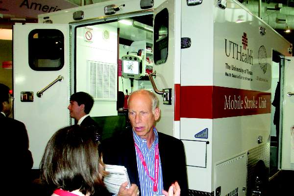
Some U.S. stroke centers now send out a team that can immediately assess and start treating stroke patients in the community. In 2014, the first two U.S. mobile stroke-treatment units began operating, one in Houston and the second in Cleveland.
Initial reports show both programs were successful in cutting the time to deliver thrombolytic treatment with intravenous tissue plasminogen activator (TPA) to appropriate patients.
In Houston, the active phase of the program started in May 2014, and by October 2014, 47 acute ischemic stroke patients had been treated with TPA. The mobile-unit crews started 43% of eligible patients on thrombolysis within 60 minutes of their symptom onset and another 31% were treated starting 61-80 minutes after symptom onset, said Stephanie A. Parker at the International Stroke Conference.

The unit also treats patients diagnosed with hemorrhagic stroke with intravenous nicardipine for rapid blood pressure reduction, said Ms. Parker, a critical care and emergency medicine–trained registered nurse who is project manager for the Houston mobile unit.
The Cleveland program began in July 2014; of the first 100 stroke patients seen by the mobile unit 16 of 19 eligible patients received tPA, with an average time of 56 minutes from symptom onset to treatment. This compared with an average 94 minutes to tPA onset in patients brought conventionally last year to a Cleveland-area hospital, Dr. M. Shazam Hussain said in a report at the meeting, sponsored by the American Heart Association.

The clinical impact and cost effectiveness of the pilot programs using the mobile units have not yet been assessed from the data, Dr. Hussain and Ms. Parker emphasized. Funding for the Cleveland and Houston vehicles came from local donors; the Houston program also received equipment donations from manufacturers.
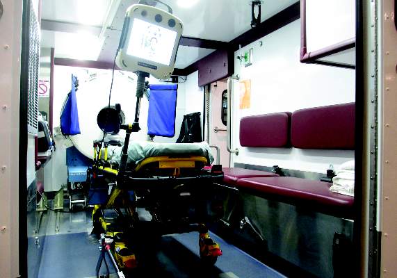
The two mobile units are standard 12-foot, box-shaped ambulances outfitted with a CT scanner, a point-of-care lab, and telemedicine components as well as more standard emergency-vehicle equipment. The Houston vehicle contains “all the diagnostic equipment that is in our emergency room,” Ms. Parker said.
The concept behind both the Cleveland unit, operated by the Cleveland Clinic, and the Houston unit, operated by the University of Texas, Houston, is that the mobile stroke unit arrives to a patient with a suspected stroke, the unit is stationary while a CT scan and other diagnostic tests are run, diagnosis occurs with telemedicine assistance. If the patient is cleared for TPA treatment, the infusion starts and the vehicle carries the patient to an appropriate stroke center.
Currently, the Houston unit goes out with a vascular neurologist and a telemedicine physician on board, but plans are in place to test the feasibility of relying entirely on telemedicine when making diagnostic and treatment decisions. The Cleveland mobile unit already operates in this fashion, with no physician on board, and was the first mobile stroke unit in the world to depend completely on telemedicine, according to Dr. Hussain, a neurologist and head of the stroke program at the Cleveland Clinic.
The world’s first mobile stroke unit began operating in Saarland, Germany, in 2008 (Lancet Neurology 2012;11:397-404), and a second unit began running in Berlin after that, Dr. Hussain noted. Because of limited funding, the service he directs in Cleveland has been operating from 8 a.m.-8 p.m., 7 days a week. The program plans to expand to 24-hour coverage. The Houston mobile unit operates 24/7; it averages two runs per day and administers TPA on 1 of every 10 runs, Ms. Parker said. Both the Houston and Cleveland units tie into the local 911 emergency activation systems for their respective regions.
On Twitter @mitchelzoler
NASHVILLE, TENN. – Bringing a CT scanner and thrombolytic treatment directly to stroke patients in the field sped the time to thrombolysis, compared with waiting for the patient to arrive at the hospital.
Some U.S. stroke centers now send out a team that can immediately assess and start treating stroke patients in the community. In 2014, the first two U.S. mobile stroke-treatment units began operating, one in Houston and the second in Cleveland.
Initial reports show both programs were successful in cutting the time to deliver thrombolytic treatment with intravenous tissue plasminogen activator (TPA) to appropriate patients.
In Houston, the active phase of the program started in May 2014, and by October 2014, 47 acute ischemic stroke patients had been treated with TPA. The mobile-unit crews started 43% of eligible patients on thrombolysis within 60 minutes of their symptom onset and another 31% were treated starting 61-80 minutes after symptom onset, said Stephanie A. Parker at the International Stroke Conference.
The unit also treats patients diagnosed with hemorrhagic stroke with intravenous nicardipine for rapid blood pressure reduction, said Ms. Parker, a critical care and emergency medicine–trained registered nurse who is project manager for the Houston mobile unit.
The Cleveland program began in July 2014; of the first 100 stroke patients seen by the mobile unit 16 of 19 eligible patients received tPA, with an average time of 56 minutes from symptom onset to treatment. This compared with an average 94 minutes to tPA onset in patients brought conventionally last year to a Cleveland-area hospital, Dr. M. Shazam Hussain said in a report at the meeting, sponsored by the American Heart Association.
The clinical impact and cost effectiveness of the pilot programs using the mobile units have not yet been assessed from the data, Dr. Hussain and Ms. Parker emphasized. Funding for the Cleveland and Houston vehicles came from local donors; the Houston program also received equipment donations from manufacturers.
The two mobile units are standard 12-foot, box-shaped ambulances outfitted with a CT scanner, a point-of-care lab, and telemedicine components as well as more standard emergency-vehicle equipment. The Houston vehicle contains “all the diagnostic equipment that is in our emergency room,” Ms. Parker said.
The concept behind both the Cleveland unit, operated by the Cleveland Clinic, and the Houston unit, operated by the University of Texas, Houston, is that the mobile stroke unit arrives to a patient with a suspected stroke, the unit is stationary while a CT scan and other diagnostic tests are run, diagnosis occurs with telemedicine assistance. If the patient is cleared for TPA treatment, the infusion starts and the vehicle carries the patient to an appropriate stroke center.
Currently, the Houston unit goes out with a vascular neurologist and a telemedicine physician on board, but plans are in place to test the feasibility of relying entirely on telemedicine when making diagnostic and treatment decisions. The Cleveland mobile unit already operates in this fashion, with no physician on board, and was the first mobile stroke unit in the world to depend completely on telemedicine, according to Dr. Hussain, a neurologist and head of the stroke program at the Cleveland Clinic.
The world’s first mobile stroke unit began operating in Saarland, Germany, in 2008 (Lancet Neurology 2012;11:397-404), and a second unit began running in Berlin after that, Dr. Hussain noted. Because of limited funding, the service he directs in Cleveland has been operating from 8 a.m.-8 p.m., 7 days a week. The program plans to expand to 24-hour coverage. The Houston mobile unit operates 24/7; it averages two runs per day and administers TPA on 1 of every 10 runs, Ms. Parker said. Both the Houston and Cleveland units tie into the local 911 emergency activation systems for their respective regions.
On Twitter @mitchelzoler
AT THE INTERNATIONAL STROKE CONFERENCE
Key clinical point: Dedicated stroke ambulances that bring a CT scanner and thrombolytic treatment to patients in the field speed thrombolytic therapy.
Major finding: In Cleveland, stroke patients received thrombolysis an average of 38 minutes sooner from the CT-equipped ambulance, compared with standard protocols.
Data source: Prospectively collected data on time-to-treatment from case series in Houston and in Cleveland.
Disclosures: Dr. Hussain and Ms. Parker had no disclosures.

Vagal stimulation may help upper limb stroke recovery
NASHVILLE, TENN. – Patients with upper limbs affected by ischemic stroke who paired traditional rehabilitation exercises with pulsed vagus nerve stimulation boosted functional scores significantly higher than did those who performed exercises alone in a small, randomized pilot trial.
Taken together with the low rate of adverse events associated with device implantation, the study suggests that coupling the interventions is feasible and likely to be beneficial, Dr. Jesse Dawson of the University of Glasgow, Scotland, said at the International Stroke Conference, sponsored by the American Heart Association.
The vagus nerve stimulator (VNS) is typically used to suppress epileptiform discharges and circumvent seizures. The usual stimulation pattern is continuous cycles of 30 seconds on and 5 minutes off. In his randomized, controlled trial, Dr. Dawson set the device to deliver 0.5-second pulses that coincided with each repetition of a rehabilitative movement. When simulated, the nerve releases two proneuroplastic neurotransmitters, acetylcholine and norepinephrine, which then disperse over the cerebral cortex.
“Our theory was that if we timed these releases at specific periods during rehabilitation therapy, we might be able to drive neuroplasticity toward those specific tasks,” Dr. Dawson said at the conference. The technique has proved effective in both aged rats and rat stroke models, he added.
The trial comprised 20 patients who had experienced an ischemic stroke about 2 years prior. Each was left with residual dysfunction in an upper extremity; seven had a paretic limb. The mean Action Research Arm Test (ARAT) score was 33, and the mean upper extremity Fugl-Meyer score was 43, indicating moderate impairment.
Ten patients underwent VNS implantation. Nine completed the trial. One withdrew after 2 weeks because of a transient vocal cord palsy. This later resolved spontaneously.
Other adverse events related to the VNS were also transient. They included taste disturbance, chest pain, mild dysphagia, and nausea after a therapy session.
The 6-week intervention consisted of 18 sessions, each lasting 2 hours. In each, the rehabilitative movement was repeated 300-400 times.
In a per-protocol analysis, there was no significant difference in the upper extremity Fugl-Meyer score at the study’s end. However, when the patient who had withdrawn was excluded from the analysis, the results did become statistically significant. Patients in the dual-therapy group gained almost 10 points, compared with a 3-point gain in the exercise-only group. The ARAT scores were not different at study’s end.
In light of the positive initial results, a sham-controlled, randomized trial is in the works. The trial will randomize 20-25 patients to either the VNS-paired exercise or exercise-only interventions. All participants will receive the VNS device, but only the paired intervention group will receive actual stimuli.
Patricia Smith, Ph.D., the Doris E. Porter Professor in Physical Therapy at the University of Texas Southwestern, Dallas, is the lead investigator.
MicroTransponder, which makes the VNS unit, is sponsoring both the studies. Neither Dr. Dawson nor Dr. Smith have any financial ties to the company.
On Twitter @alz_gal
AT THE INTERNATIONAL STROKE CONFERENCE
Key clinical point: Vagus nerve stimulation paired with physical therapy may improve functional recovery in upper limbs after stroke.
Major finding: The paired intervention boosted the upper extremity Fugl-Meyer score by 10 points in the intervention group and 3 points in the control group.
Data source: A randomized trial of 20 patients about 2 years post stroke who had residual dysfunction in an upper extremity.
Disclosures: MicroTransponder, which makes the VNS unit, is sponsoring both the studies. Dr. Dawson has no financial ties to the company.
Vertebrobasilar disease, low distal flow triggers strokes
NASHVILLE, TENN. – In patients with symptomatic vertebrobasilar disease, low distal flow measured noninvasively predicted a patient’s subsequent risk for stroke in a multicenter, prospective study of 72 patients.
The finding “has implications for investigating interventional or aggressive medical treatments,” which should be aimed at this high-risk subgroup of patients, Dr. Sepideh Amin-Hanjani said at the International Stroke Conference, sponsored by the American Heart Association.

Patients with “the highest risk for recurrence have the best chance to benefit from intervention,” said Dr. Amin-Hanjani, professor of neurosurgery and codirector of neurovascular surgery at the University of Illinois at Chicago. For the time being, no interventions for vertebrobasilar disease have proven efficacy and safety, but the new finding provides a way to identify the highest risk patients who stand to gain the most from intervention and should serve as the target population for future trials.
The VERiTAS (Vertebrobasilar Flow Evaluation and Risk of Transient Ischemic Attack and Stroke) trial enrolled 72 adults at any of five U.S. centers or at one in Canada. Enrolled patients had a recent stroke or transient ischemic attack in their vertebrobasilar territory plus angiographic evidence of at least 50% stenosis in an extra- or intracranial vertebral or basilar artery. All patients underwent quantitative MR angioplasty of their large vertebrobasilar arteries using software, Noninvasive Optimal Vessel Analysis (NOVA), that measures volumetric flow rates through vessels. Eighteen patients (25%) had low distal flow, defined as a greater than 20% reduction in flow, compared with normal, and 54 patients (75%) who had normal flow.
The study’s primary endpoint was an incident ischemic stroke in the vertebrobasilar territory during 12 months of follow-up. During a median follow-up of 23 months, 10 patients had this type of new stroke.
Among the 18 low-flow patients, four (22%) had a primary endpoint after 12 months, and among the 54 normal-flow patients, 2 (4%) had a primary endpoint after 12 months, a statistically significant difference, Dr. Amin-Hanjani reported at the conference.
In a multivariate analysis, low-flow at baseline linked with a significant, ninefold increased risk for incident stroke, compared with normal-flow patients. The location of the blockage – in the basilar region, vertebral region, or both – had no apparent impact on outcome.
About 30% of ischemic strokes occur in the posterior circulation, and about a third of those are caused by vertebrobasilar disease secondary to atherosclerosis. Overall patients who have had strokes of this type face a 10%-15% rate of new stroke annually despite receiving standard medical treatment, Dr. Amin-Hanjani said.
Dr. Amin-Hanjanihas received research grants from GE Healthcare and VasSol, the company that markets the NOVA software used in VERiTAS. A coauthor on the report has a significant ownership interest in VasSol.
On Twitter @mitchelzoler
The main message from VERiTAS is that low blood flow distal to vertebrobasilar arterial stenosis matters. When a patient with a posterior-circulation stroke has an occlusion or high-grade stenosis causing poor distal flow they have an increased risk for recurrent stroke.
 |
| Mitchel L. Zoler/Frontline Medical News Dr. Colin P. Derdeyn |
The VERiTAS results move the field forward by providing another brick in the wall of evidence that reduced perfusion in patients with occlusive cerebrovascular disease identifies a group of patients who are at high risk for a future stroke. I believe that this sort of disease is fairly common, but clinicians often do not actively look for it because no treatment for it has been proven effective. Recent trial results failed to show benefit from either angioplasty or stenting.
The next step will be a trial focused on with low-flow patients that treats them with more aggressive medical management or with an intervention to try to identify some treatment that produces incremental benefit in this target population.
Until we see positive results in such a trial, the practical implications of the VERiTAS results are unclear. VERiTAS provides a good indication of the natural history of vertebrobasilar disease when patients receive today’s standard treatment. Patients with low distal flow had about a 20% rate of new strokes during 12 months of follow-up; outcomes were much better among patients with normal distal flow.
Some clinicians will see the high risk among low-flow patients as justification for some sort of intervention even though nothing has been proven to work. Others will take a more conservative approach and treat these patients with standard medical treatment for atherosclerotic disease, or enroll them into an intervention trial. Arguably, there is no reason to even measure distal flow on a routine basis right now because there is no proven way to act on this information.
The method used in VERiTAS to noninvasively measure distal flow – quantitative MR angiography with the NOVA software – is probably not widely used today, but the VERiTAS results might change that. The study’s findings show that this type of imaging can produce clinically meaningful measurements; not many other imaging technologies can say that as of now.
Dr. Colin P. Derdeyn is professor of neurology and director of the Center for Stroke and Cerebrovascular Disease at Washington University in St. Louis. He was a coinvestigator on VERiTAS. He has received research support from MicroVention and has a modest ownership interest in Pulse Therapeutics. He made these comments in an interview.
The main message from VERiTAS is that low blood flow distal to vertebrobasilar arterial stenosis matters. When a patient with a posterior-circulation stroke has an occlusion or high-grade stenosis causing poor distal flow they have an increased risk for recurrent stroke.
|
| Mitchel L. Zoler/Frontline Medical News Dr. Colin P. Derdeyn |
The VERiTAS results move the field forward by providing another brick in the wall of evidence that reduced perfusion in patients with occlusive cerebrovascular disease identifies a group of patients who are at high risk for a future stroke. I believe that this sort of disease is fairly common, but clinicians often do not actively look for it because no treatment for it has been proven effective. Recent trial results failed to show benefit from either angioplasty or stenting.
The next step will be a trial focused on with low-flow patients that treats them with more aggressive medical management or with an intervention to try to identify some treatment that produces incremental benefit in this target population.
Until we see positive results in such a trial, the practical implications of the VERiTAS results are unclear. VERiTAS provides a good indication of the natural history of vertebrobasilar disease when patients receive today’s standard treatment. Patients with low distal flow had about a 20% rate of new strokes during 12 months of follow-up; outcomes were much better among patients with normal distal flow.
Some clinicians will see the high risk among low-flow patients as justification for some sort of intervention even though nothing has been proven to work. Others will take a more conservative approach and treat these patients with standard medical treatment for atherosclerotic disease, or enroll them into an intervention trial. Arguably, there is no reason to even measure distal flow on a routine basis right now because there is no proven way to act on this information.
The method used in VERiTAS to noninvasively measure distal flow – quantitative MR angiography with the NOVA software – is probably not widely used today, but the VERiTAS results might change that. The study’s findings show that this type of imaging can produce clinically meaningful measurements; not many other imaging technologies can say that as of now.
Dr. Colin P. Derdeyn is professor of neurology and director of the Center for Stroke and Cerebrovascular Disease at Washington University in St. Louis. He was a coinvestigator on VERiTAS. He has received research support from MicroVention and has a modest ownership interest in Pulse Therapeutics. He made these comments in an interview.
NASHVILLE, TENN. – In patients with symptomatic vertebrobasilar disease, low distal flow measured noninvasively predicted a patient’s subsequent risk for stroke in a multicenter, prospective study of 72 patients.
The finding “has implications for investigating interventional or aggressive medical treatments,” which should be aimed at this high-risk subgroup of patients, Dr. Sepideh Amin-Hanjani said at the International Stroke Conference, sponsored by the American Heart Association.
Patients with “the highest risk for recurrence have the best chance to benefit from intervention,” said Dr. Amin-Hanjani, professor of neurosurgery and codirector of neurovascular surgery at the University of Illinois at Chicago. For the time being, no interventions for vertebrobasilar disease have proven efficacy and safety, but the new finding provides a way to identify the highest risk patients who stand to gain the most from intervention and should serve as the target population for future trials.
The VERiTAS (Vertebrobasilar Flow Evaluation and Risk of Transient Ischemic Attack and Stroke) trial enrolled 72 adults at any of five U.S. centers or at one in Canada. Enrolled patients had a recent stroke or transient ischemic attack in their vertebrobasilar territory plus angiographic evidence of at least 50% stenosis in an extra- or intracranial vertebral or basilar artery. All patients underwent quantitative MR angioplasty of their large vertebrobasilar arteries using software, Noninvasive Optimal Vessel Analysis (NOVA), that measures volumetric flow rates through vessels. Eighteen patients (25%) had low distal flow, defined as a greater than 20% reduction in flow, compared with normal, and 54 patients (75%) who had normal flow.
The study’s primary endpoint was an incident ischemic stroke in the vertebrobasilar territory during 12 months of follow-up. During a median follow-up of 23 months, 10 patients had this type of new stroke.
Among the 18 low-flow patients, four (22%) had a primary endpoint after 12 months, and among the 54 normal-flow patients, 2 (4%) had a primary endpoint after 12 months, a statistically significant difference, Dr. Amin-Hanjani reported at the conference.
In a multivariate analysis, low-flow at baseline linked with a significant, ninefold increased risk for incident stroke, compared with normal-flow patients. The location of the blockage – in the basilar region, vertebral region, or both – had no apparent impact on outcome.
About 30% of ischemic strokes occur in the posterior circulation, and about a third of those are caused by vertebrobasilar disease secondary to atherosclerosis. Overall patients who have had strokes of this type face a 10%-15% rate of new stroke annually despite receiving standard medical treatment, Dr. Amin-Hanjani said.
Dr. Amin-Hanjanihas received research grants from GE Healthcare and VasSol, the company that markets the NOVA software used in VERiTAS. A coauthor on the report has a significant ownership interest in VasSol.
On Twitter @mitchelzoler
AT THE INTERNATIONAL STROKE CONFERENCE
Key clinical point: Following posterior-circulation ischemic stroke, patients with vertebrobasilar disease and low distal blood flow had significantly more subsequent strokes, compared with normal-flow patients.
Major finding: Low distal flow patients had ninefold more strokes, compared with normal-flow patients during a 1-year follow-up period.
Data source: VERiTAS, a prospective, multicenter observational study of 72 patients with a prior stroke or transient ischemic attack in the vertebrobasilar territory.
Disclosures: Dr. Amin-Hanjani has received research grants from GE Healthcare and VasSol, the company that markets the NOVA software used in VERiTAS. A coauthor on the report has a significant ownership interest in VasSol.

Thrombolysis gap for stroke octogenarians disappears
NASHVILLE, TENN. – By 2010, U.S. octogenarians with acute ischemic stroke received intravenous thrombolytic treatment about as often as younger patients, showing that a sharp, age-related disparity in thrombolytic use a decade before had disappeared, based on comprehensive national data.
The 2010 data from the Nationwide Inpatient Sample further showed that the sex-based disparity in treatment with intravenous tissue plasminogen activator (TPA) seen in 2000 resolved as well from 2000 to 2010, but other disparities worsened with declines during the period in use of TPA at rural hospitals relative to urban hospitals and at nonteaching hospitals, compared with teaching hospitals, Dr. Michelle P. Lin said at the International Stroke Conference.

Perhaps most importantly, the statistics showed a “dramatic” increase in TPA use among all age groups during the decade ending in 2010, when thrombolytic therapy was administered to 3.5%-3.9% of adult patients regardless of their age, said Dr. Lin of the department of neurology at the University of Southern California in Los Angeles. In 2000, U.S. patients received TPA at less than a third of that rate.
Low TPA use in 2000 among patients aged 80 or older in part reflected the low number of octogenarians enrolled in the trials that documented the safety and efficacy of TPA for acute ischemicstroke patients, Dr. Lin said at the International Stroke Conference, sponsored by the American Heart Association.
Data from the National Inpatient Sample included information on the treatment received by 5,932,175 patients with acute ischemic stroke at more than 1,000 U.S. hospitals during 2000-2010. The age breakdown of the nearly 6 millionpatients showed 28% were aged 18-64 years, 37% were65-79, and 35% were 80 years or older.
In 2000, medical staffs administered intravenous treatment with TPA to 1.02% of these patients aged 18-64 years, 0.92% of patients aged 65-79 years, and 0.47% of patients aged 80 or older. By 2010, the annual rates of TPA use ran 3.61% in those 18-64 years, 3.87% among those 65-79 years, and 3.55% in patients 80 years or older. In an adjusted analysis, this translated into a greater than threefold increase in TPA use among the 18- to 64-year-olds, a nearly fourfold rise in patients 65-79 years, and a nearly sevenfold jump among those 80 or older, a 24% average annual increased rate among the oldest patients, who averaged 86 years old, Dr. Lin reported.
The data also showed that among the octogenarians during 2000-2005, TPA use in women lagged behind use in men by a relative 15%, but this completely disappeared during 2006-2010, when usage rates in men and women evened out. TPA use among African Americans, Hispanics, and Asians, compared with whites, remained significantly below the rate in whites throughout the decade, although the extent of the disparity narrowed for African Americans and Asians during the second half of the decade, compared with the first half.
Dr. Lin and her associates also analyzed TPA use relative to the demographic setting of the hospital and its teaching status. During 2000-2010, the relative usage of TPA at rural hospitals, compared with urban hospitals, fell from 65% of the comparator level to 36%. Among nonteaching hospitals, the rate of TPA use dropped from 52% of the teaching hospitals’ level to 49%.
Dr. Lin said she had no relevant financial disclosures.
mzoler@frontlinemedcom.com
On Twitter @mitchelzoler
NASHVILLE, TENN. – By 2010, U.S. octogenarians with acute ischemic stroke received intravenous thrombolytic treatment about as often as younger patients, showing that a sharp, age-related disparity in thrombolytic use a decade before had disappeared, based on comprehensive national data.
The 2010 data from the Nationwide Inpatient Sample further showed that the sex-based disparity in treatment with intravenous tissue plasminogen activator (TPA) seen in 2000 resolved as well from 2000 to 2010, but other disparities worsened with declines during the period in use of TPA at rural hospitals relative to urban hospitals and at nonteaching hospitals, compared with teaching hospitals, Dr. Michelle P. Lin said at the International Stroke Conference.
Perhaps most importantly, the statistics showed a “dramatic” increase in TPA use among all age groups during the decade ending in 2010, when thrombolytic therapy was administered to 3.5%-3.9% of adult patients regardless of their age, said Dr. Lin of the department of neurology at the University of Southern California in Los Angeles. In 2000, U.S. patients received TPA at less than a third of that rate.
Low TPA use in 2000 among patients aged 80 or older in part reflected the low number of octogenarians enrolled in the trials that documented the safety and efficacy of TPA for acute ischemicstroke patients, Dr. Lin said at the International Stroke Conference, sponsored by the American Heart Association.
Data from the National Inpatient Sample included information on the treatment received by 5,932,175 patients with acute ischemic stroke at more than 1,000 U.S. hospitals during 2000-2010. The age breakdown of the nearly 6 millionpatients showed 28% were aged 18-64 years, 37% were65-79, and 35% were 80 years or older.
In 2000, medical staffs administered intravenous treatment with TPA to 1.02% of these patients aged 18-64 years, 0.92% of patients aged 65-79 years, and 0.47% of patients aged 80 or older. By 2010, the annual rates of TPA use ran 3.61% in those 18-64 years, 3.87% among those 65-79 years, and 3.55% in patients 80 years or older. In an adjusted analysis, this translated into a greater than threefold increase in TPA use among the 18- to 64-year-olds, a nearly fourfold rise in patients 65-79 years, and a nearly sevenfold jump among those 80 or older, a 24% average annual increased rate among the oldest patients, who averaged 86 years old, Dr. Lin reported.
The data also showed that among the octogenarians during 2000-2005, TPA use in women lagged behind use in men by a relative 15%, but this completely disappeared during 2006-2010, when usage rates in men and women evened out. TPA use among African Americans, Hispanics, and Asians, compared with whites, remained significantly below the rate in whites throughout the decade, although the extent of the disparity narrowed for African Americans and Asians during the second half of the decade, compared with the first half.
Dr. Lin and her associates also analyzed TPA use relative to the demographic setting of the hospital and its teaching status. During 2000-2010, the relative usage of TPA at rural hospitals, compared with urban hospitals, fell from 65% of the comparator level to 36%. Among nonteaching hospitals, the rate of TPA use dropped from 52% of the teaching hospitals’ level to 49%.
Dr. Lin said she had no relevant financial disclosures.
mzoler@frontlinemedcom.com
On Twitter @mitchelzoler
AT THE INTERNATIONAL STROKE CONFERENCE
Key clinical point: By 2010, U.S. ischemic stroke patients aged 80 or older received thrombolytic treatment as often as younger patients.
Major finding: In 2010, thrombolysis was used to treat 3.55% of U.S. stroke patients 80 years or older, 3.87% of those 65-79, and 3.61% of those 18-64.
Data source: The U.S. National Inpatient Sample for 2000-2010, which included 5,932,175 adults with acute ischemic stroke.
Disclosures: Dr. Lin said she had no relevant financial disclosures.

General anesthesia linked to worsened stroke outcomes
NASHVILLE, TENN. – When acute ischemic stroke patients undergo an emergency endovascular procedure is it best done with general anesthesia or nongeneral anesthesia?
A post hoc analysis of data collected by a Dutch randomized, controlled trial of intra-arterial therapy suggested that nongeneral anesthesia was associated with substantially better patient outcomes, and the findings convinced the Dutch investigators who ran the study to stick with nongeneral anesthesia as their default approach.
In MR CLEAN (Multicenter Randomized Clinical Trial of Endovascular Treatment for Acute Ischemic Stroke in the Netherlands) (N. Engl. J. Med. 2015;372:11-20), 216 acute ischemic stroke patients underwent intra-arterial treatment following randomization. Among these patients, 79 were treated with general anesthesia and 137 with nongeneral anesthesia. The anesthesia choice was made on a case-by-case basis by each participating interventionalist.
The study’s primary endpoint – 90-day status rated as a good function based on a modified Rankin sale score of 0-2 – occurred in 38% of the intra-arterial patients treated with nongeneral anesthesia, 23% of the intra-arterial patients treated with general anesthesia, and 19% among the control patients who received standard treatment without intra-arterial intervention.

“The effect on outcome that we found with intra-arterial treatment in MR CLEAN was not observed in the subgroup of patients treated with general anesthesia,” said Dr. Olvert A. Berkhemer at the International Stroke Conference. The analysis also showed that patients in the general and nongeneral anesthesia subgroups had similar stroke severity as measured by their National Institutes of Health Stroke Scale score, said Dr. Berkhemer, a researcher at the Academic Medical Center in Amsterdam.
But U.S. stroke specialists who heard the report cautioned that unidentified confounders might explain the results, and they also expressed skepticism that the Dutch observations would deter U.S. interventionalists from continuing to use general anesthesia when they perform endovascular procedures.
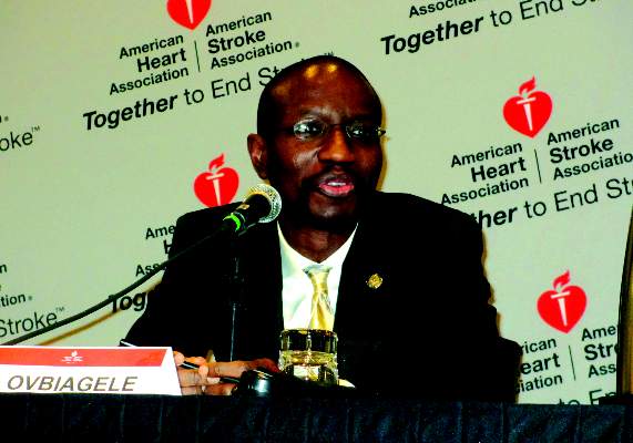
“The big concern [about this analysis] is that there may have been some things about the general anesthesia patients that they did not account for. I suspect there is a huge bias, that general anesthesia patients were sicker,” said Dr. Bruce Ovbiagele, professor and chief of neurology at the Medical University of South Carolina in Charleston. “At my institution they have used nongeneral anesthesia, but I have been at other places where they usually use general anesthesia; it is variable,” Dr. Ovbiagele added.
Dr. Berkhemer’s analysis also showed that general anesthesia linked with a delayed start to treatment, but without resulting in a significant difference in time to reperfusion. He noted that “sometimes you cannot do the procedure without general anesthesia,” and in MR CLEAN 6 of the 137 intra-arterial patients who started with nongeneral anesthesia eventually received general anesthesia because of discomfort and pain, Dr. Berkhemer said at the conference, sponsored by the American Heart Association.
He speculated that patients who did not receive general anesthesia did better because they did not undergo acute episodes of reduced blood pressure caused by the hypotensive effect of general anesthesia.

The findings reinforced the approach already used in most of the Dutch centers that participated in MR CLEAN, where nongeneral anesthesia is preferred when possible. “In our center we always use nongeneral anesthesia, we are happy with that and we are not going to change,” said Dr. Diederik W.J. Dippel, lead investigator of MR CLEAN and professor of neurology at Erasmus University Medical Center in Rotterdam, The Netherlands. This prejudice against general anesthesia would make it hard to run a trial in The Netherlands that matched the two anesthesia approaches against each other, Dr. Dippel added.
On Twitter @mitchelzoler
Results from prior analyses had also shown better outcomes of acute ischemic stroke patients undergoing endovascular intervention when they avoided general anesthesia. A notable feature of Dr. Berkhemer’s analysis was that the stroke severity levels were well balanced between the patients who received general anesthesia and those who did not. But many other factors aside from stroke severity can affect whether or not a patient receives general anesthesia.
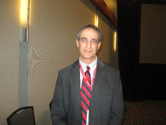 |
Dr. Larry B. Goldstein |
Although the outcome differences seen in this analysis were striking, many factors could have contributed. Patients received different drugs, patients may have had widely divergent clinical states despite their similar stroke severity, and the people performing the procedures were different. The many variables make it very hard to pinpoint the cause of the different outcomes.
At my institution, Duke, the interventionalists who work on acute ischemic stroke patients almost exclusively use general anesthesia. That’s because of their concern about how patients will act during a very delicate procedure. For example, stroke patients can have an aphasia that makes it hard for them to respond to requests to do something specific during the procedure. I am very skeptical that my colleagues will decide to switch to using no general anesthesia based on the results of this new analysis.
I agree with the Dutch investigators that a randomized, controlled trial of general anesthesia or no general anesthesia would be very hard to perform because individual interventionalists would need to believe there is equipoise between the two anesthesia approaches. Most interventionalists right now probably believe the approach they have always used remains best and so would be unwilling to participate in a randomized controlled trial.
Dr. Larry B. Goldstein is professor of neurology and chief of the stroke center at Duke University in Durham, N.C. He had no relevant disclosures. He made these comments in an interview.
Results from prior analyses had also shown better outcomes of acute ischemic stroke patients undergoing endovascular intervention when they avoided general anesthesia. A notable feature of Dr. Berkhemer’s analysis was that the stroke severity levels were well balanced between the patients who received general anesthesia and those who did not. But many other factors aside from stroke severity can affect whether or not a patient receives general anesthesia.
|
Dr. Larry B. Goldstein |
Although the outcome differences seen in this analysis were striking, many factors could have contributed. Patients received different drugs, patients may have had widely divergent clinical states despite their similar stroke severity, and the people performing the procedures were different. The many variables make it very hard to pinpoint the cause of the different outcomes.
At my institution, Duke, the interventionalists who work on acute ischemic stroke patients almost exclusively use general anesthesia. That’s because of their concern about how patients will act during a very delicate procedure. For example, stroke patients can have an aphasia that makes it hard for them to respond to requests to do something specific during the procedure. I am very skeptical that my colleagues will decide to switch to using no general anesthesia based on the results of this new analysis.
I agree with the Dutch investigators that a randomized, controlled trial of general anesthesia or no general anesthesia would be very hard to perform because individual interventionalists would need to believe there is equipoise between the two anesthesia approaches. Most interventionalists right now probably believe the approach they have always used remains best and so would be unwilling to participate in a randomized controlled trial.
Dr. Larry B. Goldstein is professor of neurology and chief of the stroke center at Duke University in Durham, N.C. He had no relevant disclosures. He made these comments in an interview.
NASHVILLE, TENN. – When acute ischemic stroke patients undergo an emergency endovascular procedure is it best done with general anesthesia or nongeneral anesthesia?
A post hoc analysis of data collected by a Dutch randomized, controlled trial of intra-arterial therapy suggested that nongeneral anesthesia was associated with substantially better patient outcomes, and the findings convinced the Dutch investigators who ran the study to stick with nongeneral anesthesia as their default approach.
In MR CLEAN (Multicenter Randomized Clinical Trial of Endovascular Treatment for Acute Ischemic Stroke in the Netherlands) (N. Engl. J. Med. 2015;372:11-20), 216 acute ischemic stroke patients underwent intra-arterial treatment following randomization. Among these patients, 79 were treated with general anesthesia and 137 with nongeneral anesthesia. The anesthesia choice was made on a case-by-case basis by each participating interventionalist.
The study’s primary endpoint – 90-day status rated as a good function based on a modified Rankin sale score of 0-2 – occurred in 38% of the intra-arterial patients treated with nongeneral anesthesia, 23% of the intra-arterial patients treated with general anesthesia, and 19% among the control patients who received standard treatment without intra-arterial intervention.
“The effect on outcome that we found with intra-arterial treatment in MR CLEAN was not observed in the subgroup of patients treated with general anesthesia,” said Dr. Olvert A. Berkhemer at the International Stroke Conference. The analysis also showed that patients in the general and nongeneral anesthesia subgroups had similar stroke severity as measured by their National Institutes of Health Stroke Scale score, said Dr. Berkhemer, a researcher at the Academic Medical Center in Amsterdam.
But U.S. stroke specialists who heard the report cautioned that unidentified confounders might explain the results, and they also expressed skepticism that the Dutch observations would deter U.S. interventionalists from continuing to use general anesthesia when they perform endovascular procedures.
“The big concern [about this analysis] is that there may have been some things about the general anesthesia patients that they did not account for. I suspect there is a huge bias, that general anesthesia patients were sicker,” said Dr. Bruce Ovbiagele, professor and chief of neurology at the Medical University of South Carolina in Charleston. “At my institution they have used nongeneral anesthesia, but I have been at other places where they usually use general anesthesia; it is variable,” Dr. Ovbiagele added.
Dr. Berkhemer’s analysis also showed that general anesthesia linked with a delayed start to treatment, but without resulting in a significant difference in time to reperfusion. He noted that “sometimes you cannot do the procedure without general anesthesia,” and in MR CLEAN 6 of the 137 intra-arterial patients who started with nongeneral anesthesia eventually received general anesthesia because of discomfort and pain, Dr. Berkhemer said at the conference, sponsored by the American Heart Association.
He speculated that patients who did not receive general anesthesia did better because they did not undergo acute episodes of reduced blood pressure caused by the hypotensive effect of general anesthesia.
The findings reinforced the approach already used in most of the Dutch centers that participated in MR CLEAN, where nongeneral anesthesia is preferred when possible. “In our center we always use nongeneral anesthesia, we are happy with that and we are not going to change,” said Dr. Diederik W.J. Dippel, lead investigator of MR CLEAN and professor of neurology at Erasmus University Medical Center in Rotterdam, The Netherlands. This prejudice against general anesthesia would make it hard to run a trial in The Netherlands that matched the two anesthesia approaches against each other, Dr. Dippel added.
On Twitter @mitchelzoler
AT THE INTERNATIONAL STROKE CONFERENCE
Key clinical point: Post hoc analysis of acute ischemic stroke patients treated intra-arterially suggests that avoiding general anesthesia produces better long-term outcomes.
Major finding: After 90 days, 38% of patients treated without general anesthesia and 23% of those who received general anesthesia had good outcomes.
Data source: Analysis of 216 patients in the Dutch MR CLEAN trial who underwent treatment for acute ischemic stroke.
Disclosures: Dr. Berkhemer, Dr. Dippel, and Dr. Ovbiagele had no disclosures.
Better stroke treatment moves tantalizingly within reach
Stroke is one of the most feared medical conditions, with the specter of suddenly finding oneself unable to talk, eat, walk, or live independently, according to study results.
In mid-February, results from three trials reported at the International Stroke Conference in Nashville, Tenn., changed the face of ischemic stroke treatment by proving that emergency endovascular catheterization to remove the embolus blocking cerebral blood flow produced better long-term outcomes than standard treatment with intravenous thrombolysis.

It wasn’t just that patients did better with endovascular embolectomy; it was how much they did better. In the two trials run in the United States and abroad, SWIFT PRIME and ESCAPE, the percentage of patients rated as not disabled (a modified Rankin Scale score of 0-1) when assessed after 90 days was 36% and 42% for patients treated with endovascular therapy in the two studies, compared with 17% and 19% in the two control arms. Embolectomy boosted the fraction of patients having the best stroke outcomes more than twofold, a breathtaking leap in efficacy.
Dr. Jeffrey L. Saver from UCLA, lead investigator for SWIFT PRIME, called it a “once-in-a-field” result, meaning that never again will stroke clinicians see this degree of incremental improvement by adding a new intervention.
The frustrating irony is how challenging delivery of this disease-altering treatment will be on a national scale. One problem is that it didn’t result from a single change in treatment, but from a careful mix of new diagnostic techniques with sophisticated CT imaging, new systems for expediting diagnosis, triage, transport, and treatment, in combination with new technology in the form of emboli-retrieving stents.
Stroke management specialists see a daunting series of issues to tackle as they attempt to roll out emergency endovascular interventions on a routine scale throughout much of the United States. Many more centers must open, modeled on the ones that succeeded in the trials. The centers need to be rationally positioned so they are close to patients but also give each center enough case volume to foster high interventional-skill levels. Staffing must be found for fast-moving stroke response teams that can make the diagnostics and interventions available around the clock and interpret the images to select appropriate patients. Ambulance systems have to be set up that take likely stroke patients to the centers that will best meet their treatment needs.
The stroke and public health communities will need to invest a lot of time, money, and leadership to make this happen, but it’s a clear mandate, given the promise endovascular treatment now holds to blunt the impact of one of medicine’s most feared maladies.
On Twitter @mitchelzoler
Stroke is one of the most feared medical conditions, with the specter of suddenly finding oneself unable to talk, eat, walk, or live independently, according to study results.
In mid-February, results from three trials reported at the International Stroke Conference in Nashville, Tenn., changed the face of ischemic stroke treatment by proving that emergency endovascular catheterization to remove the embolus blocking cerebral blood flow produced better long-term outcomes than standard treatment with intravenous thrombolysis.
It wasn’t just that patients did better with endovascular embolectomy; it was how much they did better. In the two trials run in the United States and abroad, SWIFT PRIME and ESCAPE, the percentage of patients rated as not disabled (a modified Rankin Scale score of 0-1) when assessed after 90 days was 36% and 42% for patients treated with endovascular therapy in the two studies, compared with 17% and 19% in the two control arms. Embolectomy boosted the fraction of patients having the best stroke outcomes more than twofold, a breathtaking leap in efficacy.
Dr. Jeffrey L. Saver from UCLA, lead investigator for SWIFT PRIME, called it a “once-in-a-field” result, meaning that never again will stroke clinicians see this degree of incremental improvement by adding a new intervention.
The frustrating irony is how challenging delivery of this disease-altering treatment will be on a national scale. One problem is that it didn’t result from a single change in treatment, but from a careful mix of new diagnostic techniques with sophisticated CT imaging, new systems for expediting diagnosis, triage, transport, and treatment, in combination with new technology in the form of emboli-retrieving stents.
Stroke management specialists see a daunting series of issues to tackle as they attempt to roll out emergency endovascular interventions on a routine scale throughout much of the United States. Many more centers must open, modeled on the ones that succeeded in the trials. The centers need to be rationally positioned so they are close to patients but also give each center enough case volume to foster high interventional-skill levels. Staffing must be found for fast-moving stroke response teams that can make the diagnostics and interventions available around the clock and interpret the images to select appropriate patients. Ambulance systems have to be set up that take likely stroke patients to the centers that will best meet their treatment needs.
The stroke and public health communities will need to invest a lot of time, money, and leadership to make this happen, but it’s a clear mandate, given the promise endovascular treatment now holds to blunt the impact of one of medicine’s most feared maladies.
On Twitter @mitchelzoler

Stroke thrombolysis achieved inside an hour works best
NASHVILLE, TENN. – Fast thrombolytic treatment of acute ischemic stroke clearly helps patients, but being really, really fast is even better.
Patients treated with intravenous tissue plasminogen activator (tPA) within the first 60 minutes of their stroke onset, the putative “golden hour,” had significantly better outcomes at hospital discharge, compared with patients treated just an hour later, based on U.S. data from more than 65,000 acute ischemic stroke patients.
“In national U.S. clinical practice, treatment with intravenous tPA in the golden hour, compared with later, is associated with more frequent independent ambulation at discharge, discharge to home, and freedom from disability or dependence at discharge,” compared with patients treated at 61-180 minutes or later, Dr. Jeffrey L. Saver said at the International Stroke Conference.
“These findings support intensive efforts to accelerate patient presentation and treatment initiation, such as Target: Stroke Phase II and mobile CT ambulances, to maximize benefit of thrombolytic therapy for acute ischemic stroke,” said Dr. Saver, professor of neurology and director of the stroke center at the University of California, Los Angeles.
His study used data collected from 65,348 patients with acute ischemic stroke treated at any of 1,456 U.S. hospitals participating in the Get With the Guidelines–Stroke program during 2009-2013. Of those patients, 878 (1.3%) received tPA within the first hour following onset of their stroke, 10% within 61-90 minutes, 71% within 91-180 minutes, and 18% within 181-270 minutes.
Although the 878 patients treated within an hour of symptom onset constituted little more than 1% of all patients, the series was 10-fold larger than any prior report, which allowed a venture into “terra incognita” for insight into thrombolysis efficacy when used so early during a stroke, Dr. Saver noted. “Innovations in prehospital and emergency department systems increasingly enable intravenous tPA delivery in the first 60 minutes,” he said at the meeting, which was sponsored by the American Heart Association.
In a multivariate analysis that adjusted for many potential confounders, treatment in the first 60 minutes linked with statistically significant improvements, compared with patients treated at 1-4.5 hours, for several outcome measures at hospital discharge, including a 72% relative increase in being nondisabled – a modified Rankin score of 0 – and a 58% relative increase in being independent – a modified Rankin scale score of 0-2.
Dr. Saver highlighted how even a 30-minute drop in the time to treatment produced substantively better outcomes. The percentage of patients with a modified Rankin score of 0-1 at discharge was 38% in the 0- to 60-minute patients, 33% in those treated after 91-120 minutes had elapsed, and 28% in those treated 121-180 minutes after symptom onset.
Dr. Saver has been a consultant to BrainGate, Covidien, Grifols, and Stryker and has received research support from Covidien, Lundbeck, and Stryker.
On Twitter @mitchelzoler
AT THE INTERNATIONAL STROKE CONFERENCE
Key clinical point: Acute ischemic stroke patients who started intravenous tPA within an hour of onset had the best outcomes.
Major finding: Stroke thrombolysis within an hour produced 72% more nondisabled patients at hospital discharge, compared with treatment after 1-4.5 hours.
Data source: Review of 65,348 U.S. acute ischemic patients who arrived at hospitals participating in Get With the Guidelines during 2009-2013.
Disclosures: Dr. Saver has been a consultant to BrainGate, Covidien, Grifols, and Stryker and has received research support from Covidien, Lundbeck, and Stryker.

Faster intra-arterial stroke treatment improved outcomes
NASHVILLE, TENN.– Results from several trials recently showed that endovascular therapy can improve long-term stroke outcome substantially, compared with conventional intravenous thrombolytic therapy. A new analysis from one of those studies now shows how time sensitive this benefit is, further underscoring the importance of rapid treatment for patients with acute ischemic stroke.
A delay of just an hour or 2 in the time to reperfusion of the blocked cerebral blood supply had a substantial impact on the added efficacy of intra-arterial therapy for improving long-term clinical outcome in MR CLEAN, a Dutch study with 500 acute ischemic stroke patients randomized to intra-arterial therapy or intravenous thrombolysis.
When only 2 hours elapsed from the time of stroke onset to the time of reperfusion, patients enrolled in the MR CLEAN (Multicenter Randomized Clinical Trial of Endovascular Treatment for Acute Ischemic Stroke in the Netherlands) study who received intra-arterial therapy had a 33% higher rate of scoring 0-2 on the modified Rankin scale at 90 days after stroke onset (an indication of either no or limited long-term disability), compared with patients on intravenous thrombolytic therapy. In contrast, when 6 hours elapsed from the time of stroke onset to reperfusion the incremental benefit from intra-arterial treatment dropped to 6.5%, a statistically significant decline in the treatment effect, Dr. Puck S.S. Fransen said at the International Stroke Conference.

In other words, for every 1-hour delay in time to reperfusion after 2 hours out from stroke onset, there was about a 7% drop in added advantage of intra-arterial therapy over standard therapy, reinforcing the need to start intra-arterial therapy and produce reperfusion as quickly as possible in qualifying patients, said Dr. Fransen, a researcher in the department of neurology at Erasmus University in Rotterdam, the Netherlands.
The overall benefit from intra-arterial therapy over standard treatment in MR CLEAN was a 13.5% improvement in patients having a good outcome by modified Rankin score after 90 days. By adjusted odds ratio analysis intra-arterial treatment produced a good outcome about twice as often as conventional treatment with intravenous thrombolysis.
Her analysis also showed that when patients started receiving intra-arterial therapy after more than 6 hours had elapsed from stroke onset its added benefit compared with thrombolysis became statistically insignificant, essentially disappearing.
“Delay to reperfusion is costly to the individual patient; that is the message,” commented Dr. Joseph P. Broderick, professor of neurology and director of the Neuroscience Institute at the University of Cincinnati.
MR CLEAN’s primary aim compared the efficacy and safety of intra-arterial therapy (with intra-arterial thrombolysis, intra-arterial thrombectomy, or both) vs. standard treatment with intravenous thrombolysis in 500 patients with a proximal occlusion in a large intracerebral artery seen at any of 16 Dutch centers. The primary endpoint showed a statistically significant improvement in outcomes with interventional therapy as measured by the modified Rankin scale at 90 days (N. Engl. J. Med. 2015;372:11-20).
Dr. Fransen’s new analysis divided the 500 patients in the study into tertiles based on their time to treatment, and in a second analysis based on their time to reperfusion, defined as achieving a Thrombolysis in Cerebral Infarction blood flow score of 2b or 3 or the end of the procedure when a TICI at this level did not occur.
Among MR CLEAN patients, the median time to reperfusion was 332 min (about 5.5 hours), with only about 1% of patients reperfused less than 3 hours after stroke onset, 22% reperfused 3-4.5 hours after onset, 40% reperfused 4.5-6 hours after onset, and 37% reperfused after more than 6 hours.
While the relationship between time to reperfusion and a higher rate of benefit from intra-arterial treatment reached statistical significance, the analysis of time to treatment and 90-day outcome failed to show a significant difference, Dr. Fransen said. But the trend went in the same direction. Patients treated in the first 2 hours after stroke onset had a 19% added benefit from intra-arterial therapy, while those not treated until 6 hours out from onset received a 3% incremental benefit from intra-arterial therapy compared with intravenous thrombolysis.
On Twitter @mitchelzoler
The finding reported by Dr. Fransen and her associates – that there is a strong relationship between the time it takes to open an artery and outcome – is nothing new for stroke. But it is still an important finding, and an important message. The time to reperfusion makes all the difference.
The importance of speed when treating stroke is still not fully embraced by everyone. Some clinicians continue to believe that they can take their time as long as they get an image of the patient’s brain. Any delay in the time to reperfusion can be disastrous to the individual patient with acute ischemic stroke, be it delay to intra-arterial therapy or even delay to intravenous thrombolytic therapy.
Dr. Joseph P. Broderick is professor of neurology and director of the Neuroscience Institute at the University of Cincinnati. Dr. Broderick has received research support from Genentech. He made these comments in an interview.
NASHVILLE, TENN.– Results from several trials recently showed that endovascular therapy can improve long-term stroke outcome substantially, compared with conventional intravenous thrombolytic therapy. A new analysis from one of those studies now shows how time sensitive this benefit is, further underscoring the importance of rapid treatment for patients with acute ischemic stroke.
A delay of just an hour or 2 in the time to reperfusion of the blocked cerebral blood supply had a substantial impact on the added efficacy of intra-arterial therapy for improving long-term clinical outcome in MR CLEAN, a Dutch study with 500 acute ischemic stroke patients randomized to intra-arterial therapy or intravenous thrombolysis.
When only 2 hours elapsed from the time of stroke onset to the time of reperfusion, patients enrolled in the MR CLEAN (Multicenter Randomized Clinical Trial of Endovascular Treatment for Acute Ischemic Stroke in the Netherlands) study who received intra-arterial therapy had a 33% higher rate of scoring 0-2 on the modified Rankin scale at 90 days after stroke onset (an indication of either no or limited long-term disability), compared with patients on intravenous thrombolytic therapy. In contrast, when 6 hours elapsed from the time of stroke onset to reperfusion the incremental benefit from intra-arterial treatment dropped to 6.5%, a statistically significant decline in the treatment effect, Dr. Puck S.S. Fransen said at the International Stroke Conference.
In other words, for every 1-hour delay in time to reperfusion after 2 hours out from stroke onset, there was about a 7% drop in added advantage of intra-arterial therapy over standard therapy, reinforcing the need to start intra-arterial therapy and produce reperfusion as quickly as possible in qualifying patients, said Dr. Fransen, a researcher in the department of neurology at Erasmus University in Rotterdam, the Netherlands.
The overall benefit from intra-arterial therapy over standard treatment in MR CLEAN was a 13.5% improvement in patients having a good outcome by modified Rankin score after 90 days. By adjusted odds ratio analysis intra-arterial treatment produced a good outcome about twice as often as conventional treatment with intravenous thrombolysis.
Her analysis also showed that when patients started receiving intra-arterial therapy after more than 6 hours had elapsed from stroke onset its added benefit compared with thrombolysis became statistically insignificant, essentially disappearing.
“Delay to reperfusion is costly to the individual patient; that is the message,” commented Dr. Joseph P. Broderick, professor of neurology and director of the Neuroscience Institute at the University of Cincinnati.
MR CLEAN’s primary aim compared the efficacy and safety of intra-arterial therapy (with intra-arterial thrombolysis, intra-arterial thrombectomy, or both) vs. standard treatment with intravenous thrombolysis in 500 patients with a proximal occlusion in a large intracerebral artery seen at any of 16 Dutch centers. The primary endpoint showed a statistically significant improvement in outcomes with interventional therapy as measured by the modified Rankin scale at 90 days (N. Engl. J. Med. 2015;372:11-20).
Dr. Fransen’s new analysis divided the 500 patients in the study into tertiles based on their time to treatment, and in a second analysis based on their time to reperfusion, defined as achieving a Thrombolysis in Cerebral Infarction blood flow score of 2b or 3 or the end of the procedure when a TICI at this level did not occur.
Among MR CLEAN patients, the median time to reperfusion was 332 min (about 5.5 hours), with only about 1% of patients reperfused less than 3 hours after stroke onset, 22% reperfused 3-4.5 hours after onset, 40% reperfused 4.5-6 hours after onset, and 37% reperfused after more than 6 hours.
While the relationship between time to reperfusion and a higher rate of benefit from intra-arterial treatment reached statistical significance, the analysis of time to treatment and 90-day outcome failed to show a significant difference, Dr. Fransen said. But the trend went in the same direction. Patients treated in the first 2 hours after stroke onset had a 19% added benefit from intra-arterial therapy, while those not treated until 6 hours out from onset received a 3% incremental benefit from intra-arterial therapy compared with intravenous thrombolysis.
On Twitter @mitchelzoler
AT THE INTERNATIONAL STROKE CONFERENCE
Key clinical point: A delayed start to intra-arterial treatment of acute ischemic stroke blunted its benefit.
Major finding: For every 1-hour delay in starting intra-arterial treatment, its incremental benefit over standard treatment fell by about 7%.
Data source: MR CLEAN, a multicenter, randomized trial with 500 Dutch patients.
Disclosures: MR CLEAN received modest grants from several companies. Dr. Fransen had no additional disclosures.

Mediterranean diet linked with fewer strokes
NASHVILLE, TENN. – A diet that at least partially resembled the Mediterranean diet appeared able to forestall a significant number of strokes in results from a retrospective, epidemiologic study of more than 100,000 Californian women.
“Greater adherence to a Mediterranean diet pattern was associated with a 10%-18% reduced risk of total and ischemic stroke,” Dr. Ayesha Z. Sherzai said at the International Stroke Conference.

“Our finding emphasizes the importance of addressing diet as an important, modifiable risk factor for stroke,” said Dr. Sherzai, a neurologist at Columbia University in New York.
Although the findings Dr. Sherzai reported came entirely from women, “I believe that most of the reported data show that a Mediterranean diet protects against stroke in men as well as in women,” commented Dr. Ralph L. Sacco, professor and chairman of neurology at the University of Miami, in a video statement provided by the American Heart Association, which sponsored the conference.
Dr. Sherzai and her associates used data collected by the California Teachers Study, which enrolled and followed more than 130,000 women who worked as teachers or school administrators in the state during 1995. Their analysis focused on 104,268 of these women with complete data for the diet and stroke analysis and who remained California residents; data on incident ischemic and hemorrhagic strokes came from California’s hospital-discharge database for 1996-2011. During these years, the 104,268 women had 2,270 incident ischemic strokes and 895 incident hemorrhagic strokes.
The researchers obtained baseline diet data from a food frequency questionnaire each participant completed at enrollment in 1995. They rated each participant’s diet by its resemblance to the Mediterranean diet using a validated, 10-point scale first reported in 2003 (N. Engl. J. Med. 2003:348:2599-608). They then divided the women into five subgroups based on the extent to which their reported diet resembled a classic Mediterranean diet, with women who scored 0-2 least adherent (16% of the studied population) and women who scored 6-9 most adherent (24% of the population). Remaining women in the study included 18% with a score of 3, 21% who scored 4, and 20% who scored 5 (total equals 99% because of rounding). The Mediterranean diet is plant based, with high reliance on unrefined cereals, fruits, vegetables, and monounsaturated fats like olive oil with low consumption of meat, sugar/sweeteners, and saturated fat, Dr. Sherzai said.

In an analysis that adjusted for a variety of sociodemographic and clinical variables, women with a diet score of 6-9 has a statistically significant 17% reduced rate of all strokes, compared with women with a diet score of 0-2, Dr. Sherzai reported. Women with a diet score of 5 also had significantly fewer strokes, at a rate 12% below that of the women who scored 0-2.
For ischemic stroke only, women who scored 6-9 had 18% fewer strokes than women who scored 0-2, those who scored a 5 on their diet had 15% fewer strokes, and those whose diet scored a 4 had 16% fewer strokes than the comparator group, also statistically significant differences.
For the outcome of hemorrhagic stroke, none of the diet subgroups showed a statistically significant difference relative to the women who scored 0-2. But the trends went in the same direction, with women scoring 6-9 having 18% fewer strokes and women scoring a 5 having 12% fewer strokes.
On Twitter @mitchelzoler
NASHVILLE, TENN. – A diet that at least partially resembled the Mediterranean diet appeared able to forestall a significant number of strokes in results from a retrospective, epidemiologic study of more than 100,000 Californian women.
“Greater adherence to a Mediterranean diet pattern was associated with a 10%-18% reduced risk of total and ischemic stroke,” Dr. Ayesha Z. Sherzai said at the International Stroke Conference.
“Our finding emphasizes the importance of addressing diet as an important, modifiable risk factor for stroke,” said Dr. Sherzai, a neurologist at Columbia University in New York.
Although the findings Dr. Sherzai reported came entirely from women, “I believe that most of the reported data show that a Mediterranean diet protects against stroke in men as well as in women,” commented Dr. Ralph L. Sacco, professor and chairman of neurology at the University of Miami, in a video statement provided by the American Heart Association, which sponsored the conference.
Dr. Sherzai and her associates used data collected by the California Teachers Study, which enrolled and followed more than 130,000 women who worked as teachers or school administrators in the state during 1995. Their analysis focused on 104,268 of these women with complete data for the diet and stroke analysis and who remained California residents; data on incident ischemic and hemorrhagic strokes came from California’s hospital-discharge database for 1996-2011. During these years, the 104,268 women had 2,270 incident ischemic strokes and 895 incident hemorrhagic strokes.
The researchers obtained baseline diet data from a food frequency questionnaire each participant completed at enrollment in 1995. They rated each participant’s diet by its resemblance to the Mediterranean diet using a validated, 10-point scale first reported in 2003 (N. Engl. J. Med. 2003:348:2599-608). They then divided the women into five subgroups based on the extent to which their reported diet resembled a classic Mediterranean diet, with women who scored 0-2 least adherent (16% of the studied population) and women who scored 6-9 most adherent (24% of the population). Remaining women in the study included 18% with a score of 3, 21% who scored 4, and 20% who scored 5 (total equals 99% because of rounding). The Mediterranean diet is plant based, with high reliance on unrefined cereals, fruits, vegetables, and monounsaturated fats like olive oil with low consumption of meat, sugar/sweeteners, and saturated fat, Dr. Sherzai said.
In an analysis that adjusted for a variety of sociodemographic and clinical variables, women with a diet score of 6-9 has a statistically significant 17% reduced rate of all strokes, compared with women with a diet score of 0-2, Dr. Sherzai reported. Women with a diet score of 5 also had significantly fewer strokes, at a rate 12% below that of the women who scored 0-2.
For ischemic stroke only, women who scored 6-9 had 18% fewer strokes than women who scored 0-2, those who scored a 5 on their diet had 15% fewer strokes, and those whose diet scored a 4 had 16% fewer strokes than the comparator group, also statistically significant differences.
For the outcome of hemorrhagic stroke, none of the diet subgroups showed a statistically significant difference relative to the women who scored 0-2. But the trends went in the same direction, with women scoring 6-9 having 18% fewer strokes and women scoring a 5 having 12% fewer strokes.
On Twitter @mitchelzoler
AT THE INTERNATIONAL STROKE CONFERENCE
Key clinical point: Californian women who at least partially followed a Mediterranean diet had significantly fewer strokes, compared with women with other dietary patterns.
Major finding: Women with a Mediterranean-like diet had 17% fewer strokes than did women whose diet least resembled a Mediterranean diet.
Data source: Retrospective analysis of stroke incidence and diet in 104,268 women enrolled in the California Teachers Study starting in 1995 and followed through 2011.
Disclosures: Dr. Sherzai and Dr. Sacco had no disclosures.