User login
European Committee for Treatment and Research in Multiple Sclerosis - ECTRIMS 2018
Real-world data reveal long-lasting effects achieved with RRMS treatments
BERLIN – Real-world data from six postmarketing surveillance studies suggest that currently available disease-modifying treatments (DMTs) for relapsing-remitting multiple sclerosis (RRMS) have long-lasting effects that are matched by reasonable tolerability.
Long-term efficacy and safety data on natalizumab (Tysabri), fingolimod (Gilenya), alemtuzumab (Lemtrada), dimethyl fumarate (Tecfidera), and teriflunomide (Aubagio) from four Swedish studies, one French study, and one international study were reported during a poster session on long-term treatment monitoring at the annual congress of the European Committee for Treatment and Research in Multiple Sclerosis (ECTRIMS).
The IMSE 1 study with natalizumab
The Immunomodulation and Multiple Sclerosis Epidemiology (IMSE) studies are Swedish postmarketing surveillance studies that were started with the launch of various DMTs in Sweden: natalizumab since 2006 (IMSE 1), fingolimod in 2015 (IMSE 2), alemtuzumab in 2014 (IMSE 3), and dimethyl fumarate in 2014 (IMSE 5).
“Postmarketing surveillance is important for determination of long-term safety and effectiveness in a real-world setting,” Stina Kågström and her associates observed in their poster reporting some findings of the IMSE 1 study with natalizumab (Mult Scler. 2018;24[S2]:699-700, Abstract P1232).
Ms. Kågström of the department of clinical neuroscience at the Karolinska Institute in Stockholm and her colleagues reported that data on 3,108 patients who were seen at 54 Swedish clinics had been collated via the nationwide Swedish Quality Registry for Neurological Care (NEUROreg). NEUROreg started out as an MS register but has since widened its remit to include other neurologic diagnoses.
For the IMSE 1 study, prospectively recorded data regarding natalizumab treatment, adverse events, JC-virus (JCV) status and clinical effectiveness measures were obtained from NEUROreg for 2,225 women and 883 men. Just over one-third (37%, n = 1,150) were still receiving natalizumab at the time of the analysis.
The mean age at which natalizumab was started was 39 years, with treatment primarily given for RRMS (81% of patients) and less often for secondary progressive multiple sclerosis (SPMS, 15%) and rarely for other types of progressive MS. The mean treatment duration was just under 4 years (47.6 months).
JCV testing was introduced in 2011 in Sweden, and this “has led to fewer treated JCV-positive patients,” the IMSE 1 study investigators reported. “This likely explains a reduced incidence of PML [progressive multifocal leukoencephalopathy],” they suggested. There were nine PML cases diagnosed in Sweden from 2008 to the data cut-off point in 2018, one of which was fatal.
JCV status from 2011 onward was available for 1,269 patients, of whom 39% were JCV positive and 61% were JCV negative. The overall drug survival rate was 72% for JCV-negative and 14% for JCV-positive patients. Improved health status was seen, as measured by the Expanded Disability Status Scale (EDSS), the Multiple Sclerosis Severity Score (MSSS), and the physical and psychological Multiple Sclerosis Impact Scale–29 (MSIS-29) components.
A total of 644 of 1,269 patients discontinued treatment with natalizumab at some point, of whom 67% discontinued because of being JCV positive. The main reason for discontinuation in JCV-negative patients was pregnancy or planning a pregnancy (38%), with lack of effect (10%) and adverse events (11%) as other key reasons for stopping natalizumab.
Ms. Kågström and her associates concluded that natalizumab was “generally well tolerated with sustained effectiveness.”
The IMSE 2 study with fingolimod
Data on the long-term safety and efficacy of fingolimod were reported from the IMSE 2 study (Mult Scler. 2018;24[S2]:696-7, Abstract P1228). Lead author Anna Fält, also of the Karolinska Institute, and her associates analyzed data for 1,634 patients who had been treated with fingolimod from June 2015 to September 2018.
Most patients were older than 30 years (79%), and those aged 30 and older were predominantly female (69%), had an RRMS diagnosis (88%), and been treated for a mean of about 3 years (37 months). A total of 829 were being treated with fingolimod at the time of the analysis, with 844 having discontinued treatment at some point. The main reason for discontinuing treatment with fingolimod was a lack of effect (42% of cases) or an adverse effect (34%). The IMSE 2 study authors reported in their abstract that most patients were switched to rituximab after discontinuing fingolimod.
The number of relapses per 1,000 patient-years was reduced by fingolimod treatment from 280 to 82, comparing before and during treatment for all age groups studied. Relapse rate dropped from 694 per 1,000 patient-years before treatment to 138 during treatment in patients aged 20 years or younger, from 454 to 122 in those aged 21-30 years, and from 257 to 72 in those older than 31 years.
After 1 year of treatment, improvements were seen in the health status of patients as measured by various scales, including the EDSS, MSSS, MSIS-29 Physical, and MSIS-29 Psychological. When the researchers analyzed data by age groups, significant improvements were seen in patients aged 21-30 years and older than 30 years.
Ninety nonserious and 62 serious adverse events were reported in fingolimod-treated patients during the time of analysis. Of the latter, 13 serious adverse events involved cardiac disorders, 12 neoplasms, and 10 infections and infestations.
Overall, the IMSE 2 study investigators said that fingolimod was generally tolerable and reduced disease activity in MS.
French experience with fingolimod: The VIRGILE study
Real-world data on the long-term safety and efficacy of fingolimod in France from the VIRGILE study were reported by Christine Lebrun-Frenay, MD, PhD, and her associates (Mult Scler. 2018;24[S2]:698-9, Abstract P1231).
Dr. Lebrun-Frenay of Pasteur 2 Hospital in Nice and her coauthors noted that VIRGILE study included patients starting treatment with fingolimod between January 2014 and February 2016. A total of 1,047 patients were included, and another 330 patients treated with natalizumab were included at the behest of the French health authorities.
The annualized relapse rate after 2 years of follow-up was 0.30 in the fingolimod group. Dr. Lebrun-Frenay and her colleagues noted: “The 3-year data from this interim analysis provide evidence for sustained efficacy of fingolimod.” Indeed, they report that almost 60% of patients did not relapse and 64% had no worsening of disease. On average, EDSS was stable during the 3-year follow-up period.
“Safety and tolerability profiles of fingolimod were in line with previous clinical experience, with lymphopenia being the most frequent AE [adverse event] reported,” they added.
The IMSE 3 study with alemtuzumab
Long-term experience with alemtuzumab as a treatment for RRMS in the real-world setting is more limited as it only became available for use for this indication in 2014, but some insight is provided by the results of the IMSE 3 study (Mult Scler. 2018;24[S2]:706-7, Abstract P1240).
In total, there were 113 patients treated with alemtuzumab; the vast majority (94%) had RRMS and were aged a mean of 34 years at the start of treatment. Treatment was for more than 12 months in 101 patients, more than 24 months in 86 patients, and more than 36 months in 36 patients.
“In patients treated for at least 12 months, significant improvements were seen in several clinical parameters,” Dr. Fält and her associates observed in their poster at ECTRIMS. The mean baseline and 12-month values for the EDSS were 2.0 and 1.6, and for the MSSS they were 3.46 and 2.61. The mean baseline and 12-month values for the MSIS-29 Psychological subscale were 35.1 and 30.8, respectively, and for MSIS-29 Physical they were 22.7 and 17.7.
Overall, there were 14 nonserious and 11 serious adverse events, the most common of which were infections and infestations, metabolism and nutrition disorders, and immune system disorders.
“A longer follow-up period is needed to assess the real-world effectiveness and safety of alemtuzumab,” the IMSE 3 study authors noted.
The IMSE 5 study with dimethyl fumarate
Similarly, the authors of the IMSE 5 study (Mult Scler. 2018;24[S2]:701-2, Abstract 1234) concluded that a longer follow-up period is need to assess the real-world effectiveness of dimethyl fumarate. Selin Safer Demirbüker, also of the Karolinska Institute, and her associates looked at data on 2,108 patients treated with dimethyl fumarate between March 2014 and April 2018, of whom 1,150 were still receiving treatment at the time of their assessment.
The mean age of patients at the start of treatment was 41 years, 91% had RRMS, and 73% were female. The mean treatment duration was 22.3 months. The majority of patients (n = 867) had been previously treated with interferon and glatiramer acetate (Copaxone) prior to dimethyl fumarate, with 538 being naive to treatment.
“Dimethyl fumarate seems to have a positive effect for patients remaining on treatment,” wrote Ms. Safer Demirbüker and her colleagues. The overall 1-year drug survival reported in their abstract was 74%. Their poster showed a lower 2-year drug survival rate of 63.5% for men and 56.4% for women.
“Swedish patients show cognitive, psychological, and physical benefits after 2 or more years of treatment,” the IMSE 5 study authors further noted. Mean EDSS, MSSS, and MSIS-29 Psychological values all fell from baseline to 2 years.
Overall, 958 (47%) of patients discontinued treatment with dimethyl fumarate at some point, primarily (in 52% of cases) because of adverse events or lack of effect (29% of cases). Most patients (39%) switched to rituximab (15% had no new treatment registered), but 35% of patients continued treatment for 3 or more years.
Twelve-year follow-up of teriflunomide shows continued efficacy, safety
Mark Freedman, MD, of the University of Ottawa and the Ottawa Hospital Research Institute and his associates reported long-term follow-up data on the efficacy and safety of teriflunomide (Aubagio) in relapsing forms of MS (Mult Scler. 2018;24[S2]:700-1, Abstract P1233). After up to 12 years’ follow up, teriflunomide 14 mg was associated with an overall annualized relapse rate of 0.228.
Yearly annualized relapse rates were “low and stable,” Dr. Freedman and his coauthors from the United States, Spain, Italy, France, Germany, England, the Republic of Korea, and Australia noted in their poster.
“As of August 2018, over 93,000 patients were being treated with teriflunomide,” the authors stated. This represented a real-world exposure of approximately 186,000 patient-years up to December 2017, they added.
For the analysis, data from one phase 2 study and three phase 3 studies (TEMSO, TOWER, and TENERE) and their long-term extension studies were pooled. In all, there were 1,696 patients treated with 14 mg of teriflunomide in these studies.
Annualized relapse rates ranged from 0.321 in the first year of follow-up in the studies to 0.080 by the 12th year. The proportions of patients remaining relapse free “were high and stable (ranging from 0.75 in year 1 to 0.93 in years 8 and 9).” EDSS scores were 2.57 at baseline and 2.27 at year 12.
Importantly, no new safety signals were reported, Dr. Freedman and his colleagues wrote, adding that most adverse events were mild to moderate in severity.
Taken together, “these data demonstrate the long-term efficacy and safety of teriflunomide,” they concluded.
Study and author disclosures
The teriflunomide analysis was supported by Sanofi. Dr. Freedman disclosed receiving research or educational grant support from Bayer and Genzyme; honoraria/consulting fees from Bayer, Biogen, EMD Canada, Novartis, Sanofi, and Teva; and membership on company advisory boards/boards of directors/other similar groups for Bayer, Biogen, Chugai, Merck Serono, Novartis, Opexa Therapeutics, Sanofi, and Teva.
The IMSE 1 and 5 studies were supported by Biogen and the IMSE 2 and 3 studies by Novartis. The lead study authors for the IMSE studies – Dr. Kågström, Dr. Fält, and Dr. Safer Demirbüker – had nothing personal to disclose. Other authors included employees of the sponsoring companies or those who had received research funding or honoraria for consultancy work from the companies.
The VIRGILE study was supported by Novartis Pharma AG, Switzerland. Dr. Lebrun-Frenay disclosed receiving consultancy fees from Merck, Novartis, Biogen, MedDay, Roche, Teva, and Genzyme. Coauthors included Novartis employees.
REPORTING FROM ECTRIMS 2018
Dietary sodium still in play as a potential MS risk factor
BERLIN – Among a host of potential risk factors for multiple sclerosis (MS), one emerging risk factor – dietary sodium – has accumulating evidence, bolstered by new imaging techniques and emerging research about the mediating effect of the gut microbiome.
“The word is still out on salt – there’s still some work to do; we are not where we stand with smoking or obesity” and the association with MS, said Ralf Linker, MD, speaking at the annual congress of the European Committee for Treatment and Research in Multiple Sclerosis.
For all potential emerging risk factors for MS, there’s an attractive hypothesis and, often, epidemiologic data, Dr. Linker said. “There are probably good [epidemiologic] data in multiple sclerosis for vitamin D, smoking, obesity, and probably also alcohol,” he said. “The therapeutic consequence, however, is much less clear, the best example of that being, of course, vitamin D.”
“The attractive risk factor is not enough to be a hypothesis, although some people seem to believe that nowadays,” said Dr. Linker, chair of the department of neurology at Friedrich-Alexander University, Erlangen, Germany. “Probably it’s better to start with some basic science and some experimental data to get an idea of the mechanism.”
“Today, we need clear associations with clear markers, well-defined cohorts, and proper epidemiological data telling us whether this is a real risk factor. ... If you look at the clinicians – and there are many among us in the room here – your ultimate goal, of course, is to use this as an intervention.”
For salt intake, there’s a clear overlap between high salt consumption in Westernized diets and increasing incidence of MS. The association also holds for many other autoimmune diseases, Dr. Linker added.
“The next step is experimental evidence,” Dr. Linker said. In a rodent model of experimental autoimmune encephalomyelitis (EAE), rats with high salt intake had a worse clinical course, compared with control rats fed a usual diet (Nature. 2013 Apr 25;496[7446]:518-22).
“There were a lot of follow-up studies on that,” with identification of multiple immune cells that are up- or down-regulated via distinct pathways in a high-salt environment, Dr. Linker said.
More recently, Dr. Linker was a coinvestigator in work showing that healthy humans placed on a high-salt diet had significant increases in T-helper 17 (Th17) cells after just 2 weeks of an additional 6 g of table salt daily over a baseline 2-4 g/day (Nature. 2017 Nov 30;551[7682]:585-9).
“You can also translate it to a more realistic setting,” where individuals who ate fast food four or more times weekly had significantly higher Th17 cell counts than did those who ate less fast food, he noted in reference to unpublished data.
Looking specifically at MS, a single-center study found that increased sodium intake correlated with increased MS clinical disease activity, with the highest sodium intake (more than 4.8 g/day) associated with higher incidence of MS exacerbations. Those in the highest tier of sodium intake also had a higher lesion load, with 3.65 more T2 lesions seen on MRI scans for each gram of salt consumed above average amounts of 2-4.8 g/day (J Neurol Neurosurg Psychiatry. 2015 Jan;86[1]:26-31).
On the other hand, Dr. Linker said, “There have been recent very well-conducted studies in very well-defined cohorts casting doubt on this translation to multiple sclerosis.” In particular, an examination of data from over 70,000 participants in the Nurse’s Health Study showed no association between MS risk and dietary salt assessed by a nutritional questionnaire (Neurology. 2017 Sep 26;89[13]:1322-9). “There was no hint that the diagnosis was linked in any way with salt exposure,” Dr. Linker said.
In the BENEFIT study, both a spot urine sample and a food questionnaire were used, and patients were grouped into quintiles of sodium intake. For demyelinating events and MS diagnosis, the curves for all quintiles were “completely overlapping,” with no sign of increased risk of MS with higher sodium intake (Ann Neurol. 2017;82:20-9).
An important caveat to the null findings in these analyses is the known poor agreement between self-report of salt intake and actual sodium load, Dr. Linker noted. Renal sodium excretion can vary widely despite fixed salt intake, so spot urine and even 24-hour urine collection don’t guarantee accuracy, he said, citing studies from space travel emulations that show wide day-to-day excursions in sodium excretion with a fixed diet.
A promising tool for accurate assessment of sodium load may be sodium-23 skin spectroscopy using MRI, because skin tissue binds sodium in a stable, nonosmotic fashion. Dr. Linker and his colleagues have recently found that skin sodium levels, measured at the calf, are higher in individuals with relapsing-remitting MS. “Indeed, the sodium level in the skin of the MS patients was significantly higher than in the controls” who did not have MS, he said of the study that matched 20 patients with MS with 20 healthy controls. The MRI studies were assessed by radiologists blinded to the disease status of participants.
The increase is seen in only free sodium and seen in skin, but not muscle tissue, Dr. Linker said.
Using MRI spectroscopy with a powerful 7-Tesla magnet, Dr. Linker and his colleagues returned to the rodent EAE model, also finding increased sodium in the skin. Mass spectrometry findings were similar in other rodent autoimmune models, he said. The differences were not seen for sodium in other organs, or for potassium levels in the skin.
“The most difficult point,” Dr. Linker said, is “can we use this therapeutically somehow? Of course, you can put your patients on a salt-free or very low-salt diet, but it’s not very tasty, of course, and adherence would be probably very, very low.”
The microbiome may play a modulating role that adds to the sodium-MS story and provides a potential therapeutic option. In mice, a high-salt diet was associated with marked and rapid depletion of Lactobacillus species in the mouse gut microbiome. In healthy humans as well, the drop in lactobacilli was quick and profound when 6 g/day of salt was added to the diet, Dr. Linker said (P = .0053 versus the control diet of 2-4 g sodium/day).
Working backward with the same healthy cohort, repletion of Lactobacillus by probiotic supplementation normalized systolic blood pressure, which had become elevated with increased dietary sodium. Further, Lactobacillus repletion downregulated Th17 cells to levels seen before the high-sodium diet, even when dietary sodium stayed high.
“This was even transferred to multiple sclerosis,” in work recently published by another group, Dr. Linker said. For patients with MS who consumed a Lactobacillus-containing probiotic, investigators could “clearly show, besides effects on the microbiome itself, that there were effects on antigen-presenting cells in MS patients.” Intermediate monocytes decreased, as did dendritic cells, in the small study that involved both healthy controls and MS patients who received a probiotic and then underwent a washout period. Stool and blood samples were collected in both groups to compare values with and without probiotic administration.
A question from the audience looked back at historic data: 100 or more years ago, salt was used extensively for food preservation in many parts of the world, so dietary sodium intake is thought to have been higher. The incidence of MS, though, was lower then. Dr. Linker pointed out that food preservation practices varied widely, and that a host of other variables make assessment of past or present associations difficult. “It’s hard to argue that salt is the one and only risk factor; I would strongly doubt that.”
Still, he said, this early work invites more study, with a target of establishing whether probiotic supplementation could be used as “add-on therapy to established immune drugs.”
Dr. Linker has received honoraria and research support from Bayer, Biogen, Genzyme, Merck Serono, Novartis, and TEVA.
SOURCE: Linker R. ECTRIMS 2018, Scientific Session 7.
EXPERT ANALYSIS FROM ECTRIMS 2018
Breastfeeding with MS: Good for mom, too
BERLIN – In the changing multiple sclerosis landscape, more women are having babies, and more are asking questions. With these women, what’s the best way to address the complicated interplay among pregnancy, relapse risk, breastfeeding, and medication resumption? A starting point is to recognize that “women with MS are very different today than they were 25 years ago,” said Annette Langer-Gould, MD, PhD. Not only have diagnostic criteria changed but also highly effective treatments now exist that were not available when the first pregnancy cohorts were studied, she pointed out, speaking at the annual congress of the European Committee on Treatment and Research in Multiple Sclerosis.
The existing literature, said Dr. Langer-Gould, has addressed one controversy: “Most women with MS can have normal pregnancies – and breastfeed – without incurring harm,” though it’s true that severe rebound relapses are possible if natalizumab (Tysabri) or fingolimod (Gilenya) are stopped before pregnancy. In any case, new small-molecule MS medications need to be stopped during pregnancy and breastfeeding, she pointed out. “We didn’t have to worry about that too much when we only had injectables and monoclonal antibodies because they were larger and didn’t cross the placenta.”
Since the 1980s, the conversation about pregnancy and MS has moved from asking “Is pregnancy bad for women with MS?” to the current MS landscape, in which sicker women are able to become pregnant, Dr. Langer-Gould said, adding that how women with MS fare through pregnancy and in the postpartum period is changing over time as well. She and her colleagues’ experience with pregnancy in a cohort of women with MS in the Kaiser Permanente care system, where she is a clinical neurologist and regional research lead, revealed a relapse rate of 8.4%. “So it was pretty rare for a woman to have a relapse during pregnancy,” Dr. Langer-Gould said.
Most women with MS who become pregnant, whether their care is received in a referral center or is community based, are now doing so while on a disease-modifying therapy (DMT), Dr. Langer-Gould said. On these highly effective treatments, “women who were too sick to get pregnant are now well controlled and having babies.”
As more women with MS become pregnant, more conversations about breastfeeding will inevitably crop up, she said. And the discussion about breastfeeding has now begun to acknowledge the “strong benefits to mom and the baby of not just breastfeeding, but longer breastfeeding,” as well.
“Because of this baby-friendly push in a lot of hospitals in the United States, where they’re trying to encourage all women to breastfeed,” a full 87% of women breastfed their infants at least some of the time, and over a third of women (35%) breastfed exclusively for at least 2 months, Dr. Langer-Gould said.
“There’s no one clear explanation of why the women seem to be healthier and doing better through pregnancy as a group, but it’s probably a combination of having milder disease, breastfeeding more, and they’ve got better controlled disease before pregnancy,” she said.
At least eight studies to date have examined the relationship between postpartum MS relapses and breastfeeding, Dr. Langer-Gould said.
“The thing to take away ... is that, even though we’ve studied this many, many times, no one can show that it’s harmful,” she said. For mothers who want to breastfeed, “you can support them in the breastfeeding choice, because they are not going to have more severe disease because of that.”
Whether breastfeeding is exclusive or not has not always been tracked in studies of childbearing women with MS, but when it was captured in the data, exclusive breastfeeding has exerted a protective effect, with about a 50% reduction in risk for postpartum relapse seen in one study (JAMA Neurol. 2015 Oct;72[10]:1132-8).
There is a hormonal rationale for exclusive breastfeeding exerting a protective effect on MS: With exclusive breastfeeding comes more frequent, intense suckling, with more profound elevations in prolactin, and larger drops in follicle-stimulating hormone, luteinizing hormone, progesterone, and estradiol. All these hormonal changes work together to produce more prolonged amenorrhea and anovulation, Dr. Langer-Gould said, with potentially beneficial immunologic effects.
When other, more general maternal and infant health benefits of breastfeeding also are taken into account, there’s strong evidence for the benefits of breastfeeding for women with MS whose medication profile allows them to breastfeed, she said.
However, the “treatment” effect of exclusive breastfeeding is only effective until the infant starts taking regular supplemental feedings, including the introduction of table food at around 6 months of age. “Once regular supplemental feedings are introduced, relapses return,” Dr. Langer-Gould said.
There is some suggestion that, in women without MS, prolonged breastfeeding may be associated with reduced risk of MS. In the MS Sunshine study, breastfeeding for 15 months or longer decreased the risk of later MS by 23%-53% (Nutrients. 2018 Feb 27;10[3]:268). The investigators, led by Dr. Langer-Gould, summed the total months of breastfeeding across all children, so that the 15-month threshold could be reached by breastfeeding one child for 15 months, or three children for 5 months each. “It’s a single study; I wouldn’t make too much out of it,” Dr. Langer-Gould said.
Open questions still remain, she said: “So far, no one has been able to demonstrate a clear beneficial effect in reducing the risk of postpartum relapse if they resume their DMT early in the postpartum period.” Dr. Langer-Gould noted that the literature in this area is hampered by heterogeneity and by the fact that newer, more highly active DMTs have not been well studied.
Also, the link between postpartum relapses and long-term prognosis is not completely delineated. Indirect evidence, she said, points to a postpartum relapse as being “overall, a low-impact event.”
Dr. Langer-Gould reported that she has been the site principal investigator for clinical trials sponsored by Roche and Biogen.
SOURCE: Langer-Gould A. ECTRIMS 2018, Abstract 5.
REPORTING FROM ECTRIMS 2018
Fatigue in MS: Common, often profound, tough to treat
BERLIN – In addition to the pain, motor and sensory impairments, and cognitive dysfunction that can stalk multiple sclerosis (MS) patients, for many, there’s also the challenge of an invisible, tough-to-quantify entity: fatigue.
“Approximately 80% of patients suffer from fatigue, so it’s an immense problem in MS. There’s no real clear relationship with disease severity,” Vincent de Groot, MD, said at the annual congress of the European Committee for Treatment and Research in Multiple Sclerosis. “Despite what a lot of people think, there’s no clear or strong relationship between fatigue and the amount of physical activity people undertake daily,” he noted.
“Patients all know what we are talking about when we ask about fatigue,” but there are a variety of definitions of fatigue used in research, a fact that has limited progress in the field, said Dr. de Groot.
Primary fatigue is related to the pathophysiology of MS itself, while secondary fatigue can result from MS symptoms, such as poor sleep from spasms. Secondary fatigue can also be a side effect of MS medications; baclofen, used for spasticity, is a good example, said Dr. de Groot. “We must not underestimate how many problems these drugs can give people.”
What’s the mechanism by which MS causes primary fatigue? “The simple answer is that we do not know,” said Dr. de Groot, a physiatrist and researcher at Vrije University, Amsterdam.
Though immune-mediated fatigue had been proposed as a factor for patients with MS, Dr. de Groot said that his own lab’s work has not found any connection between fatigue levels and any immune-related biomarkers. “So I don’t think the immune hypothesis has a lot of evidence.”
Similarly, though there might be mechanistic reasons to suspect the hypothalamic-pituitary-adrenal (HPA) axis as a culprit for MS fatigue, no consistent association has been found between any markers for HPA axis disruption and fatigue ratings, Dr. de Groot said.
Newer theories center on MS-related disruptions in brain connectivity, with imaging studies now able to detect some of these disruptions in functional connectivity that correlate with fatigue. “Right now, I think this is the hypothesis to bet money on,” Dr. de Groot said.
Many factors come into play, including environmental and psychological factors, he said.
“What can we do to treat MS-related fatigue?” Though several drugs have been used, “if you carefully look at the systematic reviews, the evidence is very, very disappointing,” Dr. de Groot said. For both amantadine and modafinil, “there is no evidence that these drugs are effective,” he said, citing a systematic review and meta-analysis that found a pooled effect size of 0.07 (95% confidence interval, –0.22 to 0.37) for medications (Mult Scler Int. 2014 May; doi: 10.1155/2014/798285).
Only two trials have looked at multidisciplinary rehabilitation for MS-related fatigue, Dr. de Groot said. Two studies that looked at multidisciplinary strategies that pulled in a variety of disciplines to help develop tailored fatigue management strategies saw no between-group effect when the multidisciplinary intervention was compared with nurse-provided information or with non–fatigue-related rehabilitation.
In an effort to determine whether MS-related fatigue is truly refractory to treatment, Dr. de Groot said that he and his colleagues decided to take “three steps back” to look at the individual interventions that make up a multidisciplinary approach to tackling fatigue. “So, we looked at exercise therapy, energy conservation management, and cognitive-behavioral therapy,” beginning with a literature review, he said.
Members of his research group found that effect sizes ranged from small to moderate for the three approaches, but there were methodologic problems with many of the studies; in the case of cognitive-behavioral therapy (CBT), the effect size waned over time, Dr. de Groot said. A newer randomized, controlled trial showed a relatively robust effect size of 0.52 for Internet-delivered CBT, which may provide a promising and practical approach (J Neurol Neurosurg Psychiatry. 2018 Sep;89[9]:970-6. doi: 10.1136/jnnp-2017-317463).
Looking at fatigue and societal participation, Dr. de Groot and his colleagues examined what effect aerobic training, energy conservation management, and CBT had on the two outcome measures. The three interventions were studied in three stand-alone trials, he said.
Patients were assessed at baseline, and at 8, 16, 26, and 52 weeks. The assessments were performed by a blinded researcher and were the same across trials: For fatigue, researchers used the Checklist Individual Strength–fatigue (CIS20R-fatigue), and for societal participation, they administered the Impact on Participation and Autonomy (IPA).
Each trial included 90 patients, randomized 1:1 to receive high- or low-intensity treatment. Patients had to have MS with no exacerbations within the prior 6 months and an Expanded Disability Status Scale score of 6 or less. However, the included patients had severe fatigue, with a CIS20R-fatigue subscore of 35 or higher, and the fatigue could not be attributable to such secondary causes as infection, depression, or thyroid or sleep problems. Finally, patients could not have been treated for fatigue in the 3 months prior to enrollment.
Those in the high-intensity treatment group received 12 sessions focused on the particular intervention over 4 months, provided by an expert therapist. Each type of intervention had a treatment protocol that was followed over the 4 months. Patients receiving low-intensity treatment saw an MS-specialized nurse three times over 4 months.
The aerobic training intervention had patients performing high-intensity exercise on a cycle ergometer for 30 minutes, three times weekly for 16 weeks. In addition to the 12 supervised sessions, patients also completed 36 home-based sessions. The level of intensity for each patient was personalized based on their baseline cardiopulmonary exercise test, Dr. de Groot said.
At the end of 1 year, patients in the high-intensity group and those in the low-intensity group reported virtually the same fatigue scores. Though there was an initial drop in fatigue for those in the high-intensity group, compared with baseline and with the low-intensity participants, values on the CIS20R never dropped below 35, the “severe fatigue” cutoff.
And, Dr. de Groot said, there was no effect on societal participation or in other fatigue scores. In sum, the effect size was barely significant at –0.54 (95% CI, –1.00 to –0.06), with a number needed to treat of 9.
Adherence to attempting the workouts was fairly good for participants in the high-intensity group; 74% completed the sessions, with 71% doing so at the prescribed workload. The median rate of perceived exertion on a 1-20 scale was 14.
However, the thrice-weekly exercise bouts didn’t improve aerobic fitness parameters: Neither V02peak, V02peak adjusted for body mass, nor anaerobic threshold changed for those in the high-intensity group. Peak power did increase by 11.7 watts (P = .048).
Energy conservation education, whether delivered in high- or low-intensity format, had almost no effect on fatigue scores, with a number needed to treat of 158 – a figure that is “neither significant nor clinically meaningful,” Dr. de Groot said. Other fatigue scores and societal participation levels also went unchanged.
However, CBT delivered in a series of 10 modules to address various beliefs and coping mechanisms about MS, fatigue, pain, and activity regulation did have a positive effect on fatigue. Here, the effect size was –0.79 (95% CI, –1.26 to –0.32). The number needed to treat was 3, and CIS20R values did dip below the “severe fatigue” threshold during treatment. A similar effect, Dr. de Groot said, was seen for other fatigue and quality of life measures, though societal participation scores didn’t change. No significant improvement was seen for the low-intensity CBT group.
“Severe MS-related fatigue can be reduced effectively with CBT in the short term. More research is needed on how to maintain this effect in the long term,” Dr. de Groot said. Still, “it’s currently the best treatment option,” he said.
The fact that patients reverted to their preintervention fatigue levels regardless of the intervention shows that effective treatment for MS-related fatigue should probably be ongoing, viewed more as a process than an occurrence, Dr. de Groot said.
To that end, Dr. de Groot and his colleagues are conducting a randomized, controlled trial that includes 166 MS patients with fatigue. The study has two arms: The first is a noninferiority trial comparing face-to-face CBT with e-learning delivery of the content, and the second looks at the efficacy of ongoing booster sessions after initial CBT.
An online database of randomized, controlled trials of rehabilitation for MS patients can be found at www.appeco.net.
The study was funded by Fonds NutsOhra, a private Dutch foundation. Dr. de Groot reported no relevant conflicts of interest.
SOURCE: de Groot V. Mult Scler. 2018;24(S2):83, Abstract 225.
EXPERT ANALYSIS FROM ECTRIMS 2018
Particular lesions early after CIS predict long-term MS disability
BERLIN – The presence of infratentorial and deep white matter lesions early in the course of relapse-onset multiple sclerosis was associated with high levels of disability 30 years later in a study looking at MRI predictors.
Univariate predictors of an Expanded Disability Status Scale (EDSS) score of more than 3.5, which is indicative of impaired mobility after 30 years, were the presence of an IT lesion at baseline, with an odds ratio (OR) of 12.4 (95% confidence interval [CI], 3.35-3.46; P less than .001), the presence of a deep white matter (DWM) lesion 1 year after presenting with clinically-isolated syndrome (CIS; OR = 10.65; 95% CI, 2.84-38.84; P less than .001), and the presence of an infratentorial (IT) lesion 1 year post CIS presentation (OR = 11.1; 95% CI, 3.31-37.22; P less than .001). At 5 years after a CIS presentation, the EDSS score, EDSS score change, and a DWM lesion score of more than 5 were indicative of worse disability after 30 years.
There was no significant association with age of onset, gender, CIS type, baseline EDSS, or disease duration, study investigator Karen Chung, MBBS, reported at the annual congress of the European Committee for Treatment and Research in Multiple Sclerosis.
“As we know, approximately 85% of people with MS initially present with a clinically isolated syndrome,” said Dr. Chung, a clinical research associate at the Queen Square Multiple Sclerosis Centre in the UCL Institute of Neurology in London. She added that the long-term prognosis after CIS is very variable, with some patients developing little detectable disability over time, while others may experience considerable decline.
There have been few studies examining whether there are any MRI parameters that might help predict patients’ long-term outcomes, so the aim of the study Dr. Chung presented was to see if there were any MRI parameters that might be predictive of clinical outcome 30 years after the onset of CIS.
Dr. Chung and her coauthors examined data on 120 of 132 individuals from the First London CIS Cohort who were prospectively recruited between 1984 and 1987 and had known outcomes. They looked at prospectively gathered MRI data and EDSS data at baseline, 5, 10, 14, 20, and 30 years. MRI data were obtained for 1 year after the CIS event, and data on the lowest EDSS score after the CIS event were retrospectively determined from patient notes or recall. The cohort was predominantly female (61%), with a mean of 31.5 years at CIS onset. Around half (52%) presented with optic neuritis, 27% with a spinal cord syndrome, and 20% with a brainstem syndrome. The high percentage of patients presenting with optic neuritis may be due to the fact that one of the recruiting centers was a specialist eye hospital, Dr. Chung noted later during discussion.
The 2010 McDonald Criteria were used to determine whether patients had CIS, relapsing-remitting MS (RRMS), secondary progressive MS (SPMS), or death related to MS.
“We looked at all the MRIs available to us and quantified the T2 lesion count for whole brain as well as by location,” Dr. Chung explained. The locations considered were juxtacortical, periventricular, infratentorial, and deep white matter.
“I think it is important to remind ourselves that we have come a long way with MRI technology in the 30 years timespan,” she added, noting that there was “clearly a difference in the quality.”
Clinical outcomes at 30 years were as follows: 80 patients (67%) developed MS, of whom 35 (44%) had RRMS, 26 (33%) had SPMS, 15 (20%) had died as a result of MS, and 3 (4%) had died for unknown or unrelated causes. Of the 40 patients (33%) who remained with CIS, 10 (25%) died without developing MS.
“This is a largely untreated cohort where, within the 80 people with MS, 11 (14%) were treated with a DMT [disease-modifying treatment] at some point,” Dr. Chung reported. All DMTs used were first-line injectable agents, she observed.
EDSS scores could be obtained for 107 patients. At 30 years, people with low EDSS scores were those who remained with CIS or RRMS, and as EDSS scores increased, the severity of MS increased.
“Overall, T2 lesions were better predictors of 30-year outcome than EDSS,” Dr. Chung said. For combinations of predictors, patients who had at least one IT and one DWM lesion within 1 year of a CIS had a higher probability (94%) of having an EDSS score of more than 3.5 at 30 years than when compared with those with neither lesion (13%) and those with one DWM but no IT lesions (49%).
Looking at the best independent predictors up to 5 years, the predicted probability of an EDSS score of more than 3.5 if there were no IT lesions and fewer than five DWM lesions was 18%. But if there were no IT but more than 5 DWM lesions, the probability of disability at 30 years rose to 52%. The probabilities rose even higher to 63% if there was one or more IT and five or less DWM lesions and 90% if there was one or more IT and more than five DWM lesions.
“In this cohort, the presence of early infratentorial and deep white matter lesions following a CIS are associated with higher level of disability 30 years later,” Dr. Chung concluded. “Early predictive models can add information to risk-benefit stratification.”
During discussion, one delegate expressed concerns that these data were “not generalizable to the current situation.” This was a cohort of patients that largely wasn’t treated or if they were, treatment was delayed by more than 10 years. These data were interesting “from a historical perspective,” he argued, “but I don’t understand, how in the absence of contemporary therapies this is applicable in a way that will allow us to use this information to make prognoses for the future.”
Dr. Chung agreed, noting that this was more of a natural history study. “However, I think it is applicable in clinical practice in my opinion.
“When you have a patient presenting with a CIS, at the time of diagnosis, especially now when we can diagnose patients earlier with the new 2017 criteria, it will be helpful for the patient and ourselves to apply some of the information I presented to help perhaps in aiding decisions regarding treatment.”
The study was funded by a grant from the MS Society of Great Britain. Dr. Chung disclosed receiving honoraria from Teva, Biogen, and Roche.
SOURCE: Chung K et al. Mult Scler. 2018;24(S2):58-59, Abstract 157.
REPORTING FROM ECTRIMS 2018
Key clinical point: Early magnetic resonance parameters can provide information that can help risk-stratify patients.
Major finding: Infratentorial and deep white matter lesions early in the course of relapse-onset multiple sclerosis were associated with high levels of disability 30 years later.
Study details: Data on 120 patients with clinically isolated syndrome recruited as part of the First London CIS Cohort between 1984 and 1987.
Disclosures: The MS Society of Great Britain funded the study. Dr. Chung disclosed receiving honoraria from Teva, Biogen, and Roche.
Source: Chung K et al. Mult Scler. 2018;24(S2):58-9, Abstract 157.
MS disease activity returns in one-third after fingolimod withdrawal
BERLIN – At least one-third of patients being treated with fingolimod (Gilenya) for multiple sclerosis (MS) will relapse after stopping the drug, according to data from two “real-world” studies reported at the annual congress of the European Committee for Treatment and Research in Multiple Sclerosis, one of which was undertaken in pregnant women.
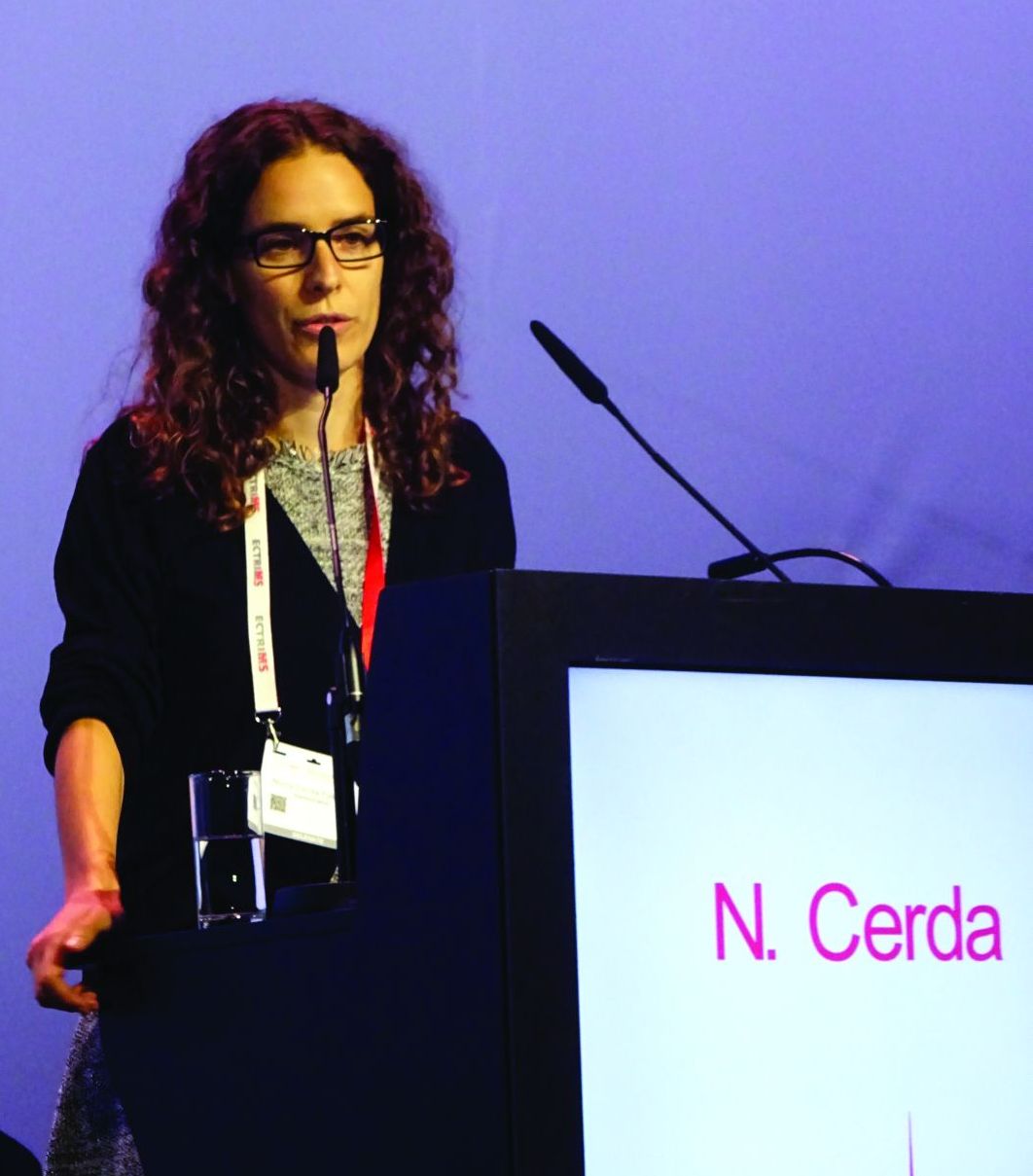
In a retrospective analysis of 110 MS patients who discontinued fingolimod from a 433-patient cohort, around 35% experienced a recurrence of disease activity after fingolimod withdrawal. Study investigator Nuria Cerda, MD, of the Neurologic Clinic and Polyclinic at University Hospital Basel (Switzerland), reported that risk factors for increased disease activity were younger rather than older age at MS diagnosis (P = .01) and at discontinuation (P less than .0001), having had a shorter rather than longer disease duration (P = .01), and evidence of MRI-confirmed disease activity occurring while on treatment with fingolimod (P = .02).
“We aimed to describe the frequency of recurrence of disease activity after fingolimod discontinuation in a real-world cohort of MS patients,” Dr. Cerda said. She noted that there were conflicting data on reappearing disease activity after stopping the drug. Case reports and observational data suggest a frequency of 10%-53%, but post hoc analyses of phase 3 trials with fingolimod have not found any higher risk for recurrence versus placebo.
The study also compared “demographic and clinical characteristics in patients with and without recurrence of disease activity, in order to identify possible risk factors for reactivation of the disease.”
The patients included in the study had been treated with fingolimod for a median of 24 months (interquartile range, 10-43 months) and experienced 118 discontinuation events between them. The majority (n = 81) had relapsing-remitting MS, and the remainder (n = 19) were in transition from RRMS to secondary progressive MS.
The mean age at discontinuation was 40 years and, while 60% of patients switched to another disease-modifying therapy (DMT), 40% did not receive further DMT. The reasons for discontinuation were classified as being from disease activity in 44%, side effects in 21%, pregnancy or childbearing preferences in 16%, noncompliance in 2%, and other reasons in 17%.
Recurrence of disease activity was defined as clinical symptoms, gadolinium-enhancing (Gd+) lesions on MRI, or both, within 6 months of stopping fingolimod. This was seen in 38 patients with 41 events, giving a rate of recurrence of 35%. Of these: “almost 60% had clinical and MRI activity, almost 30% had clinical activity only without Gd+ lesions on MRI, and 12% had MRI activity only without ever having a relapse,” Dr. Cerda said. In those who relapsed, the recurrence took a median 8 weeks to happen after fingolimod discontinuation.
A further definition of severe recurrence of disease activity also was used, defined as either a worsening in the Expanded Disability Status Scale (EDSS) score of more than 2 points (signifying a severe relapse) with or without pronounced MRI activity with at least five cerebral Gd+ lesions within 6 months of fingolimod withdrawal. Thirteen severe events were seen in 11 patients, giving a frequency of approximately 11%. Dr. Cerda reported that patients with severe recurrence were significantly younger at MS diagnosis than were those who did not experience recurrence (P = .003). A trend toward a higher annualized relapse rate (ARR) was seen in patients with severely active versus inactive disease (0.84 vs. 0.54), but this was not statistically significant. In addition, “confirmed disability worsening of 1 EDSS point or more was observed in about 3%.”
Relapse rates coming off fingolimod before or during pregnancy
In another study, which used German registry data on 129 pregnancies that occurred around or after the time of fingolimod withdrawal, up to 56% of women who stopped fingolimod before pregnancy experienced a relapse during pregnancy versus 29% of those who stopped fingolimod upon a positive pregnancy test.
In a comparison of women who stopped fingolimod 1 year to 61 days before their last menstrual period against those who stopped less than 60 days prior to or after their last menstrual period, relapse rates were higher during the first (25.8% for early withdrawal vs. 12.5% with late withdrawal) and second trimesters (32.3% vs. 11.5%) than in the third (9.7% vs. 10.4%), Spalmai Hemat, MD, of the department of neurology at St. Josef Hospital, Ruhr University of Bochum (Germany) reported at the congress.
Relapse rates at 6 months postpartum proved to be similar at 36.7% for women who stopped fingolimod before their pregnancy and 38% in those who stopped later. Difference in the percentage of women experiencing disability progression during pregnancy did not prove to be statistically significant for stopping fingolimod before pregnancy (22.7%) vs. stopping later (11.1%; P = .283), while the 23.8% rate of disability progression after pregnancy among women who stopped the drug later versus 13.6% seen with stopping before was also not statistically significant (P = .31). Relapse during pregnancy was the only significant predictor for relapses postpartum.
“The large majority of women did not experience permanent disability,” Dr. Hemat said, but noted that as many as 10%-20% could experience substantial EDSS worsening (2 or more points) at 6 months postpartum.
Dr. Hemat concluded that more data were needed to see if reintroduction of fingolimod very soon after birth, such as within the first 2 weeks, could reduce the postpartum relapse risk.
Dr. Hemat and Dr. Cerda had no relevant disclosures.
SOURCES: Cerda N et al. Mult Scler. 2018;24(S2):73-4, Abstract 206; Hemat S et al. Mult Scler. 2018;24(S2):74-5, Abstract 207.
REPORTING FROM ECTRIMS 2018
Key clinical point:
Major finding: A higher risk of recurrent disease activity was seen in younger MS patients with shorter disease duration and evidence of disease activity while on fingolimod treatment. Pregnant women who stopped more than 2 months before a planned pregnancy also had a higher risk versus those who stopped after a positive pregnancy test.
Study details: A retrospective analysis of 110 MS patients who discontinued fingolimod from a 433-patient cohort, and German registry data on 129 pregnancies that occurred around or after the time of fingolimod withdrawal.
Disclosures: Dr. Spalmai Hemat and Dr. Nuria Cerda had no relevant disclosures.
Sources: Cerda N et al. Mult Scler. 2018;24(S2):73-4, Abstract 206; Hemat S et al. Mult Scler. 2018;24(S2):74-5, Abstract 207.
Anti-MOG antibodies associated with non-MS, monophasic demyelinating disease in young children
BERLIN – In children with an incident acquired demyelinating episode, the presence of antibodies against myelin oligodendrocyte glycoprotein (MOG) weighs against an eventual diagnosis of multiple sclerosis, especially if the child is younger than 11 years.
“Anti-MOG antibodies are present in about 30% of children with acquired demyelinating syndromes,” Giulia Fadda, MD, said at the annual congress of the European Committee for Treatment and Research in Multiple Sclerosis. “About 80% experience a monophasic disease course.”
The antibodies are found in almost all children who have a relapsing non-MS demyelinating disease, said Dr. Fadda, who is a postdoctoral research fellow at the University of Pennsylvania, Philadelphia. But when the antibodies are present in children with an MS diagnosis, they identify a very specific subset with atypical clinical and imaging features.
The large prospective Canadian Pediatric Demyelinating Disease Study provided the sample for the study she presented at ECTRIMS. Children are recruited into the study soon after a first demyelinating event, and they are followed clinically, serologically, and with regular brain MRI.
The cohort in Dr. Fadda’s study comprised 275 children who were a mean of about 11 years old at symptom onset. The mean clinical follow-up was 6.7 years; the mean serologic follow-up, 4 years, and the mean imaging follow-up, 3.5 years. The study examined 1,368 serum samples and 1,459 MRI scans.
Overall, 32% of the children were positive for anti-MOG antibodies. Positivity was not associated with sex, but children with antibodies were significantly younger than those without (7 vs. 12 years). In fact, 77% of anti-MOG–positive children were younger than 11 years; just 15% of older children were positive for the antibodies.
Anti-MOG positivity was also associated with certain clinical phenotypes. Among positive children, 40% presented with optic neuritis, 37% with acute disseminated encephalomyelitis, and 14% with transverse myelitis; the rest had other phenotypes. Optic neuritis was significantly less common among antibody-negative patients (22%), as was encephalomyelitis (17%). Transverse myelitis was significantly more common (31%).
The analysis of MRI scans was stratified according to age younger than 11 years and 11 years and older. “The first thing we noticed among the younger MOG-positive children is that they had a high number of lesions, which were more commonly ill-defined, diffuse, and bilateral,” Dr. Fadda said. “Almost all the brain areas were affected, with a slight preponderance of thalamic and juxtacortical lesions. Among the MOG-negative children, lesions were more often perpendicular to the major axis of the corpus callosum.”
Among the older MOG antibody–positive children, the diffuse pattern was rarer, and the lesions were frequently cerebellar. “But by far, the features that best differentiated positivity from negativity were black holes and enhancing lesions, which we saw in a high proportion of children without the antibodies” at 73% and 49%, respectively.
Over a mean imaging follow-up period of 4 years, lesions were more likely to resolve completely in antibody-positive children than in those without antibodies (50% vs. 21%). Serologically, children who were MOG-antibody negative at baseline were likely to stay that way, with 99% remaining seronegative. Positive children, on the other hand, were significantly more likely to change serologic status, with 56% turning negative and 8% serologically fluctuating over the follow-up period; only 36% remained persistently seropositive. Persistent positive status was significantly associated with younger age (7 vs. 9 years) and an optic neuritis presentation (62% vs. 27%).
Most children (81%) who were antibody positive at baseline experienced no relapses. Among the 16 children who did experience a clinical relapse, the mean time to a second event was about 1 year. Nine of these children stayed persistently positive, while five seroconverted and two had fluctuating status.
Of the 60 antibody-positive children who had a monophasic course, 23 were persistently positive, 34 seroconverted, and 3 had fluctuating serology.
Eventually, 54 children received an MS diagnosis. Of these, 83% were antibody negative at baseline. Ten children received a diagnosis of a relapsing non-MS demyelinating disorder; of these, 91% were antibody positive at baseline.
Dr. Fadda disclosed relationships with Atara Biotherapeutic and Sanofi-Genzyme.
SOURCE: Waters P et al. Mult Scler. 2018;24(S2):29-30, Abstract 65.
REPORTING FROM ECTRIMS 2018
Key clinical point:
Major finding: Most children (81%) who were antibody positive at baseline experienced no relapses.
Study details: A cohort of 275 children from the prospective Canadian Pediatric Demyelinating Disease Study.
Disclosures: Dr. Fadda disclosed relationships with Atara Biotherapeutics and Sanofi-Genzyme.
Source: Waters P et al. Mult Scler. 2018;24(S2):29-30, Abstract 65.
Demyelinating diseases, especially MS, disrupt normal brain development in children
BERLIN – Demyelinating diseases appear to disrupt white matter development in children, slowing the trajectory of brain growth to almost unmeasurable levels, based on results from a single-center comparison study of 213 individuals.
While children with multiple sclerosis (MS) showed the most severe slowing, even a single demyelinating event slowed white matter growth, Robert A. Brown, PhD, said at the annual congress of the European Committees for Treatment and Research in Multiple Sclerosis.
Dr. Brown of the Montreal Neurological Institute at McGill University, Montreal, employed a signal mass correction of consecutive brain MRIs enhanced with magnetization transfer. Magnetization transfer ratio (MTR) quantifies myelin more effectively than does other imaging enhancement modalities, Dr. Brown said.
“It labels the macromolecules of myelin and correlates almost perfectly with Luxol fast blue stain on histology,” he said. And by measuring myelin instead of whole-brain volume, MTR sidesteps the confounders of inflammation and edema. “When tissue swells, the water dilutes the myelin. MRI is really sensitive to density, so dilution with water lowers that signal.”
But MTR isn’t failsafe either, he said, especially in teens. “A cautionary note: In healthy adolescents, white matter MTR can actually decrease, not increase, not because they are losing myelin but because the axons in brain tissue are growing so fast that they outstrip the production of new myelin. So, we can get another dilution effect here, except that instead of water, axons are diluting the myelin. We have to take that into account when using MTR.”
A volume-corrected MTR calculates both mass and volume to give what Dr. Brown termed signal mass. “We have demonstrated previously that signal mass is about twice as powerful as volume change alone for measuring the differences [in brain volume] between adults with MS and healthy controls.”
The study he presented at ECTRIMS used this technique to examine the trajectory of white matter change in a cohort of children from the Canadian Pediatric Demyelinating Disease Study who were all scanned at the same site in the same center. He compared brain volume at baseline and 1 year in 102 children with a monophasic demyelinating disease, 87 with MS, and 24 healthy, age-matched controls.
The children with MS were a median of about 17 years old at baseline, while those with a monophasic event and healthy controls were a median of about 12 years old. Median follow-up was 1 year in the healthy controls, 2 years in the MS cohort, and 4 years in the monophasic group. The investigators adjusted their comparisons for sex, since both bioavailable testosterone and androgen-receptor activity correlate with decreased MTR in young men. This doesn’t mean, though, that testosterone decreases myelination. Rather, it’s postulated that testosterone increases axonal caliber, which would decrease the number of neurons in each imaging voxel and, thus, the MTR signal (J Neurosci. 2008 Sep 17;28[38]:9519-24).
In the volume-only assessment, white matter in healthy controls increased at a rate of about 0.5% per year. White matter growth was about 0.2% per year in children with monophasic demyelination, which was significantly lower than in healthy controls.
“The MS children had no white matter growth that we could measure,” with an annual change of about 0.01%, Dr. Brown said. “It looks like a failure of normal development and was significantly lower than what we saw in the children with a demyelination event.”
MTR showed the expected age-associated decreases, which were highest among those with MS: –0.8% per year in healthy controls, –0.6% per year in those with a monophasic event, and –0.9% per year in those with MS.
The signal mass change showed the whole picture, Dr. Brown said. Signal mass declined 0.3% per year in healthy controls, 0.5% per year in the monophasic group, and 0.9% per year in the MS group – a significantly worse trajectory than either the control subjects or those with a monophasic event.
“Signal mass puts it all together and gives us the total picture of tissue loss, with quite severe loss in children with MS. It seems as though both monophasic insult and pediatric-onset MS disrupt brain development.”
Dr. Brown has been a consultant for NeuroRx Research and Biogen.
SOURCE: Brown RA et al. Mult Scler. 2018;24(S2):27-8, Abstract 63.
REPORTING FROM ECTRIMS 2018
Key clinical point: Demyelinating disorders disrupt brain growth in children.
Major finding: Children with MS had virtually no white matter growth, and those with ADM lagged significantly behind controls.
Study details: The prospective imaging study comprised 24 controls, 102 with an ADM, and 87 with MS.
Disclosures: Dr. Brown has been a consultant for NeuroRx Research and Biogen Idec.
Source: Brown RA et al. Mult Scler. 2018;24(S2):27-8, Abstract 63
Alemtuzumab switch linked to good MS outcomes
BERLIN – Switching to alemtuzumab from fingolimod is associated with improved disease activity in patients with relapsing-remitting multiple sclerosis (RRMS), according to the results of a real-world study reported at the annual congress of the European Committee for Treatment and Research in Multiple Sclerosis.
Jessica Frau, MD, of the University of Cagliari (Italy) reported that a switch from fingolimod (Gilenya) to alemtuzumab (Lemtrada) in 77 patients treated at 11 Italian centers was able to “reduce dramatically disease activity in patients who did not respond to fingolimod.”
Dr. Frau reported: “When we compared in our cohort the last year of fingolimod with the first year after the first course of alemtuzumab, we found a significant decrease in the annualized relapse rate [ARR].” The ARRs were 0.60 for fingolimod and 0.20 after 1 year of alemtuzumab treatment.
“We found also a trend towards an improvement in the EDSS [Expanded Disability Status Scale] score (P = .23), and less evidence of disease activity on MRI, both in terms of new T2 lesions and gadolinium-enhancing (Gd+) lesions.”
The last MRI during fingolimod treatment showed new T2 and Gd+ enhancing lesions in 69.2% and 58.6% of patients, respectively. Corresponding figures for the first MRI during alemtuzumab treatment were 10.4% and 2.2% of patients.
The beneficial effects of switching to fingolimod in the Italian study “was not influenced by a shorter washout [period] or a low lymphocyte count when alemtuzumab was started,” Dr. Frau said. A shorter washout period has been hypothesized to account for recent accounts of disease flares seen when switching from fingolimod to alemtuzumab, she explained.
Indeed, Dr. Frau noted that there had been a few studies that reported MS disease reactivation soon after the switch to alemtuzumab was made, which could be because lymphocytes remain in the lymph nodes when alemtuzumab is administered, this means that potentially they could repopulate the central nervous system and reactivate the disease.
However, “when alemtuzumab is started after fingolimod it is not a risk factor for reactivation of the disease,” Dr. Frau said, based on the current study’s findings.
As expected, the frequency of relapses increased during the washout period after stopping fingolimod, going from 12.7% of patients with relapse in the first month, 18.2% at 2 months, and 22.2% at 3 months. The time to first relapse from the start of alemtuzumab treatment was 6 months for 2.9% of patients, 9 months for 10.5% of patients, and 1 year for 20.7% of patients.
Asked to comment on when the optimal time to switch from fingolimod to alemtuzumab might be, Dr. Frau said: “The optimal time could be 1 month when the lymphocyte count is not too low.” However, lymphocyte counts were not measured in the entire cohort, so “these data perhaps need to have more strength.”
The switch from natalizumab to alemtuzumab
Other data on switching to alemtuzumab, this time from natalizumab (Tysabri), in the ANSWERS MS study were presented by Paul Gallagher, MBChB, of Queen Elizabeth University Hospital, London, and the University of Glasgow (Scotland).
ANSWERS MS (Alemtuzumab after Natalizumab Switch in Evolving Rapidly Severe MS) is a retrospective, observational analysis of routinely collected data on the use of alemtuzumab by 13 centers the United Kingdom and Ireland. These centers have been collecting data since before alemtuzumab was licensed in 2014 for MS, Dr. Gallagher observed, with some centers having experience of making the switch for more than a decade.
ANSWERS MS addresses a common clinical question: “Is it safe and effective to switch to alemtuzumab if natalizumab fails in highly active MS?” Dr. Gallagher said. “The truth is we don’t really know the answer to this, although it’s becoming an increasingly used switch.”
Alemtuzumab was developed in Cambridge, England, in 1983, originally as an anticancer agent, and first started being used in MS patients in the 1990s. Natalizumab was first licensed in the United Kingdom in 2007.
The aim of the study was mainly to look at safety, but also examine efficacy, and to offer advice on how to best manage the switch. A total of 79 patients formed the safety cohort; 51 of these patients had more than 2 years of follow-up after their first infusion of alemtuzumab and formed the efficacy cohort.
Data were examined in five phases: before natalizumab, during natalizumab, during the switchover period, during alemtuzumab treatment, and after alemtuzumab treatment, with the latter starting 2 years after the first alemtuzumab infusion.
Dr. Gallagher noted that 43% started natalizumab as a first-line therapy, and almost half (49%) of patients stopped taking natalizumab because of breakthrough disease, making this a bit of an unusual cohort with highly active disease, although other cohort characteristics were pretty typical of an MS population.
“The headline is that there are no new safety concerns identified from this cohort,” Dr. Gallagher reported. “Most [61%] patients had infusion reactions with alemtuzumab as expected, but this gradually reduced with subsequent courses.”
Fewer than 20% of patients developed autoimmune thyroid disease, he added, and there were no cases of idiopathic thrombocytopenic purpura.
Infections were seen in nine patients, including three cases of shingles, two urinary tract infections – one of which was classed as a severe adverse event – and one case each of oral thrush, fungal skin infection, tonsillitis, and norovirus.
There was also one cytomegalovirus infection and one death from sepsis unrelated to alemtuzumab; both of these were classed as serious adverse events.
In terms of efficacy, mean ARRs were 2.3 before and 0.8 during natalizumab treatment, decreasing to 0.4 during alemtuzumab treatment and 0.5 post alemtuzumab. A “spike” in relapses was seen, however, during the switch period.
“There was a similar story with MRI imaging,” Dr. Gallagher said. “The profile suggests high disease activity during the switch phase in comparison to everything else.” The mean number of new or worsened MRI lesions was 4.32 per scan per year during the switch period. This fell, however, during alemtuzumab treatment to 0.006 per MRI scan per year and remained low after the end of alemtuzumab treatment at 0.017 per scan per year.
There was no real benefit to switching on the EDSS, with scores increasing from 3.4 in the pre-natalizumab period to 4.7 during the switch period, but then plateauing out to 4.4. and 4.3 after the initiation of alemtuzumab and in the post-alemtuzumab phase.
“These data were based on medical records, often incomplete, and so not all patients had an EDSS in every phase, for example,” Dr. Gallagher noted. He said an analysis was done to try to account for the missing information. This showed that there was an improvement in EDSS while on alemtuzumab, but the effect was not maintained.
It was evident in looking at the switch period that a shorter time between natalizumab and alemtuzumab was associated with the best outcomes, with the optimum time being around 2-4 months. Bridging therapy with fingolimod did not reduce disease activity during the switch, Dr. Gallagher said.
ANSWERS MS was funded by Sanofi-Genzyme. Paul Gallagher disclosed that he had received salary payment and travel funding for educational events from Sanofi-Genzyme and travel funding from Novartis and Biogen.
Dr. Frau disclosed that she serves on scientific advisory boards for Biogen, Merck, and Genzyme and that she has received honoraria for speaking from Merck Serono, Genzyme, Biogen, and Teva.
SOURCE: Frau J et al. Mult Scler. 2018;24(S2):100-1, Abstract 265; Gallagher P et al. Mult Scler. 2018;24(S2):99-100, Abstract 264.
REPORTING FROM ECTRIMS 2018
Key clinical point: Good results can be achieved by switching from fingolimod or natalizumab to alemtuzumab in relapsing-remitting multiple sclerosis (RRMS).
Major finding: Annualized relapse rates were 0.60 for fingolimod at the time of the switch and 0.20 after 1 year of alemtuzumab treatment in one real-world study. In another, alemtuzumab was effective in reducing inflammatory disease activity when natalizumab failed.
Study details: Two real-world, observational studies: one with 77 RRMS patients treated at 11 Italian centers and the other a retrospective analysis of routinely collected data on 79 patients.
Disclosures: Dr. Frau disclosed that she serves on scientific advisory board for Biogen, Merck, and Genzyme and that she has received honoraria for speaking from Merck Serono, Genzyme, Biogen, and Teva. ANSWERS MS was funded by Sanofi-Genzyme. Dr. Gallagher disclosed he had received salary payment and travel funding for educational events from Sanofi-Genzyme and travel funding from Novartis and Biogen.
Source: Frau J et al. Mult Scler. 2018;24(S2):100-1, Abstract 265; Gallagher P et al. Mult Scler. 2018;24(S2):99-100, Abstract 264.
Delay in plasma exchange increases chance of poor outcomes in NMOSD
BERLIN – A delay in undertaking plasma exchange may predict a poorer outcome after a first attack of neuromyelitis optica spectrum disorder, while antibodies to myelin oligodendrocyte glycoprotein (MOG) appear to predict a more positive outcome.
“We saw that for each day of delay in plasma exchange, the Expanded Disability Status Scale [EDSS] at 6 months increased by about 0.028 points, indicating a worse prognosis,” Maxime Guillaume, MD, said at the annual congress of the European Committee for Treatment and Research in Multiples Sclerosis.
However, said Dr. Guillaume, a resident at Rouen University Hospital, France, steroids are still a reasonable first-line therapy as long as they are discontinued quickly if they don’t appear to be helping. Plasma exchange is most effective if administered less than 2 weeks after symptom onset.
His study examined 6-month outcomes among 214 attacks in 188 patients; some patients had several first attacks in different areas. Response was defined in two ways. First, patients were clinically classified as having a good response, a bad response, or no response to treatment. The second definition was based on the EDSS. Good response was an EDSS decrease of at least 2 points for an initial score of 3 or higher, or a decrease of 1 point if the initial score was less than 3. Poor response was an EDSS that decreased without reaching these thresholds.
The cohort was largely female, with a mean age of 38 years. Most (55%) were positive for antibodies against aquaporin-4. Anti-MOG antibodies were present in 30%. A total of 7.5% were negative for both antibodies, and the remainder had an undetermined serotype.
The clinical presentations varied. Most frequently, patients presented with myelitis only (44%). This was followed by optic neuritis only (34%), both myelitis and optic neuritis (8%), and myelitis plus brainstem involvement (5%). Other clinical manifestations were acute demyelinating encephalomyelitis, and encephalitis alone.
The most common treatment was methylprednisolone (73%), followed by plasma exchange (25%), which occurred at a median of 9 days after symptom onset.
Outcomes varied according to the definition of response. By clinical characteristics, there was a complete response in 41, a partial response in 122, and no response in 51. By change in EDSS, 136 had a good response and 27 a partial response; 51 were still considered nonresponders.
Dr. Guillaume conducted a multivariate analysis to determine predictive factors. In both definitions, anti-MOG antibodies nearly quadrupled the chance of a good treatment response, and delaying plasma exchange was associated with a significantly increased chance of a poor response. When judged by the clinical response definition, multiple lines of treatment also were associated with a poor response. This, he said, was another reflection of plasma exchange delay.
Dr. Guillaume had no financial disclosures.
REPORTING FROM ECTRIMS 2018
Key clinical point:
Major finding: Each day’s delay in receiving plasma exchange increased the mean 6-month EDSS by about 0.028 points.
Study details: The retrospective study comprised 214 attacks in 188 patients.
Disclosures: Dr. Guillaume had no financial disclosures.
Source: Guillaume M. et al. ECTRIMS 2018, Abstract 211.
