User login
Jeff Evans has been editor of Rheumatology News/MDedge Rheumatology and the EULAR Congress News since 2013. He started at Frontline Medical Communications in 2001 and was a reporter for 8 years before serving as editor of Clinical Neurology News and World Neurology, and briefly as editor of GI & Hepatology News. He graduated cum laude from Cornell University (New York) with a BA in biological sciences, concentrating in neurobiology and behavior.
Model may predict prolonged status epilepticus outcomes
Two clinical parameters measurable at seizure onset appear to predict a return to baseline after prolonged status epilepticus (SE), based on a study of patients who presented to a single, tertiary academic medical center over a 12-year period.
Absence of nonconvulsive SE with coma and a decreasing Charlson Comorbidity Index were the only independent predictors for return to baseline in patients with SE duration greater than 48 hours, the researchers found. However, the research fell short of developing a model for identifying patients at risk for prolonged SE.
“These findings are of great clinical importance, as up to now, clinicians have had no reliable prediction tools to direct decisions regarding the level of care with progressive SE duration. Early and reliable identification of patients with potential favorable outcome despite having SE for several days is of utmost clinical importance, as this insight may urge clinicians to intensify treatment rather than consider care withdrawal as systemic and neurologic sequelae increase, and chances of SE termination decrease over time,” first author Raoul C. Sutter, MD, of University Hospital Basel (Switzerland), and his colleagues wrote about their findings in Epilepsia.
The researchers identified 467 adult patients with prolonged SE at University Hospital Basel during 2005-2016 – excluding those with SE as a consequence of hypoxic‐ischemic brain injury – who had a median age of 66.7 years and median SE duration of 1 day. While 11.8% of patients died in the hospital and 12.4% at 30 days after SE onset, 40.9% made a complete neurologic and functional recovery to their premorbid status.
There were significant differences in in-hospital outcomes between patients with different SE durations. For example, rates of returning to baseline differed significantly at 55.6% of those with a SE duration of 0-12 hours, 36.8% with 12-24 hours’ duration, 34.6% with 24-48 hours’ duration, and 25.5% with more than 48 hours.
A multivariable regression model identified absence of nonconvulsive SE with coma and a decreasing Charlson Comorbidity Index as the only independent predictors for return to baseline in patients with SE duration greater than 48 hours, and both remained significant predictors after adjustment for use of anesthetics and vasopressors. These predictors of a return to baseline after prolonged SE remained significant after excluding patients who died. This two-variable prediction model had an area under the receiver operating curve (AUROC) of 0.82, “indicating good discrimination,” and an AUROC of 0.76 following cross-validation.
The investigators also sought to develop a model to identify patients at risk for prolonged SE, but the model showed relatively poor discriminative ability with AUROCs of just 0.67-0.72 for predicting no termination of SE within 12, 24, or 48 hours. “Our attempt to generate a highly reliable prediction model for early recognition of patients at increased risk for developing prolonged SE failed, as demonstrated by the rather small AUROC and the fact that sensitivity analyses after exclusion of patients who died revealed inconsistent association of the identified predictors,” they wrote.
Prior reports identified younger age, absence of acute brain lesions at presentation, and the absence of multiple concomitant medical problems as factors associated with favorable outcome after prolonged SE, but “none of the studies performed multivariable regression models and generated or tested predictions models in this context,” they noted.
The authors cautioned that “although internal cross-validation of the final prediction model indicated adequate performance [based on an AUROC of 0.76], further external validation of our prediction model is warranted before our prediction model can be implemented and used for decision making in daily clinical practice.”
Some authors reported receiving research, travel, and/or personal grants or speaker fees from companies marketing antiepileptic drugs, such as UCB, Eisai, and GlaxoSmithKline.
SOURCE: Sutter RC et al. Epilepsia. 2018 Nov 22. doi: 10.1111/epi.14603
FROM Epilepsia
Key clinical point:
Major finding: A two-variable prediction model had an AUROC of 0.82.
Study details: A single-center study of 467 adult patients treated for status epilepticus during 2005-2016.
Disclosures: Some authors reported receiving research, travel, and/or personal grants or speaker fees from companies marketing antiepileptic drugs, such as UCB, Eisai, and GlaxoSmithKline.
Source: Sutter RC et al. Epilepsia. 2018 Nov 22. doi: 10.1111/epi.14603
ACR forges ahead with physician-focused APM for RA
CHICAGO – with hopes to send the fully developed model to the Physician-Focused Payment Model Technical Advisory Committee once financial data gathering and analysis is complete, followed by pilot testing once it is accepted by the Centers for Medicare & Medicaid Services, speakers said at the annual meeting of the ACR.
The “RA Care Team” APM is meant to be versatile and work across various practice settings, from rural Alaska and the Southwest to urban Chicago and Boston, and would consist of a rheumatologist or a nurse practitioner or physician assistant working with a rheumatologist in some areas, while in others a patient would be managed by a primary care physician who has a formal arrangement with a rheumatologist to provide early treatment of RA. Participation in the model would use a standard treatment approach pathway that follows ACR treatment guidelines and saves money by reducing the variability in initiation of expensive medications and would be versatile enough to allow for unique patients by requiring only 75% adherence to the pathway across a practice’s patients, said Kwas Huston, MD, cochair of the ACR’s APM work group and a rheumatologist with Kansas City (Mo.) Physician Partners.
The model covers four phases of care, including diagnosis and treatment planning for patients with potential RA, support for primary care practices in evaluating joint symptoms, the initial treatment of patients with RA, and the continued care of RA. For instance, a rheumatologist could receive payment for an e-consult with a primary care provider to determine if a patient has symptoms requiring an evaluation for RA. A rheumatologist could also receive a one-time payment for treatment and planning services when a diagnosis of RA cannot be established in a patient suspected of having RA, whereas the rheumatologist or a primary care provider with rheumatology support would receive monthly payments for 6 months for the initial treatment of an RA patient and then thereafter would receive monthly payments for continued care of RA. The payments would not be dependent on the number of visits or face-to-face time, would be stratified based on patient characteristics, and would include some lab testing and imaging.
Currently, the RA APM work group is analyzing financial data gathered from two large rheumatology practices for specific CPT codes matched to specific patients based on their clinical characteristics “to get a sense of how much revenue is coming in right now for practices taking care of patients with, let’s say, moderate disease activity and two comorbidities or low disease activity and no comorbidities,” Dr. Huston said. The work group is also surveying practices to estimate additional costs required to participate in the APM and thereby develop a financial model to adequately pay for APM services. They additionally plan to develop a tool kit that is designed to help individual practices determine the economic impact that the APM would have.
Notably, the cost of medications is not included in the APM. “That would be too much of a risk for small practices to take on,” Dr. Huston said.
“This model is a work in progress. We still have a lot of additional work to validate the data,” he added.
Providers who wish to participate in an advanced APM in 2019 need to have at least 25% of their Medicare Part B payments or have at least 20% of Medicare patients in their practice come through the APM in order to avoid having to submit data to comply with the performance criteria requirements of the Merit-Based Incentive Payment System, said Angus Worthing, MD, chair of the ACR’s Government Affairs Committee and a practicing rheumatologist in the Washington area.
“The ACR has advocated to reduce these thresholds over the years, but unfortunately that has not happened yet,” Dr. Worthing said.
jevans@mdedge.com
REPORTING FROM THE ACR ANNUAL MEETING
Funding for NIH BRAIN Initiative reaches new heights
The National Institutes of Health’s Brain Research through Advancing Innovative Neurotechnologies (BRAIN) Initiative will finish 2018 with its largest round of grant funding ever, giving $220 million to more than 200 research awards, and bringing this year’s total to more than $400 million, according to an announcement from the agency.
The BRAIN Initiative began in 2013 with the objective of revolutionizing our understanding of the human brain by accelerating the development and application of innovative technologies that will allow researchers to show how individual cells and complex neural circuits interact in both time and space and thereby seek new ways to treat, cure, and prevent brain disorders.
In the current round of funding that was authorized by Congress through the regular appropriations process and the 21st Century Cures Act, new projects include the creation of a wireless optical tomography cap for scanning human brain activity; the development of a noninvasive brain-computer interface system for improving the lives of paralysis patients; and the testing of noninvasive brain stimulation devices for treating schizophrenia, attention deficit disorders, and other brain diseases; the development of self-growing biological electrodes for recording brain activity; and the creation of an indestructible hydrogel system to help map neural circuits, according to the announcement.
Not all of the research involves technological advancement. In fact, one line of funding involves neuroethics. For instance, for epilepsy syndromes in the latest round of funding for 2018, researchers aim to explore ethical issues confronting families and clinicians when considering new treatment options for drug-resistant epilepsy in children.
The NIH is also leveraging some of the BRAIN Initiative funding toward finding new, nonaddictive pain treatments as part of the its HEAL (Helping to End Addiction Long-term) Initiative, such as support for research on the fundamental neurobiology of endogenous opioid systems.
Bills affecting rheumatologists make way to Capitol Hill
CHICAGO – Rheumatology advocates have been working to get a variety of bills passed in Congress, including efforts on creating exemptions to step therapy or “fail first” insurer policies, increasing osteoporosis screening payment, and raising research dollars for arthritis in the Department of Defense, Dr. Angus Worthing said at the annual meeting of the American College of Rheumatology.
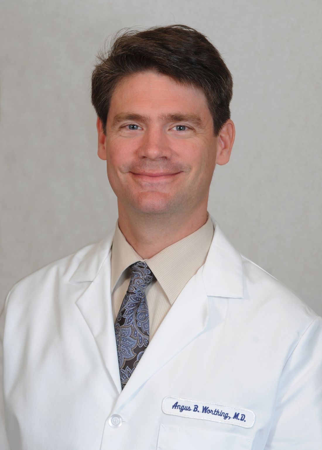
Visits by rheumatologist advocates to receptive members of the House and Senate have been fruitful for these issues, said Dr. Worthing, who is chair of the ACR’s Government Affairs Committee and a practicing rheumatologist in the Washington area.
“There’s a bill in the House [H.R. 2077, Restoring the Patient’s Voice Act], reforming step therapy that would institute appropriate exemptions if a doctor can predict that a drug that’s the first step in a step therapy will cause side effects, will be ineffective, or has already been used by a patient, as well as other appropriate guardrails,” so that the policy can be circumvented and the patient can get the prescription that the doctor ordered, he said.
The ACR is working with coalition partners such as the National Psoriasis Foundation and other patient organizations to get a companion bill to H.R. 2077 introduced in the Senate, Dr. Worthing noted.
The ACR is also working to advance separate bills in the House (H.R. 1898) and Senate (S. 3160) to increase payment levels for dual x-ray absorptiometry scans, which were cut by 60%-70% about 10 years ago.
The two bills have broad bipartisan support in both the House and Senate. “Watch for that to hopefully become law,” Dr. Worthing said.
The ACR has been advocating for the last 4 years to increase research dollars specifically for arthritis and rheumatology at the Department of Defense. After years of trying, rheumatologist advocates were able to get funding for studying trauma-related osteoarthritis and military serology data banks added as an amendment in the most recent appropriations bill. However, it was pulled at the last minute because of a lack of support.
“But with that advancement we’ll be able to go into the next Congress in January and either get it into the bill or hopefully have the amendment voted on next year,” he said.
Dr. Worthing called for those interested in advocating for rheumatology to visit the ACR’s Legislative Action Center.
CHICAGO – Rheumatology advocates have been working to get a variety of bills passed in Congress, including efforts on creating exemptions to step therapy or “fail first” insurer policies, increasing osteoporosis screening payment, and raising research dollars for arthritis in the Department of Defense, Dr. Angus Worthing said at the annual meeting of the American College of Rheumatology.
Visits by rheumatologist advocates to receptive members of the House and Senate have been fruitful for these issues, said Dr. Worthing, who is chair of the ACR’s Government Affairs Committee and a practicing rheumatologist in the Washington area.
“There’s a bill in the House [H.R. 2077, Restoring the Patient’s Voice Act], reforming step therapy that would institute appropriate exemptions if a doctor can predict that a drug that’s the first step in a step therapy will cause side effects, will be ineffective, or has already been used by a patient, as well as other appropriate guardrails,” so that the policy can be circumvented and the patient can get the prescription that the doctor ordered, he said.
The ACR is working with coalition partners such as the National Psoriasis Foundation and other patient organizations to get a companion bill to H.R. 2077 introduced in the Senate, Dr. Worthing noted.
The ACR is also working to advance separate bills in the House (H.R. 1898) and Senate (S. 3160) to increase payment levels for dual x-ray absorptiometry scans, which were cut by 60%-70% about 10 years ago.
The two bills have broad bipartisan support in both the House and Senate. “Watch for that to hopefully become law,” Dr. Worthing said.
The ACR has been advocating for the last 4 years to increase research dollars specifically for arthritis and rheumatology at the Department of Defense. After years of trying, rheumatologist advocates were able to get funding for studying trauma-related osteoarthritis and military serology data banks added as an amendment in the most recent appropriations bill. However, it was pulled at the last minute because of a lack of support.
“But with that advancement we’ll be able to go into the next Congress in January and either get it into the bill or hopefully have the amendment voted on next year,” he said.
Dr. Worthing called for those interested in advocating for rheumatology to visit the ACR’s Legislative Action Center.
REPORTING FROM THE ACR ANNUAL MEETING
3-D model neurovascular unit developed with working blood-brain barrier
The development of a 3-D brain organoid that contains all six of the major cell types found in adult human brain cortex will be the featured topic of a presentation at the Young Research Forum of the International Conference on Parkinson’s Disease and Movement Disorders in New York on Oct. 20.
Goodwell Nzou, a doctoral student at Wake Forest University, Winston-Salem, N.C., will give the presentation, titled “The development of a multicellular three dimensional neurovascular unit model with a functional blood-brain barrier” at 1:45 p.m.
In the study that Mr. Nzou and his colleagues at the Wake Forest Institute for Regenerative Medicine published in May in Scientific Reports, they noted that, in addition to the model’s use of all six of the major cell types found in adult human brain cortex (human brain microvascular endothelial cells, pericytes, astrocytes, microglia, oligodendrocytes, and neurons), they found that it also promotes the formation of a blood-brain barrier that mimics normal human anatomy.
The researchers said their 3-D in vitro system could have potential applications in drug discovery, toxicity screening, and disease modeling for neurologic diseases such as Alzheimer’s disease, multiple sclerosis, and amyotrophic lateral sclerosis. They demonstrated the model’s use in toxicity assessment studies for molecules that have the potential to cross or open the blood-brain barrier and also reported establishing a model of the blood-brain barrier during clinical ischemia “showing physiologic responses under hypoxic conditions.”
REPORTING FROM ICPDMD 2018
Studies reveal pregnancy trends in American women with MS
New evidence provides estimates of the pregnancy rates for American women with multiple sclerosis (MS), their complication rates, and the rates of relapse and disease-modifying drug treatment during different phases before and after pregnancy.
The two new studies, conducted by Maria K. Houtchens, MD, of Brigham and Women’s Hospital and Harvard Medical School, Boston, and her colleagues involved retrospective mining of U.S. commercial health plan data in the IQVIA Real-World Data Adjudicated Claims–U.S. database between Jan. 1, 2006, and June 30, 2015.
The mean age of pregnant women in the nine annual cohorts during that period was just over 32 years for those with MS and just over 29 years for those without. The percentage of women without MS who had a pregnancy-related claim in the database declined from 8.83% in 2006 to 7.75% in 2014 after adjusting for age, region, payer, and Charlson Comorbidity Index score, whereas the percentage increased in women with MS during the same period, from 7.91% to 9.47%. The investigators matched 2,115 women with MS and 2,115 without MS who had live births for a variety of variables and found that women with MS had higher rates of premature labor (31.4% vs. 27.4%; P = .005), infection in pregnancy (13.3% vs. 10.9%; P = .016), maternal cardiovascular disease (3.0% vs. 1.9%; P = .028), anemia or acquired coagulation disorder (2.5% vs. 1.3%; P = .007), neurologic complications in pregnancy (1.6% vs. 0.6%; P = .005), and sexually transmitted diseases in pregnancy (0.4% vs. 0%; P = .045). During labor and delivery, women with MS who had a live birth more often had a claim for acquired damage to the fetus (27.8% vs. 23.5%; P = .002) and congenital fetal malformations (13.2% vs. 10.3%; P = .004) than did women without MS.
In the second study, Dr. Houtchens and two coauthors from the first study of the database reported on a set of 2,158 women who had a live birth during the study period and had 1 year of continuous insurance eligibility before and after pregnancy. The odds for having an MS relapse declined during pregnancy (odds ratio, 0.623; 95% confidence interval, 0.521-0.744), rose during the 6-week postpartum puerperium (OR, 1.710; 95% CI, 1.358-2.152), and leveled off during the last three postpartum quarters to remain at a higher level than before pregnancy (OR, 1.216; 95% CI, 1.052-1.406). Disease-modifying drug treatment followed the same pattern with 20% using it before pregnancy, dropping to about 2% in the second trimester, and peaking in about a quarter of all patients 9-12 months post partum.
SOURCES: Houtchens MK et al. Neurology. 2018 Sep 28. doi: 10.1212/WNL.0000000000006382; Houtchens MK et al. Neurology. 2018 Sep 28. doi: 10.1212/WNL.0000000000006384.
FROM NEUROLOGY
Hot topic explores neuropathologic differences between MS individuals
One of the first sessions to begin the annual congress of the European Committee for Treatment and Research in Multiple Sclerosis in Berlin takes a close look at how researchers are honing in on the mechanisms and cellular and molecular mediators that shape the ways in which multiple sclerosis neuropathology differs between individuals.
In the first presentation of the session “Hot Topic 1: Hot topics in MS neuropathology” at 8:00 a.m. (local time) on Oct. 10, Bruce Trapp, PhD, will present the details of the recently published study that he and his associates at the Cleveland Clinic in Ohio conducted on the brains of postmortem MS patients. They describe a new disease subtype, called myelocortical MS, that they characterized through 12 postmortem MS brains in which there was cortical neuronal loss and demyelination of spinal cord and cerebral cortex in the absence of cerebral white matter demyelination.
But what might be driving the degeneration of cerebral gray matter in MS patients, particularly in the cortex? Roberta Magliozzi, PhD, of the University of Verona (Italy), will follow Dr. Trapp’s talk with a presentation of recent neuropathologic findings describing a specific inflammatory protein profile in cerebrospinal fluid (CSF) stemming from meningeal infiltrates and circulating cells in the subarachnoid space that could account for differences in the speed of physical and cognitive disability progression between individuals with MS. Earlier research by Dr. Magliozzi and her colleagues characterized some of these CSF biomarkers. Whether the specific CSF inflammatory pattern proves to be a good surrogate for meningeal inflammation and thereby a good predictor of which MS patients may develop more severe gray matter demyelination and a higher risk of disease progression needs to be examined further.
In the final talk, Claudia Lucchinetti, MD, of the Mayo Clinic, Rochester, Minn., will describe the latest understanding of the pathology of radiologically isolated syndrome.
REPORTING FROM ECTRIMS 2018
Benign MS is real in small minority of patients
Nearly 3% of patients with multiple sclerosis (MS) are estimated to have a truly benign course of disease over at least 15 years without the use of disease-modifying therapy, based on findings from a U.K. population-based study that also showed how poorly benign disease tracks with disability measures and lacks agreement between patients and physicians.

“The study of the individuals with extremely favorable outcomes may uncover insights about disease pathogenesis or repair. However, the insensitivity of EDSS [Expanded Disability Status Scale]–based definitions of benign MS and the discrepancy between patient and clinician perception of benign MS undermine use of the term ‘benign’ in the clinical setting,” Emma Clare Tallantyre, MD, of Cardiff (Wales) University, and her colleagues wrote in the Journal of Neurology, Neurosurgery & Psychiatry.
Dr. Tallantyre and her colleagues found that, of 1,049 patients with disease duration longer than 15 years, 200 had a recent EDSS score of less than 4.0. Of those 200, 60 were clinically assessed and 9 (15%) were found to have truly benign MS, defined as having an EDSS less than 3.0 and having no significant fatigue, mood disturbance, cognitive impairment, or disruption to employment in the absence of disease-modifying therapy at least 15 years after symptom onset.
The investigators extrapolated these data to estimate that 30 patients in the study population of 1,049 had truly benign MS, for a prevalence of 2.9%. However, of the 60 patients who were clinically assessed, 39 thought they had benign MS based on the lay definition provided: “When referring to illness, ‘benign’ usually means a condition which has little or no harmful effects on a person. There are no complications and there is a good outcome or prognosis.”
Patients who self-reported benign MS had significantly lower EDSS scores, fewer depressive symptoms, lower fatigue severity, and lower reported MS impact than did patients who did not report benign MS. “Self-reported benign MS status showed poor agreement with our composite definition of benign MS status and only fair agreement with EDSS-based definitions of benign MS status,” the investigators wrote.
SOURCE: Tallantyre EC et al. J Neurol Neurosurg Psychiatry. 2018 Sep 3. doi: 10.1136/jnnp-2018-318802.
Nearly 3% of patients with multiple sclerosis (MS) are estimated to have a truly benign course of disease over at least 15 years without the use of disease-modifying therapy, based on findings from a U.K. population-based study that also showed how poorly benign disease tracks with disability measures and lacks agreement between patients and physicians.
“The study of the individuals with extremely favorable outcomes may uncover insights about disease pathogenesis or repair. However, the insensitivity of EDSS [Expanded Disability Status Scale]–based definitions of benign MS and the discrepancy between patient and clinician perception of benign MS undermine use of the term ‘benign’ in the clinical setting,” Emma Clare Tallantyre, MD, of Cardiff (Wales) University, and her colleagues wrote in the Journal of Neurology, Neurosurgery & Psychiatry.
Dr. Tallantyre and her colleagues found that, of 1,049 patients with disease duration longer than 15 years, 200 had a recent EDSS score of less than 4.0. Of those 200, 60 were clinically assessed and 9 (15%) were found to have truly benign MS, defined as having an EDSS less than 3.0 and having no significant fatigue, mood disturbance, cognitive impairment, or disruption to employment in the absence of disease-modifying therapy at least 15 years after symptom onset.
The investigators extrapolated these data to estimate that 30 patients in the study population of 1,049 had truly benign MS, for a prevalence of 2.9%. However, of the 60 patients who were clinically assessed, 39 thought they had benign MS based on the lay definition provided: “When referring to illness, ‘benign’ usually means a condition which has little or no harmful effects on a person. There are no complications and there is a good outcome or prognosis.”
Patients who self-reported benign MS had significantly lower EDSS scores, fewer depressive symptoms, lower fatigue severity, and lower reported MS impact than did patients who did not report benign MS. “Self-reported benign MS status showed poor agreement with our composite definition of benign MS status and only fair agreement with EDSS-based definitions of benign MS status,” the investigators wrote.
SOURCE: Tallantyre EC et al. J Neurol Neurosurg Psychiatry. 2018 Sep 3. doi: 10.1136/jnnp-2018-318802.
FROM THE JOURNAL OF NEUROLOGY, NEUROSURGERY & PSYCHIATRY
Noninferiority endpoint not met between Xeljanz formulations for RA
An 11-mg, once-daily, modified-release formulation of the Janus kinase inhibitor Xeljanz (tofacitinib) did not meet a noninferiority of efficacy primary endpoint when compared against a 5-mg, immediate-release formulation of the drug taken twice daily in a phase 3, randomized trial of 209 Japanese patients with rheumatoid arthritis.
Although the modified-release formulation did not achieve noninferiority with the immediate-release dosing after 12 weeks, the number of patients who achieved a clinically meaningful outcome was similar in both groups, Yoshiya Tanaka, MD, PhD, and his colleagues reported online Aug. 17 in Rheumatology.
Noninferiority between the two formulations “was to be declared if the upper bound of the two-sided 95% [confidence interval] for the difference in [Disease Activity Score in 28 Joints–C-reactive protein] between treatment groups was less than 0.6,” they said. The difference in DAS28-CRP scores of 0.43 had a 95% CI upper bound of 0.69, demonstrating a lack of noninferiority.
The percentages of patients achieving 1.2 or more points improvement in DAS28-CRP were similar at 89% of patients on the modified-release tofacitinib and 85% of those on immediate-release tofacitinib. At baseline, patients in the study were on a stable dose of methotrexate at 6-16 mg/week (mean of about 9.5 mg/week) for 6 or more weeks.
About half of patients in both groups reported adverse events, and serious adverse events occurred in 4%-5%.
Pfizer funded the study.
SOURCE: Tanaka Y et al. Rheumatology (Oxford). 2018 Aug 17. doi: 10.1093/rheumatology/key250.
FROM RHEUMATOLOGY
Cost of medical care for older adults with epilepsy steadily grows
An analysis of health care expenditures faced by elderly patients with epilepsy over a 12-year period in the United States found rising annual costs over time that approached a mean of $20,000, according to researchers from the Medical University of South Carolina, Charleston.
Alain Lekoubou, MD, and his colleagues estimated health care expenditures during 2003-2014 for patients aged 65 years and older with and without epilepsy by extrapolating data from the Medical Expenditure Panel Survey Household Component to more than 37 million elderly individuals in the United States. The investigators published their findings in Epilepsia.
The unadjusted mean direct health care expenditures for elderly people with epilepsy rose from $15,850 in 2003-2006 to $22,038 in 2007-2010 but dropped in 2011-2014 to $17,985. Figures for the same period for elderly people without epilepsy went from $10,214 to $10,358 to $9,965.
“The high prevalence of epilepsy and high health care expenditures in elderly patients should give priority to epilepsy in the elderly in the medical and public health communities’ agenda,” the authors wrote.
SOURCE: Lekoubou A et al. Epilepsia. 2018 June 19. doi: 10.1111/epi.14455.
FROM EPILEPSIA