User login
NHLBI seeks to accelerate hemostasis/thrombosis research
SAN DIEGO – The National Heart, Lung, and Blood Institute is looking to jump-start research into hemostasis and thrombosis. Donna DiMichele, MD, offered two reasons for the research push.
“The first is to stimulate research in areas that are undersubscribed through investigator-initiated research,” Dr. DiMichele, deputy director of the NHLBI’s Division of Blood Diseases and Resources, said at the biennial summit of the Thrombosis & Hemostasis Societies of North America. “We also do it sometimes to steer research in directions that are not developing organically through investigator-initiated efforts. In hemostasis and thrombosis, 50% of the investigator-led research is basic in nature. Nonetheless, there were several basic research initiatives that NHLBI released in the last few years that stimulated the field in a new direction.”
The centers will be required to look beyond current active science disciplines in this field, to include emerging sciences and technologies not currently being exploited in this research area. The initiative is also intended to cross-train the next generation of physicians/scientists with interdisciplinary skill sets. “The research teams with successful applications are going to begin their work by the summer of 2018, and hopefully we’ll soon gain some new insights into Factor VIII immunogenicity,” Dr. DiMichele said.
One novel scientific area that successful applicants will be able to take advantage of is the Trans-Omics for Precision Medicine Program (TOPMed). Launched in 2014, this program facilitates whole-genome sequencing across cohorts in heart, lung, blood, and sleep science. TOPMed now includes more than 120,000 whole genomes, with more than 5,000 of those in hemophilia.
In basic science, two NHLBI-funded initiatives aim to improve the understanding of thrombosis.
One, Sex Hormone Induced Thromboembolism in Premenopausal Women (R61/R33), is meant to elucidate the mechanisms by which female sex hormones and sex hormone-based therapies can increase the risk of venous and arterial thromboembolism in premenopausal women. The other initiative, Consortium Linking Oncology with Thrombosis (U01), is aimed at encouraging studies in individual cancer types that expand investigation into the intersection between cancer and thrombotic pathways. It also aims to help researchers identify and develop biomarkers of thrombotic risk or cancer progression, and new strategies for preventing or treating the deleterious interplay between cancer, cancer therapy, and hemostasis/thrombosis.
In the translational research arena, the NHLBI released an initiative on Perinatal Stroke (R01) in 2017. It solicits applications that propose basic and/or translational research studies related to the developing neurovascular unit, perinatal injury/repair response, and/or stroke-related etiologies and risk factors. The purpose is to stimulate research that will identify therapeutic targets in perinatal stroke.
Other NHLBI-sponsored efforts include the Trans-Agency Research Consortium for Trauma-Induced Coagulopathy (TACTIC), as well as the Translational Research Centers in Thrombotic and Hemostatic Disorders program, which set out to enhance the translation of basic research discoveries that could lead to improved prevention, diagnosis, and treatment for thrombotic and hemostatic disorders. This program ended but it led to the development of several projects that are moving into the commercialization phase.
Finally, to further stimulate research in FVIII immunogenicity, on May 15-16, 2018, the NHBLI will host a free workshop entitled “Factor VIII Inhibitors: Generating a Blueprint for Future Research.” For information, visit https://factorviiinhibitors.eventbrite.com.
Dr. DiMichele reported having no financial disclosures.
EXPERT ANALYSIS FROM THSNA 2018
Does warfarin cause acute kidney injury?
SAN DIEGO – Patients with chronic kidney disease (CKD) and those on renin angiotensin system inhibitors and/or diuretics should have their renal function monitored during periods of overanticoagulation, results from a large retrospective study suggest.
“Unfortunately, warfarin-related nephropathy is quite hard to study,” Hugh Traquair, MD, the study’s lead author, said in an interview at the biennial summit of the Thrombosis & Hemostasis Societies of North America. “The best way to establish diagnosis is with a kidney biopsy. No one is very keen to stick a needle into a kidney when someone’s overanticoagulated. It’s been observed previously that acute kidney injury related to over-anticoagulation is more common in people with CKD, but we don’t know more about risk factors.”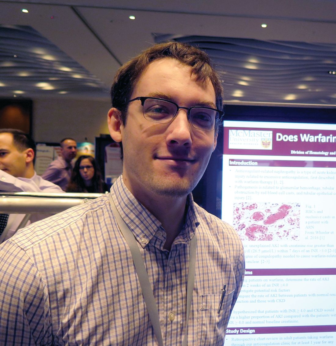
The primary outcome was AKI, defined as an acute increase in creatinine of greater than 26.5 micromol/L within 7-14 days of an INR 4 or greater. The secondary outcome was creatinine level within 3 months of the abnormal INR. The researchers excluded patients with AKI due to another cause, and those who lacked a creatinine level at baseline, within 7-14 days of an INR of 4 or greater, and/or at 3 months.
The median age of the 292 patients was 79 years, 55% were male, 30% were taking aspirin, and 77% were taking renin angiotensin inhibitors and/or diuretics. The control group consisted of 93 patients with a 12-month time in therapeutic range of 100%. The median age of controls was 68 years, 67% were male, and 9% had CKD. None of the controls had an AKI, said Dr. Traquair, a second-year internal medicine resident in the department of medicine at McMaster University.
Of the 292 patients with an INR of 4 or greater, 13% had an AKI, and the incidence of AKI was significantly higher in the CKD patients, compared with those who had a normal baseline creatinine level (19% vs. 10%; odds ratio, 2.1; P less than .05).
In a binomial logistic regression model, diuretic use was the only significant predictor of AKI (OR 3.4; P less than .05). The researchers also found that of the 52 patients with an INR of 4 or greater who did not use renin angiotensin system inhibitors and/or diuretics and did not have CKD, only 1 had an AKI (2%).
“We don’t know that all of these episodes of AKI are related to warfarin, but we do see a definite increase of AKI after an episode of overanticoagulation (an INR greater than 4),” Dr. Traquair said. “In patients who are at risk for AKI, monitoring their kidney function after an episode of overanticoagulation is probably warranted.”
Dr. Traquair reported having no financial disclosures.
SOURCE: Traquair H et al. THSNA 2018, Poster 79.
REPORTING FROM THSNA 2018
Key clinical point:
Major finding: Among patients with warfarin anticoagulation, 13% had an acute kidney injury.
Study details: A retrospective study of 292 patients with an INR of 4.0 or greater who were treated between 2007 and 2017.
Disclosures: Dr. Traquair reported having no financial disclosures.
Source: Traquair H et al. THSNA 2018, Poster 79.
Consider caregiver oral health for children with bleeding disorders
SAN DIEGO – Caregiver oral health status is an identifiable risk factor that could be used to screen for poor oral health among children and young adults with bleeding disorders, results from a single-center suggest.
“We ask parents one simple question: ‘Have you had a cavity in the last year?’ If they say yes, we would be more concerned that their children would be more likely to have poor oral health,” Elizabeth Hastie said in an interview during a poster session at the biennial summit of the Thrombosis & Hemostasis Societies of North America.
Proper oral health may prevent joint disease and other conditions that predispose patients to bleeds, according to Ms. Hastie, a fourth-year medical student at Emory University, Atlanta. However, of the 147 hemophilia treatment centers in the United States, just 30% have a dentist on staff, while 90% of centers have expressed interest in increasing patient education in oral health.
In an effort to evaluate the dental habits, needs, and oral health issues of children and young adults up to age 18 with bleeding disorders, Ms. Hastie and her associates conducted a cross-sectional study of 226 patients who were evaluated by a staff dental hygienist at Children’s Healthcare of Atlanta Comprehensive Bleeding Disorders Clinic from May 2016 to October 2017.
The evaluation consisted of a 14-question survey derived from the American Academy of Pediatric Dentistry Caries–Risk Assessment Tool completed by the primary caregiver present during the visit and oral screening. The researchers extracted demographic and clinical characteristics from the patient’s chart and included age, race, county of residence, and bleeding disorder type and severity.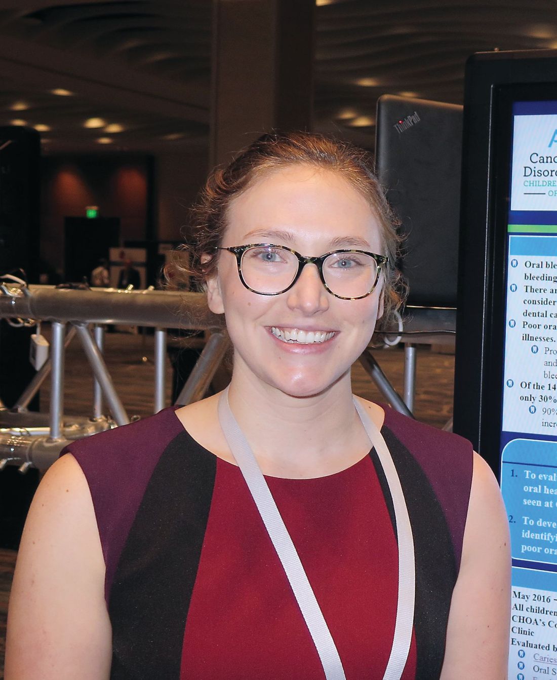
Nearly half of the patients (44%) reported they did not brush their teeth twice a day. Children younger than age 5 years were more likely to not brush their teeth twice a day, compared with children aged 5-14 years and young adults aged 15-18 years (57% vs. 44% and 31% respectively, P = .08).
More than one-quarter of patients (27%) reported not having a current dentist and 15% reported specific challenges with access to dental care including burdens related to distance, insurance coverage, and finding a provider willing to treat in the setting of their medical condition. Those who were Medicaid eligible or of low socioeconomic status were significantly more likely to report dental care access issues, compared with other patients (20% vs. 9%; P = .01).
Oral screening performed by the dental hygienist demonstrated significant oral pathology: 89% of patients had plaque accumulation, 57% had white spots or decalcifications, 37% had gingivitis, and 8% had suspicious lesions suggestive of dental caries.
The researchers also found that having a caregiver with active oral disease in the past 12 months increased the odds of suspicious lesions (odds ratio, 4.34), increased the odds of gingivitis (OR, 3.80), and decreased the odds of the patients’ brushing their teeth at least twice per day (OR, 0.17).
“Hopefully, if we can target those high-risk patients in clinic, we could reduce costs, the number of bleeds, the number of products and factor used, and potentially even morbidity in the future,” Ms. Hastie said.
She acknowledged certain limitations of the study, including its single-center design and the fact that a dental hygienist performed the majority of evaluations. She reported having no financial disclosures.
REPORTING FROM THSNA 2018
Key clinical point:
Major finding: Having a caregiver with active oral disease in the past 12 months increased the odds of the child having a suspicious lesion (OR 4.34) and gingivitis (OR 3.80).
Study details: A cross-sectional study of 226 pediatric patients who were evaluated by a dental hygienist.
Disclosures: Ms. Hastie reported having no financial disclosures.
Source: Hastie E et al. THSNA 2018, Poster 150.
Time to scrap LMWH for prevention of placenta-mediated pregnancy complications?
SAN DIEGO – Low molecular weight heparin does not appear to reduce the risk of recurrent placenta-mediated pregnancy complications in women with prior such complications, according to Marc Rodger, MD.
“It’s time to put the needles away for pregnant patients,” he said at the biennial summit of the Thrombosis & Hemostasis Societies of North America.
The pathophysiology of placenta-mediated pregnancy complications includes placental thrombosis. Thrombophilias predispose to the development of thrombosis in slow-flow circulation of the placenta. “It’s possible that the etiology mix of placental-mediated pregnancy complications includes thrombophilias, and by extension, that anticoagulants would prevent these complications,” said Dr. Rodger, a senior scientist at the hospital and professor at the University of Ottawa.
In a study from 1999, researchers demonstrated that patients with pregnancy-mediated placental complications were 8.2 times more likely to develop thrombophilia, compared with controls (N Engl J Med. 1999;340:9-13). “But as with positive initial case-control studies, subsequent work downplayed this association,” Dr. Rodger said. “Now, we’re at a point where we recognize that thrombophilias are weakly associated with recurrent early loss, late pregnancy loss, and severe preeclampsia ([odds ratio] of about 1.5-2.0 for all associations), while thrombophilias are not associated with nonsevere preeclampsia and small for gestational age.”
Currently, low-molecular-weight heparin (LMWH) is the preferred pharmacoprophylaxis in pregnancy. Unfractionated heparin, meanwhile, requires b.i.d. or t.i.d. injections, and has a 10-fold higher risk of heparin-induced thrombocytopenia and a greater than 10-fold higher risk of osteoporotic fracture. Warfarin is teratogenic antepartum and inconvenient postpartum, while direct oral anticoagulants cross the placenta and enter breast milk.
Downsides of LMWH include the burden of self-injections and costs of over $10,000 per antepartum period, Dr. Rodger said. Common side effects include minor bleeding and elevated liver function tests, and it complicates regional anesthetic options at term. Uncommon side effects include major bleeding, skin reactions, and postpartum wound complications, while rare but serious complications include heparin-induced thrombocytopenia and osteoporotic fractures.
He offered a hypothetical case. A 32-year-old woman with prior severe preeclampsia who delivered at 32 weeks asks you, “Should I be treated with LMWH in my next pregnancy?” What should you tell her? To answer this question, Dr. Rodger and his associates conducted a study-level meta-analysis of six randomized controlled trials that included 848 pregnant women with prior placenta-mediated pregnancy complications (Blood. 2014;123[6]:822-8). The primary objective was to determine the effect of LMWH in preventing placenta-mediated pregnancy complications in women with prior late placenta-mediated pregnancy complications. This included patients with or without thrombophilia who were treated with or without LMWH. The primary outcome was a composite of preeclampsia, birth of an SGA newborn, placental abruption, or pregnancy loss greater than 20 weeks. Overall, 67 (18.7%) of 358 of women being given prophylactic LMWH had recurrent severe placenta-mediated pregnancy complications, compared with 127 (42.9%) of 296 women with no LMWH (relative risk reduction, 0.52; P = .01, indicating moderate heterogeneity). They identified similar relative risk reductions with LMWH for individual outcomes, including any preeclampsia, severe preeclampsia, SGA below the 10th percentile, SGA below the 5th percentile, preterm delivery less than 37 weeks, and preterm delivery less than 34 weeks with minimal heterogeneity. They concluded that LMWH “may be a promising therapy for recurrent, especially severe, placenta-mediated pregnancy complications, but further research is required.”
At the meeting, Dr. Rodger noted that the positive studies in the analysis were single-center trials, “which are generally acknowledged to be of a lesser methodologic quality, and the majority of patients in these single-center trials are from a small area in the south of France. Multicenter trials don’t show an effect, so is it single-centeredness or is it something else? The other feature that’s distinct is that the positive trials recruited patients with prior severe complications only, while the negative trials included patients with nonsevere complications. So maybe LMWH works in patients who have a very strong phenotype that have had very bad prior complications. We can’t tease that out with a study-level meta-analysis because we’re getting average effects over heterogeneous groups of patients.”
To expand on the study-level meta-analysis, Dr. Rodger and his associates conducted a systematic review and individual patient data meta-analysis of eight randomized trials of 963 patients conducted between 2000 and 2013 of LMWH to prevent recurrent placenta-mediated pregnancy complications (Lancet. 2016;388:2629-41). “In this approach you get individual patient data from the trials, and you create a new randomized, controlled data set,” he explained. “That way we could tease out the patients who have had the prior severe complications and whether their mild or severe outcomes are being prevented or not.”
The study’s composite primary outcome was one or more of the following: early-onset or severe preeclampsia, SGA newborn below the 5th percentile, late pregnancy loss (over 20 weeks), or placental abruption. Dr. Rodger and his associates found that LMWH did not significantly reduce the risk of recurrent placenta-mediated pregnancy complications, compared with patients who did not receive LMWH (14% vs. 22%, respectively; P = .09). In subgroup analyses, however, LMWH in multicenter trials reduced the primary outcome in women with previous abruption (P = .006) but not in any of the other subgroups of previous complications. “There were small numbers of patients in this subgroup, though, so I would use caution,” Dr. Rodger said. Two recent randomized, controlled trials from separate investigators further support the overall null findings of the individual patient data meta-analysis (Obstet Gynecol. 2016;128[5]:1053-63 and Am J Obstet Gynecol. 2017 Mar;216[3]:296.e1-296.e14).
Revisiting the hypothetical case of a 32-year-old woman with prior severe preeclampsia who delivered at 32 weeks, Dr. Rodger said that he would “definitely not” recommend LMWH during her next pregnancy.
He acknowledged limitations of the systematic review, including the limited numbers of patients in subgroups and the large differences between single-center and multicenter trials. “We still can’t explain this, and it remains an open question that bugs me,” he said. “This has been seen in many disease areas. Empirically, single-centeredness leans toward positivity.”
He called for more research in women with recurrent pregnancy loss. Dr. Rodger reported having no financial disclosures.
REPORTING FROM THSNA 2018
Experts offer guidance on use of emicizumab
SAN DIEGO – Emicizumab is a safe and effective new therapy for individuals with hemophilia A and inhibitor antibodies that will likely provide a paradigm shift for managing this patient population, according to Michael U. Callaghan, MD.
“It’s a safe drug, but you do have to be cautious about treating breakthrough bleeds with activated prothrombin complex concentrate (aPCC) resistance in particular,” Dr. Callaghan, a pediatric hematologist/oncologist at Children’s Hospital of Michigan, Detroit, said in an interview at the biennial summit of the Thrombosis & Hemostasis Societies of North America. “Patients require laboratory monitoring, and you need to educate anyone who’s going to see the patient about how the drug affects laboratory tests.”
Approved in November 2017, emicizumab (Hemlibra) is a recombinant, humanized bispecific immunoglobulin G4 monoclonal antibody that mimics the cofactor function of activated factor VIII (FVIIIa) by bridging activated factor IX and factor X. After 4 weeks of a loading dose of 3 mg/kg, subcutaneous once weekly dosing at 1.5 mg/kg demonstrated significant reduction in annualized bleeding rates in patients of all ages with congenital hemophilia A and inhibitors. But treatment-related adverse events occurred during the pivotal trials.
In an effort to provide recommendations on use of emicizumab beyond information contained in the agent’s package insert, Dr. Callaghan and his associates reviewed published literature, meeting abstracts, and expert experience with emicizumab on clinical trials.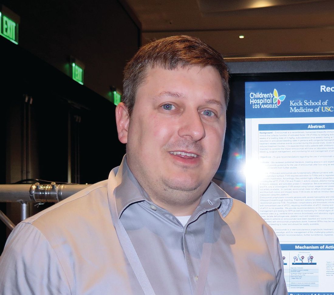
Since emicizumab is highly selective for human FIXa and FX, only chromogenic FVIII assays using human reagents can assess emicizumab activity but those assays are not widely available, the researchers noted in their abstract. “In contrast, emicizumab does not affect chromogenic assays that contain bovine reagents and thus both native and infused factor FVIII levels as well as inhibitor titers (modified Bethesda assay) can be measured using this platform.”
In a phase 3 trial of emicizumab known as HAVEN 1, serious adverse events included three cases of thrombotic microangiopathy (TMA) and two thrombotic events (TE) (N Engl J Med 2017;377:809-18).* To prevent breakthrough bleeding, aPCC should be avoided unless there are no effective alternatives to control bleeding, Dr. Callaghan said. Treatment options for bleeding include bypassing agents such as human or recombinant porcine FVIII.
To prevent, monitor, and treat TMA and TE, prior to starting emicizumab, patients should be informed that baseline hemostasis is increased with the agent and that there is an increased risk of pathologic thrombosis with bypassing agents.
Patients should also be informed about the risk of TE/TMA and the signs and symptoms of TE/TMA. “If repeated dosing of bypass agents is needed, particularly aPCC, patients should contact their hemophilia treatment center,” the researchers wrote. “If TE/TMA is suspected, platelet count, creatinine, d-dimer, and fibrinogen should be monitored. If TE/TMA occur, emicizumab should be held and aPCC discontinued until resolution. Upon resolution of TE/TMA, consideration should be given to restarting emicizumab on a case-by-case basis.”
As for laboratory considerations, the researchers noted that results of activated partial prothrombin time (aPTT) will be shortened in patients on emicizumab, often into the normal range even at low concentrations. In addition, one-stage aPTT based factor VIII activity assays will yield high factor VIII activities, even at low concentrations of the drug. “Health care providers including dentists, surgeons, and emergency room staff need to be informed of the effects of emicizumab on laboratory tests,” they wrote in a poster at THSNA 2018.
HAVEN 1 and HAVEN 2 showed that 22 patients underwent 29 surgical procedures: tooth extractions (6), CVAD procedures (9), and other procedures (14). Of the 29 surgical procedures, 9 (31%) were managed with prophylactic bypassing agents, and one treated bleed occurred. At the same time, 20 procedures (69%) were managed without prophylactic bypassing agents, and two treated bleeds occurred.
The researchers concluded that additional studies are needed to inform the use of emicizumab in people with hemophilia A, with and without inhibitor antibodies.
Dr. Callaghan reported having no financial disclosures.
*Correction, 4/26/2018: An earlier version of this story misstated the number of cases of thrombotic microangiopathy.
EXPERT ANALYSIS FROM THSNA 2018
The ‘holy grail’ of thrombosis prevention
SAN DIEGO – The “holy grail” of thrombosis prevention is the ability to determine the risk of recurrence with or without continuation of anticoagulant treatment, according to Philip S. Wells, MD.
“Very little data exists for the comparison of active treatment to placebo in the acute and long-term phases of treatment,” he said at the biennial summit of the Thrombosis & Hemostasis Societies of North America. “With low molecular weight heparin, vitamin K antagonists, and direct-acting oral anticoagulants, the relative risk reduction is about 90% in the acute phase and 80%-85% in the extended phase. After discontinuing anticoagulants, the absolute risk of recurrence varies depending on VTE category.”
Dr. Wells, chair and chief of the department of medicine at The Ottawa Hospital and the University of Ottawa, said that after 3 months of anticoagulation the chance of recurrence in postsurgical VTE patients is less than 1% per year. After 3 months of anticoagulant use in nonsurgical patients with provoked risk factors, it is around 4%. This includes medical patients, trauma victims, pregnant women, and patients wearing a plaster cast.
In patients who survive an unprovoked VTE, after 3-6 months of anticoagulant therapy their overall recurrence risk is 10% in the first year and 30% after 5 years. The risk of recurrence is 50% higher if a patient experiences a second unprovoked VTE, and the risk of fatality is 50% higher if the initial event was a pulmonary embolism (PE), he said.
According to the ongoing prospective Austrian Study on Recurrent Venous Thromboembolism, the risk of recurrent VTE is 20% in men and 6% in women (N Engl J Med. 2004 Jun 17;350[25]:2558-63). A multicenter prospective study in Canada yielded similar results. It found that the risk of recurrent VTE is 19% in men versus 9% in women (CMAJ 2008;179[5]:417-26).
That Canadian prospective study, led by Marc Rodger, MD, described the development of the HERDOO2 clinical decision rule for determining a patient’s risk for a recurrent VTE. This includes hyperpigmentation, edema, or redness in either leg (signs of postthrombotic syndrome), D-dimer level of 250 mcg/L or greater while on warfarin, body mass index of 30 kg/m2 or greater, and age of 65 years or older.
If patients have zero or one risk factor, the annual risk of VTE after 6 months of treatment is 1.6%, while two or more risk factors bumps the annual risk of VTE to 14.1%, according to the researchers.
In a subsequent study to validate the HERDOO2, researchers found that the risk of recurrent major VTE was 3.0% in low-risk women who discontinued oral anticoagulants (OACs), 8.1% in high-risk women and men who discontinued OACs, 1.6% in high-risk women and men who continued OACs, and 7.4% in high-risk women who discontinued OACs (BMJ 2017;356:j1065).
“I think the HERDOO2 rule is working pretty well to determine a low-risk group of women, and it’s not an unreasonable tool to be using,” Dr. Wells said.
Other variables that might help clinicians predict a patient’s VTE recurrence include the presence of recurrent venous obstruction (adjusted HR 1.32), and older age (HR 1.01 for every 1 year increase).
D-dimer levels can also be helpful. “If the serial D-dimers are positive, stay on anticoagulants,” Dr. Wells advised. “If they’re negative, discontinue anticoagulants and have the D-dimer levels repeated monthly for 3 months. If positive or positive conversions, return to OAC therapy.”

In one study, the annual risk of a VTE was 3% in the negative D-dimer patients, compared with 6.1% in those who had a history of an unprovoked VTE (Blood 2014;124:196-203).
In a separate study of 319 patients with two negative D-dimer results who did not restart anticoagulation therapy, the rate of recurrent VTE was 6.7% per patient-year (Ann Intern Med 2015;162:27-34). It was 9.7% per patient-year in men, compared with 5.4% per patient-year in women.
Dr. Wells emphasized the importance of shared decision-making with the patient when devising a strategy for long-term anticoagulation following a VTE. “We don’t have a lot of good tools, but [trying to elicit] patient preference is the right thing to try and do,” he said. “Physicians should present an unbiased perspective to patients regarding their treatment, including the benefits and harms, effect on quality of life, and cost.”
Dr. Wells also shared his current clinical approach. In women with an unprovoked VTE, he applies the HERDOO2 rule. If there’s a low recurrence risk, he discontinues the anticoagulant. If there’s a non-low recurrence risk he continues with the anticoagulant unless there’s a high risk for bleeding. Men with an unprovoked VTE receive indefinite anticoagulant therapy, but if the index event is a deep vein thrombosis (DVT), Dr. Wells applies a bleeding risk tool to help him determine management going forward. If the patient has a high risk of bleeding, he does not use an anticoagulant.
“If there is a high risk of bleeding it’s best of stay off anticoagulant therapy,” he said. “If there is an intermediate risk of bleeding and the index event was a DVT, the patient could stay off anticoagulants. I think we have a long way to go to developing tools that actually enable us to reach these points with each patient in discussions we have with them about continuing anticoagulants.”
Dr. Wells reported having received research support from BMS/Pfizer and honoraria from Bayer AG, Janssen, Pfizer, and Daiichi Sankyo. He is a member of the scientific advisory board for Bayer AG and Pfizer.
SAN DIEGO – The “holy grail” of thrombosis prevention is the ability to determine the risk of recurrence with or without continuation of anticoagulant treatment, according to Philip S. Wells, MD.
“Very little data exists for the comparison of active treatment to placebo in the acute and long-term phases of treatment,” he said at the biennial summit of the Thrombosis & Hemostasis Societies of North America. “With low molecular weight heparin, vitamin K antagonists, and direct-acting oral anticoagulants, the relative risk reduction is about 90% in the acute phase and 80%-85% in the extended phase. After discontinuing anticoagulants, the absolute risk of recurrence varies depending on VTE category.”
Dr. Wells, chair and chief of the department of medicine at The Ottawa Hospital and the University of Ottawa, said that after 3 months of anticoagulation the chance of recurrence in postsurgical VTE patients is less than 1% per year. After 3 months of anticoagulant use in nonsurgical patients with provoked risk factors, it is around 4%. This includes medical patients, trauma victims, pregnant women, and patients wearing a plaster cast.
In patients who survive an unprovoked VTE, after 3-6 months of anticoagulant therapy their overall recurrence risk is 10% in the first year and 30% after 5 years. The risk of recurrence is 50% higher if a patient experiences a second unprovoked VTE, and the risk of fatality is 50% higher if the initial event was a pulmonary embolism (PE), he said.
According to the ongoing prospective Austrian Study on Recurrent Venous Thromboembolism, the risk of recurrent VTE is 20% in men and 6% in women (N Engl J Med. 2004 Jun 17;350[25]:2558-63). A multicenter prospective study in Canada yielded similar results. It found that the risk of recurrent VTE is 19% in men versus 9% in women (CMAJ 2008;179[5]:417-26).
That Canadian prospective study, led by Marc Rodger, MD, described the development of the HERDOO2 clinical decision rule for determining a patient’s risk for a recurrent VTE. This includes hyperpigmentation, edema, or redness in either leg (signs of postthrombotic syndrome), D-dimer level of 250 mcg/L or greater while on warfarin, body mass index of 30 kg/m2 or greater, and age of 65 years or older.
If patients have zero or one risk factor, the annual risk of VTE after 6 months of treatment is 1.6%, while two or more risk factors bumps the annual risk of VTE to 14.1%, according to the researchers.
In a subsequent study to validate the HERDOO2, researchers found that the risk of recurrent major VTE was 3.0% in low-risk women who discontinued oral anticoagulants (OACs), 8.1% in high-risk women and men who discontinued OACs, 1.6% in high-risk women and men who continued OACs, and 7.4% in high-risk women who discontinued OACs (BMJ 2017;356:j1065).
“I think the HERDOO2 rule is working pretty well to determine a low-risk group of women, and it’s not an unreasonable tool to be using,” Dr. Wells said.
Other variables that might help clinicians predict a patient’s VTE recurrence include the presence of recurrent venous obstruction (adjusted HR 1.32), and older age (HR 1.01 for every 1 year increase).
D-dimer levels can also be helpful. “If the serial D-dimers are positive, stay on anticoagulants,” Dr. Wells advised. “If they’re negative, discontinue anticoagulants and have the D-dimer levels repeated monthly for 3 months. If positive or positive conversions, return to OAC therapy.”
In one study, the annual risk of a VTE was 3% in the negative D-dimer patients, compared with 6.1% in those who had a history of an unprovoked VTE (Blood 2014;124:196-203).
In a separate study of 319 patients with two negative D-dimer results who did not restart anticoagulation therapy, the rate of recurrent VTE was 6.7% per patient-year (Ann Intern Med 2015;162:27-34). It was 9.7% per patient-year in men, compared with 5.4% per patient-year in women.
Dr. Wells emphasized the importance of shared decision-making with the patient when devising a strategy for long-term anticoagulation following a VTE. “We don’t have a lot of good tools, but [trying to elicit] patient preference is the right thing to try and do,” he said. “Physicians should present an unbiased perspective to patients regarding their treatment, including the benefits and harms, effect on quality of life, and cost.”
Dr. Wells also shared his current clinical approach. In women with an unprovoked VTE, he applies the HERDOO2 rule. If there’s a low recurrence risk, he discontinues the anticoagulant. If there’s a non-low recurrence risk he continues with the anticoagulant unless there’s a high risk for bleeding. Men with an unprovoked VTE receive indefinite anticoagulant therapy, but if the index event is a deep vein thrombosis (DVT), Dr. Wells applies a bleeding risk tool to help him determine management going forward. If the patient has a high risk of bleeding, he does not use an anticoagulant.
“If there is a high risk of bleeding it’s best of stay off anticoagulant therapy,” he said. “If there is an intermediate risk of bleeding and the index event was a DVT, the patient could stay off anticoagulants. I think we have a long way to go to developing tools that actually enable us to reach these points with each patient in discussions we have with them about continuing anticoagulants.”
Dr. Wells reported having received research support from BMS/Pfizer and honoraria from Bayer AG, Janssen, Pfizer, and Daiichi Sankyo. He is a member of the scientific advisory board for Bayer AG and Pfizer.
EXPERT ANALYSIS FROM THSNA 2018
Two scoring systems helpful in diagnosing heparin-induced thrombocytopenia
SAN DIEGO – Both the 4Ts Score and the HIT Expert Probability (HEP) Score are useful in clinical practice for the diagnosis of heparin-induced thrombocytopenia, but the HEP score may have better operative characteristics in ICU patients, results from a “real world” analysis showed.
“The diagnosis of heparin-induced thrombocytopenia (HIT) is challenging,” Allyson M. Pishko, MD, one of the study authors, said at the biennial summit of the Thrombosis & Hemostasis Societies of North America. “The 4Ts Score is commonly used, but limitations include its low positive predictive value and significant interobserver variability.”
One external prospective study showed operating characteristics similar to those of 4Ts scores (Thromb Haemost 2015;113[3]:633-40).
The aim of the current study was to validate the HEP Score in a “real world” setting and to compare the performance of the HEP Score versus the 4Ts Score. The researchers enrolled 292 adults with suspected acute HIT who were hospitalized at the University of Pennsylvania or affiliated community hospitals, and who had HIT laboratory testing ordered.
The HEP Score and the 4Ts Score were calculated by a member of the clinical team and were completed prior to return of the HIT lab test result. The majority of scorers (62%) were hematology fellows, followed by attendings (35%), and residents/students (3%). All patients underwent testing with an HIT ELISA and serotonin-release assay (SRA). Patients in whom the optical density of the ELISA was less than 0.4 units were classified as not having HIT. The researchers used the Wilcoxon rank-sum test to compare HEP and 4Ts Scores in patients with and without HIT.
Of the 292 patients, 209 were HIT negative and 83 had their data reviewed by an expert panel. Of these 83 patients, 40 were found to be HIT negative and 43 were HIT positive, and their mean ages were 65 years and 63 years, respectively. Among the cases found to be positive for HIT, 93% had HIT ELISA optical density of 1 or greater and 69.7% were SRA positive. The median HEP Score in patients with and without HIT was 8 versus 5 (P less than .0001).
At the prespecified screening cut-off of 2 or more points, the HEP Score was 97.7% sensitive and 21.9% specific, with a positive predictive value of 17.7% and a negative predictive value of 98.2%. A cut-off of 5 or greater provided 90.7% sensitivity and 47.8% specificity with a positive predictive value of 23.1% and a negative predictive value of 96.8%. The mean time to calculate the HEP Score was 4.1 minutes.
The median 4Ts Score in patients with and without HIT was 5 versus 4 (P less than .0001), Dr. Pishko reported. A 4Ts Score of 4 or greater had a sensitivity of 97.7% and specificity of 32.9%, with a positive predictive value of 20.1% and a negative predictive value of 98.8%.
The area under the ROC curves for the HEP Score and 4Ts Score were similar (0.81 vs. 0.76; P = .121). Subset analysis revealed that compared with the 4Ts Score, the HEP Score had better operating characteristics in ICU patients (AUC 0.87 vs. 0.79; P= .029) and with trainee scorers (AUC 0.79 vs. 0.73; P = .032).
“Our data suggest that either the HEP Score or the 4Ts Score could be used in clinical practice,” Dr. Pishko said.
The National Institutes of Health funded the study. Dr. Pishko reported having no financial disclosures.
SOURCE: Pishko A et al. THSNA 2018.
REPORTING FROM THSNA 2018
Key clinical point:
Major finding: The area under the ROC curves for the HEP Score and 4Ts Score were similar (0.81 vs. 0.76; P = .121).
Study details: A prospective study of 292 adults with suspected acute HIT who were hospitalized at the University of Pennsylvania or affiliated community hospitals.
Disclosures: The National Institutes of Health funded the study. Dr. Pishko reported having no financial disclosures.
Source: Pishko A et al. THSNA 2018.
Researchers seek better understanding of von Willebrand disease
SAN DIEGO – Several groups of researchers are examining cohorts of von Willebrand disease (VWD), looking at its pathogenesis and molecular causes, as well as ways to improve treatment strategies.
At the biennial summit of the Thrombosis & Hemostasis Societies of North America, Robert F. Sidonio Jr., MD, MSc, highlighted the efforts underway, including the Zimmerman Program for the Molecular and Clinical Biology of VWD study (ZPMCB-VWD), a large grant project funded by the National Institutes of Health.
At the time of publication, there were more than 700 index cases and more than 2,200 families in the study. It includes data from 8 primary centers and 23 secondary centers. The goals are to characterize the molecular causes of VWD and the examine the fidelity of diagnosis, “which is a critical component of the study,” said Dr. Sidonio, clinical director of the hemostasis/thrombosis program at Children’s Healthcare of Atlanta.
When the ZPMCB-VWD investigators examined the correlation of bleeding phenotype and the genotype, the found that as the level of VW factor goes down, the bleeding score generally goes up, but it becomes a little bit flat in the 20-30 IU/dL range.
“Where we spend a lot of our time is with patients who have levels of 30%-50%, which can be quite heterogeneous,” Dr. Sidonio said.
Another finding made out of the ZPMCB-VWD project was the discovery of the single nucleotide polymorphism p.D1472H, which was noted to be more common in African American patients and leads to low Von Willebrand Ristocetin Cofactor (VWF:RCo) and VWF:RCo/VWF:Ag ratio, but does not increase the bleeding score.
The fidelity of diagnosis was another key finding to come out of ZPMCB-VWD. Most type 1 VWD cases were identified by low VWF:RCo. There was poor correlation between historical and current assays (r2 = 0.22), and diagnostic labs improved after central lab testing.
Next, Dr. Sidonio discussed findings from RENAWI 1 and 2, which are Italian registries of about 1,000 VWF patients that were organized by 12 centers in 2002. The goals are to evaluate the natural history of VWD in Italy and to characterize treatment strategies. According to preliminary findings from the researchers, the biological response to desmopressin (DDAVP) was 69% in those with VWD1, 26% in those with VWD2A, 20% in those with VWD2B, 33% in those with VWD2M, 71% in those with VWD2N, and 0% in those with VWD3 (Blood 2014;123:4037-44).
These researchers also found that a mean bleeding score of 3.5 corresponds to a VWF:RCo score of 30 U/dL or greater. “This indicates that there is something slightly different about patients that are above and below that threshold,” Dr. Sidonio said. “I think that’s something we’ve all been struggling with: trying to understand where the differences are and how aggressively we should be treating our patients with mild VWD.”
Another effort, The Willebrand in the Netherlands’ study (WiN), is a prospective cohort trial of about 700 patients with types 1, 2, and 3 VWD from 12 centers in that country (Blood 2008;112:4510). It was the first large study to use VWF propeptide (pp) to discriminate between severe type 1 and type 3 VWD. It also found that type 2 VWD is more characterized by increased clearance in VWF in contrast to type 1 VWD, leading to higher VWFpp/VWF:Ag ratio. In addition, in type 1 VWD, antigen rates increased about 3.5 U/dL per decade, RCo increased about 9.5 U/dL per decade, and Factor VIII: C increased about 7.1 U/dL per decade (J Thromb Haemost. 2014; 12[7]:1066-75).
“I don’t think we have a study to be able to follow patients for 20 years or so, knowing that we rely on the assays that we were using 20 years ago,” Dr. Sidonio said. “That’s a challenge at a lot of our centers, but we know that VWF generally rises with age in mild VWD patients.”
In patients with definitively diagnosed type 2 VWD, no age-related VWF or FVIII changes were observed. The researchers also observed an increase in surgical bleeding and GI bleeding in elderly VWD patients.
In the meantime, the Canadian Type 1 VWD study was one of the first to elucidate the complexity of the pathogenesis of type 1 VWD. It identified CLEC4M as playing a role in VWF clearance, with polymorphisms contributing to the variability of VWF.
More data collection is underway through a partnership between the Centers for Disease Control and Prevention and the American Thrombosis and Hemostasis Network (ATHN).
The CDC Universal Data Collection Project gathered surveillance data on bleeding disorders on patients from 1998-2011. The goals were to characterize bleeding complications, monitor safety of blood-based products to manage bleeding, identify health issues in need of additional research, evaluate bleeding disorders over the lifespan, and evaluate quality of life. In 2006, the ATHN was formed to provide stewardship of the secured national database housed at the CDC. To date, at least 34,000 patients have opted in to the data set, which includes demographic and clinical data used for research.
In a separate, phase 4 study of about 130 patients funded by Shire and led by Dr. Sidonio and Angela C. Weyand, MD, researchers will conduct a “real-world” safety and efficacy study of prophylaxis for severe VWD. Known as ATHN 9, the study includes patients currently enrolled in the ATHN data set. Treatment regimen is at the discretion of patients’ providers, and patients will be followed for up to 2 years from the start of enrollment. The study’s primary aim is to collect data on effectiveness and safety, including adverse events of various VWF regimens in adult and pediatric patients with severe congenital VWD.
Another effort is the Medical and Scientific Advisory Council (MASAC) Working Group, of which Dr. Sidonio is a member. The first meeting took place in July of 2016. The goals include making improvements to diagnostic testing and laboratory standards, assessing existing standards of care and clinical practice guidelines, developing educational programming, conducting research to better understand and develop effective treatments for VWD, and collaborating with partner organizations.
Dr. Sidonio reported that he has participated in advisory boards for Shire, CSL Behring, Biogen/Bioverativ, Pfizer, Emergent Solutions, Roche/Genentech, Aptevo, Novo Nordisk, Hema Biologics, and Octapharma. He also has received investigator-initiated grant funding from Bioverativ, Grifols, Kedrion, and Shire.
EXPERT ANALYSIS FROM THSNA 2018
Why is gene therapy for hemophilia taking so long?
SAN DIEGO – The goal of gene therapy for hemophilia and other genetic diseases is to achieve long-term expression and levels adequate to improve the phenotype of disease, according to Katherine A. High, MD.
“Sometimes people ask me, ‘Why is it taking so long to develop these therapeutics?’ ” Dr. High said at the biennial summit of the Thrombosis & Hemostasis Societies of North America. “The answer is that gene therapy vectors are arguably one of the most complex therapeutics yet developed.”
Spark Therapeutics is currently developing gene therapies for hemophilia A (SPK-8011) and hemophilia B (SPK-9001).
Hemostasis and thrombosis targets in gene therapy trials include hemophilia, as well as peripheral artery disease/claudication and congestive heart failure. In the latter, a prior phase 2b trial of adeno-associated virus (AAV) expressing SERCA2a did not support efficacy (Lancet 2016;387:1178-86), while a current trial of adenovirus 5–vector expressing adenylyl cyclase–type 6 is entering phase 3 study (NCT03360448).
To get a sense of how long it may take for a new class of therapeutics to become established, Dr. High noted that the first monoclonal antibody to be licensed was OKT3 (muromonab-CD3) in 1986, followed by abciximab in 1994, rituximab and daclizumab in 1997, and four additional products in 1998. By 2007, 8 of the top 20 biotech drugs were monoclonal antibodies.
Hemophilia has long been a favored gene therapy target because biology is in its favor. “It has a wide therapeutic window, it does not require tissue-specific expression of transgene, small and large animal models exist, and endpoints are well validated and easy to measure,” she said. “Thus, early gene-therapy clinical investigation since 1998 explored many strategies.”
There are several current investigational efforts in AAV-mediated gene transfer in hemophilia, including:
- A single-arm study to evaluate the efficacy and safety of valoctocogene roxaparvovec in hemophilia A patients at a dose of 4×10¹³ vector genome per kilogram (NCT03392974).
- A dose-ranging study of recombinant AAV2/6 human factor 8 gene therapy SB-525 in subjects with severe hemophilia A (NCT03061201).
- A safety and dose-escalation study of an adeno-associated viral vector for gene transfer in hemophilia A subjects (NCT03370172).
Other approaches in preclinical investigation include lentiviral transduction of hematopoietic stem cells with megakaryocyte-restricted expression, lentiviral transduction of liver cells and endothelial cells, and genome editing using zinc finger nucleases.
“AAV vectors are one of the smallest of all naturally occurring viruses,” said Dr. High, who is also emeritus professor of pediatrics at the University of Pennsylvania, Philadelphia. “The recombinant AAV consists of a highly ordered set of proteins [vector capsid] containing DNA [the active agent].”
Overall goals for a hemophilia gene therapy include long-term expression and levels adequate to prevent bleeds in someone with a normal active lifestyle. “We’d like to see consistency of results from one person to the next, and we’d like to use the lowest possible dose,” she said. “In the setting of gene transfer, the lower the dose, the lower the likelihood of immune responses that need to be managed. Theoretically, the lower the dose, the lower the risk of insertional mutagenesis, and the shorter-term duration of vector shedding in body fluids, including in semen.”
Going forward, a key question for researchers relates to the long-term effect of gene therapy. “How long is long enough?” Dr. High asked. “The longest reported durability is 8 years, with observation ongoing, from studies initially reported in men with severe hemophilia B. The durability in large animal models exceeds 10 years.”
Another unanswered question is what level of factor VIII to aim for in treatment. “Some data suggest that FVIII levels greater than 100 IU/dL are associated with a greater level of thrombosis,” Dr. High said. “So I think somewhere between 12% and 100% is probably the ideal level.”
EXPERT ANALYSIS FROM THSNA 2018
Few acutely ill hospitalized patients receive VTE prophylaxis
SAN DIEGO – Among patients hospitalized for acute medical illnesses, the risk of venous thromboembolism (VTE) remained elevated 30-40 days after discharge, results from a large analysis of national data showed.
Moreover, only 7% of at-risk patients received VTE prophylaxis in both the inpatient and outpatient setting.
“The results of this real-world study imply that there is a significantly unmet medical need for effective VTE prophylaxis in both the inpatient and outpatient continuum of care among patients hospitalized for acute medical illnesses,” researchers led by Alpesh Amin, MD, wrote in a poster presented at the biennial summit of the Thrombosis & Hemostasis Societies of North America.
According to Dr. Amin, who chairs the department of medicine at the University of California, Irvine, hospitalized patients with acute medical illnesses face an increased risk for VTE during hospital discharge, mainly within 40 days following hospital admission. However, the treatment patterns of VTE prophylaxis in this patient population have not been well studied in the “real-world” setting. In an effort to improve this area of clinical practice, the researchers used the Marketscan database between Jan. 1, 2012, and June 30, 2015, to identify acutely ill hospitalized patients, such as those with heart failure, respiratory diseases, ischemic stroke, cancer, infectious diseases, and rheumatic diseases. The key outcomes of interest were the proportion of patients receiving inpatient and outpatient VTE prophylaxis and the proportion of patients with VTE events during and after the index hospitalization. They used Kaplan-Meier analysis to examine the risk for VTE events after the index inpatient admission.
The mean age of the 17,895 patients was 58 years, 55% were female, and most (77%) were from the Southern area of the United States. Their mean Charlson Comborbidity Index score prior to hospitalization was 2.2. Nearly all hospitals (87%) were urban based, nonteaching (95%), and large, with 68% having at least 300 beds. Nearly three-quarters of patients (72%) were hospitalized for infectious and respiratory diseases, and the mean length of stay was 5 days.
Dr. Amin and his associates found that 59% of hospitalized patients did not receive any VTE prophylaxis, while only 7% received prophylaxis in both the inpatient and outpatient continuum of care. At the same time, cumulative VTE rates within 40 days of index admission were highest among patients hospitalized for infectious diseases and cancer (3.4% each), followed by those with heart failure (3.1%), respiratory diseases (2%), ischemic stroke (1.5%), and rheumatic diseases (1.3%). The cumulative VTE event rate for the overall study population within 40 days from index hospitalization was nearly 3%, with 60% of VTE events having occurred within 40 days.
The study was funded by Portola Pharmaceuticals. Dr. Amin reported having no financial disclosures.
SOURCE: Amin A et al. THSNA 2018, Poster 51.
REPORTING FROM THSNA 2018
Key clinical point: There is a significant unmet medical need for VTE prophylaxis in the continuum of care of patients hospitalized for acute medical illnesses.
Major finding: Of the overall study population, only 7% received both inpatient and outpatient VTE prophylaxis.
Study details: An analysis of national data from 17,895 acutely ill hospitalized patients.
Disclosures: The study was funded by Portola Pharmaceuticals. The presenter reported having no financial conflicts.
Source: Amin A et al. THSNA 2018, Poster 51.