User login
Serum Nf-L shows promise as biomarker for BMT response in MS
LOS ANGELES – Serum neurofilament light chain shows promise as a biomarker for disease severity and response to treatment with autologous bone marrow transplant in patients with multiple sclerosis, according to findings from an analysis of paired samples.

Serum neurofilament light chain (Nf-L) levels were found to be significantly elevated in both the serum and cerebrospinal fluid (CSF) of 23 patients with aggressive multiple sclerosis (MS), compared with samples from 5 controls with noninflammatory neurologic conditions such as chronic fatigue syndrome or migraine. The levels in the patients with MS were significantly reduced following autologous bone marrow transplant (BMT), Simon Thebault, MD, reported in a poster at the annual meeting of the American Academy of Neurology.
Nf-L has been shown to be a biomarker for neuronal damage and to have potential for denoting disease severity and treatment response in MS; for this analysis, samples from patients were collected at baseline and annually for 3 years after transplant. Samples from controls were obtained for comparison.
“Our objective, really, was [to determine if we could] detect a treatment response in neurofilament light chain, and secondly, did the degree of neurofilament light chain being high at baseline predict anything about subsequent disease outcome?” Dr. Thebault, a third-year resident at the University of Ottawa, reported in the poster.
Indeed, a treatment response was detected; baseline levels were high, and levels in both serum and CSF were down significantly (P = .05) at both 1 and 3 years following bone marrow transplant, and they stayed down. “In fact, they were so low, they were not significantly different from [the levels] in noninflammatory controls,” he said, noting that serum and CSF NfL levels were highly correlated (r = .833; P less than .001).
Study subjects were patients with aggressive MS, characterized by early disease onset, rapid progression, frequent relapses, and poor treatment responses. Such patients who have received autologous BMT represent an interesting group for examining treatment responses, he said.
At baseline, these patients presumably have a high burden of tissue injury, which would be representative of high levels of Nf-L; good, prospective data show these patients have no evidence of ongoing inflammatory disease following an intensive regimen that includes chemoablation and reinfusion of autologous stem cells, which is representative of a significant reduction of neurofilament levels, he explained.
Importantly, serum and CSF levels were correlated in this study, he said, noting that most prior work has involved CSF, but “the real clinical utility in neurofilament light chain is probably in the serum, because patients don’t like having lumbar punctures too often.”
With respect to the second question pertaining to disease outcomes, the study did show that patients with baseline Nf-L levels above the median for the group had worse T1 lesion volumes at baseline and after transplant (P = .0021 at 36 months), and also had worsening brain atrophy even after transplant (P = .0389 at 60 months).
The curves appeared to be diverging, suggesting that, in the absence of inflammatory disease activity, patients with high Nf-L levels at baseline continue to have ongoing atrophy at a differential rate, compared with those with low baseline levels, Dr. Thebault said.
Other findings included better Expanded Disability Status Scale outcomes after transplant in patients with lower baseline Nf-L, and a trend toward worsening outcomes among those with higher baseline Nf-L levels, although the study may have been underpowered for this. Additionally, lower baseline Nf-L predicted significantly lower T1 and T2 lesion volume, number of gadolinium-enhancing lesions, and a reduction in brain atrophy, whereas higher baseline Nf-L predicted the opposite, he noted, adding that N-acetylaspartate-to-creatinine ratios also tended to vary inversely with Nf-L levels.
The most important findings of the study are “the sustained reduction in Nf-L levels after BMT, and that higher baseline levels predicted worse MRI outcomes,” Dr. Thebault noted. “Together these data add to a growing body of evidence that suggests that serum Nf-L has a role in monitoring treatment responses and even a predictive value in determining disease severity.”
This study was supported by Quanterix, which provided Nf-L assay kits. Dr. Thebault reported having no disclosures.
SOURCE: Thebault S et al. Neurology. 2018 Apr;90(15 Suppl.):P5.036.
LOS ANGELES – Serum neurofilament light chain shows promise as a biomarker for disease severity and response to treatment with autologous bone marrow transplant in patients with multiple sclerosis, according to findings from an analysis of paired samples.
Serum neurofilament light chain (Nf-L) levels were found to be significantly elevated in both the serum and cerebrospinal fluid (CSF) of 23 patients with aggressive multiple sclerosis (MS), compared with samples from 5 controls with noninflammatory neurologic conditions such as chronic fatigue syndrome or migraine. The levels in the patients with MS were significantly reduced following autologous bone marrow transplant (BMT), Simon Thebault, MD, reported in a poster at the annual meeting of the American Academy of Neurology.
Nf-L has been shown to be a biomarker for neuronal damage and to have potential for denoting disease severity and treatment response in MS; for this analysis, samples from patients were collected at baseline and annually for 3 years after transplant. Samples from controls were obtained for comparison.
“Our objective, really, was [to determine if we could] detect a treatment response in neurofilament light chain, and secondly, did the degree of neurofilament light chain being high at baseline predict anything about subsequent disease outcome?” Dr. Thebault, a third-year resident at the University of Ottawa, reported in the poster.
Indeed, a treatment response was detected; baseline levels were high, and levels in both serum and CSF were down significantly (P = .05) at both 1 and 3 years following bone marrow transplant, and they stayed down. “In fact, they were so low, they were not significantly different from [the levels] in noninflammatory controls,” he said, noting that serum and CSF NfL levels were highly correlated (r = .833; P less than .001).
Study subjects were patients with aggressive MS, characterized by early disease onset, rapid progression, frequent relapses, and poor treatment responses. Such patients who have received autologous BMT represent an interesting group for examining treatment responses, he said.
At baseline, these patients presumably have a high burden of tissue injury, which would be representative of high levels of Nf-L; good, prospective data show these patients have no evidence of ongoing inflammatory disease following an intensive regimen that includes chemoablation and reinfusion of autologous stem cells, which is representative of a significant reduction of neurofilament levels, he explained.
Importantly, serum and CSF levels were correlated in this study, he said, noting that most prior work has involved CSF, but “the real clinical utility in neurofilament light chain is probably in the serum, because patients don’t like having lumbar punctures too often.”
With respect to the second question pertaining to disease outcomes, the study did show that patients with baseline Nf-L levels above the median for the group had worse T1 lesion volumes at baseline and after transplant (P = .0021 at 36 months), and also had worsening brain atrophy even after transplant (P = .0389 at 60 months).
The curves appeared to be diverging, suggesting that, in the absence of inflammatory disease activity, patients with high Nf-L levels at baseline continue to have ongoing atrophy at a differential rate, compared with those with low baseline levels, Dr. Thebault said.
Other findings included better Expanded Disability Status Scale outcomes after transplant in patients with lower baseline Nf-L, and a trend toward worsening outcomes among those with higher baseline Nf-L levels, although the study may have been underpowered for this. Additionally, lower baseline Nf-L predicted significantly lower T1 and T2 lesion volume, number of gadolinium-enhancing lesions, and a reduction in brain atrophy, whereas higher baseline Nf-L predicted the opposite, he noted, adding that N-acetylaspartate-to-creatinine ratios also tended to vary inversely with Nf-L levels.
The most important findings of the study are “the sustained reduction in Nf-L levels after BMT, and that higher baseline levels predicted worse MRI outcomes,” Dr. Thebault noted. “Together these data add to a growing body of evidence that suggests that serum Nf-L has a role in monitoring treatment responses and even a predictive value in determining disease severity.”
This study was supported by Quanterix, which provided Nf-L assay kits. Dr. Thebault reported having no disclosures.
SOURCE: Thebault S et al. Neurology. 2018 Apr;90(15 Suppl.):P5.036.
REPORTING FROM AAN 2018
Key clinical point:
Major finding: Serum and cerebrospinal fluid Nf-L levels declines significantly after bone marrow transplant (P less than .05) and did not differ from the levels in controls.
Study details: An analysis of paired samples from 23 patients with multiple sclerosis and 5 controls.
Disclosures: This study was supported by Quanterix, which provided Nf-L assay kits. Dr. Thebault reported having no disclosures.
Source: Thebault S et al. Neurology. 2018 Apr;90(15 Suppl.):P5.036.
Novel cEEG-based scoring system predicts inpatient seizure risk
LOS ANGELES – A novel scoring system based on six readily available seizure risk factors from a patient’s history and continuous electroencephalogram (cEEG) monitoring appears to accurately predict seizures in acutely ill hospitalized patients.

The final model of the system, dubbed the 2HELPS2B score, has an area under the curve (AUC) of 0.821, suggesting a “good overall fit,” Aaron Struck, MD, reported at the annual meeting of the American Academy of Neurology.
However, more relevant than the AUC and suggestive of high classification accuracy is the low calibration error of 2.7%, which shows that the actual incidence of seizures within a particular risk group is, on average, within 2.7% of predicted incidence, Dr. Struck of the University of Wisconsin, Madison, explained in an interview.
The use of cEEG has expanded, largely because of a high incidence of subclinical seizures in hospitalized patients with encephalopathy; EEG features believed to predict seizures include epileptiform discharges and periodic discharges, but the ways in which these variables may jointly affect seizure risk have not been studied, he said.
He and his colleagues used a prospective database to derive a dataset containing 24 clinical and electroencephalographic variables for 5,427 cEEG sessions of at least 24 hours each, and then, using a machine-learning method known as RiskSLIM, created a scoring system model to estimate seizure risk in patients undergoing cEEG.
The name of the scoring system – 2HELPS2B – represents the six variables included in the final model:
- 2 H is for frequency greater than 2.0 Hz for any periodic rhythmic pattern (1 point).
- E is for sporadic epileptiform discharges (1 point).
- L is for the presence of lateralized periodic discharges, lateralized rhythmic delta activity, or bilateral independent periodic discharges (1 point).
- P is for the presence of “plus” features, including superimposed, rhythmic, sharp, or fast activity (1 point).
- S is for prior seizure (1 point).
- 2B is for brief, potentially ictal, rhythmic discharges (2 points).
The predicted seizure risk rose with score, such that the seizure risk was less than 5% for a score of 0, 12% for 1, 27% for 2, 50% for 3, 73% for 4, 88% for 5, and greater than 95% for 6-7, Dr. Struck said. “Really, anything over 2 points, you’re at substantial risk for having seizures.”
Limitations of the study, which are being addressed in an ongoing, multicenter, prospective validation trial through the Critical Care EEG Monitoring Research Consortium, are mainly related to the constraints of the database; the duration of EEG needed to accurately calculate the 2HELPS2B score wasn’t defined, and cEEGs were of varying length.
“So in our validation study moving forward, these are two things we will address,” he said. “We also want to show that this is something that’s useful on a day-to-day basis – that it accurately gauges the degree of variability or potential severity of the ictal-interictal continuum pattern.”
With validation, Dr. Struck said that the 2HELPS2B score could ultimately be used to rapidly communicate seizure potential based on EEG severity and to guide decision making with respect to initiation of empiric antiseizure medication.
Findings from the validation study are “trending in the right direction,” but the confidence intervals are wide, as only 404 patients have been included at this point, Dr. Struck said.
This study was supported by a research infrastructure award from the American Epilepsy Society and the Epilepsy Foundation.
SOURCE: Struck A et al. Neurology. 2018 Apr 90(15 Suppl.):S11.002.
LOS ANGELES – A novel scoring system based on six readily available seizure risk factors from a patient’s history and continuous electroencephalogram (cEEG) monitoring appears to accurately predict seizures in acutely ill hospitalized patients.
The final model of the system, dubbed the 2HELPS2B score, has an area under the curve (AUC) of 0.821, suggesting a “good overall fit,” Aaron Struck, MD, reported at the annual meeting of the American Academy of Neurology.
However, more relevant than the AUC and suggestive of high classification accuracy is the low calibration error of 2.7%, which shows that the actual incidence of seizures within a particular risk group is, on average, within 2.7% of predicted incidence, Dr. Struck of the University of Wisconsin, Madison, explained in an interview.
The use of cEEG has expanded, largely because of a high incidence of subclinical seizures in hospitalized patients with encephalopathy; EEG features believed to predict seizures include epileptiform discharges and periodic discharges, but the ways in which these variables may jointly affect seizure risk have not been studied, he said.
He and his colleagues used a prospective database to derive a dataset containing 24 clinical and electroencephalographic variables for 5,427 cEEG sessions of at least 24 hours each, and then, using a machine-learning method known as RiskSLIM, created a scoring system model to estimate seizure risk in patients undergoing cEEG.
The name of the scoring system – 2HELPS2B – represents the six variables included in the final model:
- 2 H is for frequency greater than 2.0 Hz for any periodic rhythmic pattern (1 point).
- E is for sporadic epileptiform discharges (1 point).
- L is for the presence of lateralized periodic discharges, lateralized rhythmic delta activity, or bilateral independent periodic discharges (1 point).
- P is for the presence of “plus” features, including superimposed, rhythmic, sharp, or fast activity (1 point).
- S is for prior seizure (1 point).
- 2B is for brief, potentially ictal, rhythmic discharges (2 points).
The predicted seizure risk rose with score, such that the seizure risk was less than 5% for a score of 0, 12% for 1, 27% for 2, 50% for 3, 73% for 4, 88% for 5, and greater than 95% for 6-7, Dr. Struck said. “Really, anything over 2 points, you’re at substantial risk for having seizures.”
Limitations of the study, which are being addressed in an ongoing, multicenter, prospective validation trial through the Critical Care EEG Monitoring Research Consortium, are mainly related to the constraints of the database; the duration of EEG needed to accurately calculate the 2HELPS2B score wasn’t defined, and cEEGs were of varying length.
“So in our validation study moving forward, these are two things we will address,” he said. “We also want to show that this is something that’s useful on a day-to-day basis – that it accurately gauges the degree of variability or potential severity of the ictal-interictal continuum pattern.”
With validation, Dr. Struck said that the 2HELPS2B score could ultimately be used to rapidly communicate seizure potential based on EEG severity and to guide decision making with respect to initiation of empiric antiseizure medication.
Findings from the validation study are “trending in the right direction,” but the confidence intervals are wide, as only 404 patients have been included at this point, Dr. Struck said.
This study was supported by a research infrastructure award from the American Epilepsy Society and the Epilepsy Foundation.
SOURCE: Struck A et al. Neurology. 2018 Apr 90(15 Suppl.):S11.002.
REPORTING FROM AAN 2018
Key clinical point:
Major finding: The 2HELPS2B score has an AUC of 0.821 and calibration error of 2.7%.
Study details: An analysis of 5,427 cEEG sessions to develop a risk scoring system model.
Disclosures: This study was supported by a research infrastructure award from the American Epilepsy Society and the Epilepsy Foundation.
Source: Struck A et al. Neurology. 2018 Apr 90(15 Suppl.):S11.002.
Fitbit Flex is feasible, provides nuanced step count data in MS patients
LOS ANGELES – The commercially available Fitbit Flex accelerometer proved useful and feasible for longitudinal measurement of average daily steps in a prospective, 1-year study of patients with multiple sclerosis.

The wrist-worn device was well received, with 79 of 96 participants (82%) completing follow-up, and it revealed changes in daily functioning that were not captured using more traditional disability metrics, Valerie J. Block, DPTSc, reported at the annual meeting of the American Academy of Neurology.
The study involved 61 adults with relapsing multiple sclerosis (MS) and 35 with progressive MS who were recruited into the University of California, San Francisco (UCSF) FITriMS cohort. All were able to walk at least 2 minutes at baseline, and their daily physical activity was recorded continuously by the device over 1 year. Performance-based measures, including the Expanded Disability Status Scale (EDSS) and timed 25-foot walk test, were evaluated at baseline and at 1 year, and patient-reported outcomes such as the 12-item MS walking scale were completed at baseline and at 1.5, 3, 6, 9, and 12 months. Nine patients withdrew, and eight were lost to follow-up.
“This was a fairly stable, actively treated cohort, and overall, there was no significant change in average daily steps (STEPS) over 1 year. However, there was a trend toward a decrease (–315 steps/day; P = .117), and 53% of patients had a decrease of more than 800 steps per day, which is a proposed minimal clinically important difference threshold,” Dr. Block, a postdoctoral scholar at UCSF, said in an interview.
Despite the modest, gradual reduction in STEPS over the year, most participants were able to complete the study, providing at least 1 valid week of STEPS data per month throughout the year, she said, noting that there was no difference in device use between relapsing MS and progressive MS participants.
A significant association between decreasing STEPS and worsening of clinic-based and patient-reported outcomes was demonstrated, but declining STEPS occurred even when disability levels, as measured by the EDSS, remained stable.
“Participants who started off the study with lower STEPS, below the cohort median of 4,766 STEPS, had fourfold higher odds of clinically-meaningful disability worsening at 1 year after adjusting for age, sex, and disease duration,” she said.
Further, earlier data published by Dr. Block and her colleagues showed there is wide variability in the change in steps over 1 year. In that study, the change in step count in patients whose EDSS score remained unchanged ranged from +814 to –3,718, suggesting that popular mainstream wearable technology is not only feasible but also can provide a more detailed and nuanced measure of how patients are functioning in their daily lives, she said.
Together, these findings suggest that remote accelerometry provides useful continuous information about physical activity in real-world setting, she said, concluding that “these results support the possibility of using STEPS as a more sensitive and granular outcome measure in clinical trials and for targeted interventions in clinical care.”
This study was supported by the National Center for Advancing Translational Sciences. Dr. Block reported having no disclosures.
SOURCE: Block VJ et al. Neurology. 2018 Apr 10;90(15 Suppl):N5.001.
LOS ANGELES – The commercially available Fitbit Flex accelerometer proved useful and feasible for longitudinal measurement of average daily steps in a prospective, 1-year study of patients with multiple sclerosis.
The wrist-worn device was well received, with 79 of 96 participants (82%) completing follow-up, and it revealed changes in daily functioning that were not captured using more traditional disability metrics, Valerie J. Block, DPTSc, reported at the annual meeting of the American Academy of Neurology.
The study involved 61 adults with relapsing multiple sclerosis (MS) and 35 with progressive MS who were recruited into the University of California, San Francisco (UCSF) FITriMS cohort. All were able to walk at least 2 minutes at baseline, and their daily physical activity was recorded continuously by the device over 1 year. Performance-based measures, including the Expanded Disability Status Scale (EDSS) and timed 25-foot walk test, were evaluated at baseline and at 1 year, and patient-reported outcomes such as the 12-item MS walking scale were completed at baseline and at 1.5, 3, 6, 9, and 12 months. Nine patients withdrew, and eight were lost to follow-up.
“This was a fairly stable, actively treated cohort, and overall, there was no significant change in average daily steps (STEPS) over 1 year. However, there was a trend toward a decrease (–315 steps/day; P = .117), and 53% of patients had a decrease of more than 800 steps per day, which is a proposed minimal clinically important difference threshold,” Dr. Block, a postdoctoral scholar at UCSF, said in an interview.
Despite the modest, gradual reduction in STEPS over the year, most participants were able to complete the study, providing at least 1 valid week of STEPS data per month throughout the year, she said, noting that there was no difference in device use between relapsing MS and progressive MS participants.
A significant association between decreasing STEPS and worsening of clinic-based and patient-reported outcomes was demonstrated, but declining STEPS occurred even when disability levels, as measured by the EDSS, remained stable.
“Participants who started off the study with lower STEPS, below the cohort median of 4,766 STEPS, had fourfold higher odds of clinically-meaningful disability worsening at 1 year after adjusting for age, sex, and disease duration,” she said.
Further, earlier data published by Dr. Block and her colleagues showed there is wide variability in the change in steps over 1 year. In that study, the change in step count in patients whose EDSS score remained unchanged ranged from +814 to –3,718, suggesting that popular mainstream wearable technology is not only feasible but also can provide a more detailed and nuanced measure of how patients are functioning in their daily lives, she said.
Together, these findings suggest that remote accelerometry provides useful continuous information about physical activity in real-world setting, she said, concluding that “these results support the possibility of using STEPS as a more sensitive and granular outcome measure in clinical trials and for targeted interventions in clinical care.”
This study was supported by the National Center for Advancing Translational Sciences. Dr. Block reported having no disclosures.
SOURCE: Block VJ et al. Neurology. 2018 Apr 10;90(15 Suppl):N5.001.
REPORTING FROM AAN 2018
Key clinical point: Wearable accelerometers like the Fitbit Flex are feasible and useful for daily step counts in MS patients.
Major finding: 79 of 96 participants (82%) completed follow-up,
Study details: A prospective, 1-year study of 96 patients with MS
Disclosures: This study was supported by the National Center for Advancing Translational Sciences. Dr. Block reported having no disclosures.
Source: Block VJ et al. Neurology. 2018 Apr 10;90(15 Suppl):N5.001.

Midlife retinopathy predicts ischemic stroke
LOS ANGELES – The more severe retinopathy is at midlife, the greater the risk of ischemic stroke – particularly lacunar stroke – later on, according to investigators from Johns Hopkins University, Baltimore.
Retinopathy has been associated with strokes before, but the investigators wanted to see whether it could predict stroke type. The idea is that microvascular changes in the retina could mirror microvascular changes in the brain that could lead to stroke.
The positive findings mean that “retinal microvasculature may serve as a biomarker for cerebrovascular health. Retinal imaging may enable further risk stratification of cerebrovascular and neurodegenerative diseases for early, intensive preventive interventions,” said lead investigator Michelle Lin, MD, a stroke fellow at Johns Hopkins.
A full evaluation is beyond the scope of a quick ophthalmoscope check up in the office. The advent of smartphone fundoscopic cameras and optical coherence tomography – which provides images of retinal vasculature at micrometer-level resolution – will likely help retinal imaging reach its full potential in the clinic, she said at the annual meeting of the American Academy of Neurology.
Dr. Lin and her team reviewed 10,468 participants in the Atherosclerosis Risk in Communities Study database. They had baseline retinal photographs from 1993-1995 when they were 45-65 years old. The photos were checked for four types of retinopathy: arteriovenous nicking, focal arteriolar narrowing, retinal microaneurysms, and retinal hemorrhage. The presence of each one was given a score of 1, yielding a retinopathy severity score of 0-4, with 4 meaning subjects had all four types.
Over a median follow-up period of 18.8 years, 578 participants had an ischemic stroke, including 114 lacunar strokes, 292 nonlacunar strokes, and 172 cardioembolic strokes. Hemorrhagic strokes occurred in 95 subjects.
The incidence of ischemic stroke increased with the severity of baseline retinopathy, from 2.7 strokes per 1,000 participant-years among those with no retinopathy to 10.2 among those with a severity score of 3 or higher (P less than .001). The 15-year cumulative risk of ischemic stroke with any retinopathy was 3.4% versus 1.6% with no retinopathy (P less than .001).
After adjustment for age, sex, race, comorbidities, and other confounders, retinal microvasculopathy associated positively with ischemic stroke, especially lacunar stroke (adjusted hazard ratio, 1.84; 95% confidence interval, 1.23-2.74; P = .005).
Trends linking retinopathy severity to the incidence of nonlacunar, cardioembolic, and hemorrhagic strokes were not statistically significant. Factors associated with higher retinopathy grade included older age, black race, hypertension, and diabetes, among others.
There were slightly more women than men in the review. The average age at baseline was 59 years. Patients with stroke histories at baseline were excluded.
The work was funded by the National Institutes of Health. The investigators had no disclosures.
SOURCE: Lin MP et al. Neurology. 2018 Apr;90(15 Suppl.):CCI.001.
The findings are really not surprising. Retinal microvascular changes are a sign of end-organ damage. Small-vessel disease in the eye, small-vessel disease in the brain. This makes a lot of sense.
The question is: Which magic wand do we need to be able to measure and calculate these changes? These are not changes you are going to be able to detect easily when looking at the ocular fundus with your ophthalmoscope. These are very subtle changes we are taking about.
There’s new technology, like optical coherence tomography, and this is what will save us. People are working to provide us tools to automatically calculate retinal microvascular changes from fundus photographs. I have no doubt that within the next 2-3 years we will be able to use this technology. We are almost there; we are in the hands of engineers.
Valerie Biousse, MD , is a professor of neuro-ophthalmology at Emory University, Atlanta. She had no relevant disclosures.
LOS ANGELES – The more severe retinopathy is at midlife, the greater the risk of ischemic stroke – particularly lacunar stroke – later on, according to investigators from Johns Hopkins University, Baltimore.
Retinopathy has been associated with strokes before, but the investigators wanted to see whether it could predict stroke type. The idea is that microvascular changes in the retina could mirror microvascular changes in the brain that could lead to stroke.
The positive findings mean that “retinal microvasculature may serve as a biomarker for cerebrovascular health. Retinal imaging may enable further risk stratification of cerebrovascular and neurodegenerative diseases for early, intensive preventive interventions,” said lead investigator Michelle Lin, MD, a stroke fellow at Johns Hopkins.
A full evaluation is beyond the scope of a quick ophthalmoscope check up in the office. The advent of smartphone fundoscopic cameras and optical coherence tomography – which provides images of retinal vasculature at micrometer-level resolution – will likely help retinal imaging reach its full potential in the clinic, she said at the annual meeting of the American Academy of Neurology.
Dr. Lin and her team reviewed 10,468 participants in the Atherosclerosis Risk in Communities Study database. They had baseline retinal photographs from 1993-1995 when they were 45-65 years old. The photos were checked for four types of retinopathy: arteriovenous nicking, focal arteriolar narrowing, retinal microaneurysms, and retinal hemorrhage. The presence of each one was given a score of 1, yielding a retinopathy severity score of 0-4, with 4 meaning subjects had all four types.
Over a median follow-up period of 18.8 years, 578 participants had an ischemic stroke, including 114 lacunar strokes, 292 nonlacunar strokes, and 172 cardioembolic strokes. Hemorrhagic strokes occurred in 95 subjects.
The incidence of ischemic stroke increased with the severity of baseline retinopathy, from 2.7 strokes per 1,000 participant-years among those with no retinopathy to 10.2 among those with a severity score of 3 or higher (P less than .001). The 15-year cumulative risk of ischemic stroke with any retinopathy was 3.4% versus 1.6% with no retinopathy (P less than .001).
After adjustment for age, sex, race, comorbidities, and other confounders, retinal microvasculopathy associated positively with ischemic stroke, especially lacunar stroke (adjusted hazard ratio, 1.84; 95% confidence interval, 1.23-2.74; P = .005).
Trends linking retinopathy severity to the incidence of nonlacunar, cardioembolic, and hemorrhagic strokes were not statistically significant. Factors associated with higher retinopathy grade included older age, black race, hypertension, and diabetes, among others.
There were slightly more women than men in the review. The average age at baseline was 59 years. Patients with stroke histories at baseline were excluded.
The work was funded by the National Institutes of Health. The investigators had no disclosures.
SOURCE: Lin MP et al. Neurology. 2018 Apr;90(15 Suppl.):CCI.001.
REPORTING FROM AAN 2018
Key clinical point:
Major finding: The incidence of ischemic stroke increased with the severity of baseline retinopathy, from 2.7 strokes per 1,000 participant-years with no retinopathy to 10.2 with a severity score of 3 or higher (P less than .001).
Study details: Review of 10,468 subjects in a population-based cohort study.
Disclosures: The work was funded by the National Institutes of Health. The investigators had no disclosures.
Source: Lin MP et al. Neurology. 2018 Apr;90(15 Suppl.):CCI.001.
Value of alemtuzumab demonstrated in RRMS patients with prior IFNB-1a treatment
LOS ANGELES – Patients with multiple sclerosis in the CARE-MS II study who switched from interferon beta-1a therapy to the humanized monoclonal antibody alemtuzumab experienced continued reductions in brain volume loss and lesions on MRI through 5 years, according to follow-up data from the CARE-MS II extension study known as TOPAZ.
These outcomes support core findings from the CARE-MS II study, and suggest that alemtuzumab (Lemtrada) provides a unique treatment approach for patients with prior subcutaneous interferon beta-1a (IFNB-1a) treatment, Daniel Pelletier, MD, reported in a poster discussion session at the annual meeting of the American Academy of Neurology.
In CARE-MS II, relapsing-remitting multiple sclerosis patients with inadequate response to prior therapy experienced improvements in MRI lesions and brain volume loss with two courses of alemtuzumab versus IFNB-1a through 2 years, and in a 4-year extension in which participants discontinued subcutaneous IFNB-1a and initiated alemtuzumab at 12 mg/day, they experienced durable efficacy in the absence of continuous treatment, explained Dr. Pelletier, a professor of neurology at the University of Southern California, Los Angeles.
In the extension, patients could receive alemtuzumab retreatment as needed for relapse/MRI activity or receive other disease-modifying therapies at the investigator’s discretion. Patients completing the extension could enter the 5-year TOPAZ study for further evaluation, he said.
Of 119 patients who completed TOPAZ year 1, and thus had 5 years of follow-up after initiating alemtuzumab, 78% were free of new, gadolinium-enhancing lesions in IFNB-1a year 2; this increased significantly to 92% in post-alemtuzumab year 2, and remained high at 85%-89% in years 3-5. Additionally, 48% of patients were free of new/enlarging T2 lesions in IFNB-1a year 2; this increased significantly to 81% in post-alemtuzumab year 2 and remained high in years 3-5.
Further, 47% of the TOPAZ patients were MRI disease activity–free in IFNB-1a year 2; this increased significantly to 81% in post-alemtuzumab year 2, and remained high at 67%-72% in years 3-5.
“Perhaps for me what makes even more sense is looking at the yearly brain parenchymal fraction change,” Dr. Pelletier said, noting that the median annual reductions in years 1-5, respectively, were 0.02%, 0.04%, 0.15%, 0.14%, and 0.08%, compared with –0.33% for subcutaneous IFNB-1a at year 2 in CARE-MS II. The median brain parenchymal fraction change from baseline to post-alemtuzumab year 5 was –1.40%.
In 61% of patients, no further treatment was given after the second course of alemtuzumab.
The findings suggest that alemtuzumab provides durable efficacy without continuous treatment, he concluded.
This study was supported by Sanofi and Bayer Healthcare Pharmaceuticals. Dr. Pelletier has received personal compensation for consulting, serving on a scientific advisory board, speaking, or other activities with Biogen, Merck Serono, Novartis, Roche, and Sanofi, as well as research support from Biogen, Merck Serono, Novartis, Roche, and Sanofi.
SOURCE: Pelletier D et al. Neurology. 2018 Apr 10. 90(15 Suppl.):P5.031.
REPORTING FROM AAN 2018
Key clinical point: Alemtuzumab appears to provide durable efficacy without continuous treatment.
Major finding: Brain parenchymal fraction reductions with alemtuzumab in years 1-5, respectively, were 0.02%, 0.04%, 0.15%, 0.14%, and 0.08%.
Study details: A total of 119 patients who completed CARE-MS II and its 4-year extension, as well as 1 year of the TOPAZ study.
Disclosures: This study was supported by Sanofi and Bayer Healthcare Pharmaceuticals. Dr. Pelletier has received personal compensation for consulting, serving on a scientific advisory board, speaking, or other activities with Biogen, Merck Serono, Novartis, Roche, and Sanofi, as well as research support from Biogen, Merck Serono, Novartis, Roche, and Sanofi.
Source: Pelletier D et al. Neurology. 2018 Apr 10. 90(15 Suppl.):P5.031.
Sulforaphane for autism? Maybe
LOS ANGELES –
Sulforaphane is a compound in cruciferous vegetables, especially 3-day-old broccoli sprouts. It’s sold widely online and in stores, often as broccoli sprout extract, for anticancer and other effects.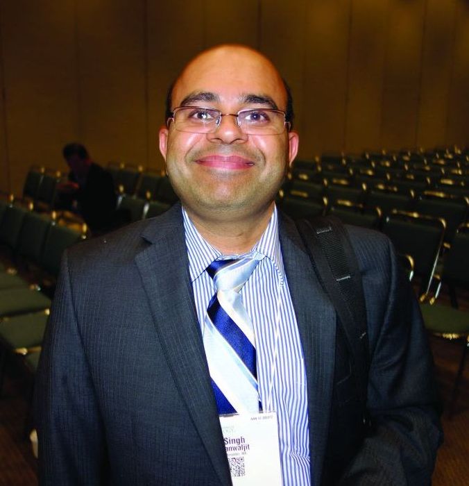
The idea of using it for autism came about after investigators noticed that, in the lab, it induced some of the cellular changes associated with fever, including upregulation of heat shock proteins, according to Kanwaljit Singh, MD, a pediatrics instructor at the University of Massachusetts, Worcester.
Fever has been reported to improve autism symptoms. So, several years ago, “we decided to do a pilot study with sulforaphane” to see if it had a similar effect, Dr. Singh said at the annual meeting of the American Academy of Neurology.
At 18 weeks, 29 young autistic men randomized to the supplement outperformed 15 randomized to placebo on the Aberrant Behavior Checklist and other measures. It was the first study of sulforaphane for autism, and it got a good deal of press attention when it was published in 2014; Dr. Singh was the lead author (Proc Natl Acad Sci U S A. 2014 Oct 28;111[43]:15550-5).
“Because we had a very good signal in our pilot study, we decided to do a slightly larger, slightly more complex clinical trial, which is ongoing right now,” he said. The results aren’t due until the second half of 2018, but he gave an interim report at the meeting.
There are 50 children with autism in the new study, aged 3-12 years. Half are randomized to sulforaphane, half to placebo, for the first 15 weeks, then all are switched to open-label sulforaphane for 15 weeks more, followed by a 6-week washout period.
The randomized portion is still blinded. But so far, 31% have responded positively at 15 weeks, meaning a much or very much improved score in at least two domains on the Ohio Autism Clinical Global Impressions Scale; domains cover social interaction, violent behavior, communication, and other areas.
Among the patients who have completed the study, the response rate at week 30 almost doubled, to 56%. “We don’t know which patients were on sulforaphane and which were on placebo” in the randomized phase, Dr. Singh said. “But we think because the response doubled” when the second half of the children were switched to sulforaphane, “there should probably not be a very large placebo effect here.”
Meanwhile, after the washout period, “some patients still do well, but many more [go] back to baseline,” added Dr. Singh, the senior investigator in the new trial.
The most common side effects are insomnia (28%), vomiting (19%), flatulence (17%), diarrhea (15%), and constipation (13%). A few patients have dropped out because of insomnia and diarrhea; more have dropped out because they simply didn’t want to take the pills – 125 mg of broccoli seed powder three to eight times a day, depending on weight.
Other groups are looking into sulforaphane, too – not just for autism, but also for schizophrenia, prostate cancer, and other indications.
The U.S. Department of Defense is funding the research. The investigators said they have no relevant disclosures.
SOURCE: Zimmerman A et al. Neurology. 2018 Apr 22; 90(15 Suppl.):N1.002.
REPORTING FROM AAN 2018
Key clinical point: The dietary supplement is showing benefit in an ongoing trial, but there are a lot of pills.
Major finding: The response rate at 30 weeks was 56%.
Study details: A randomized trial involving 50 children with autism.
Disclosures: The U.S. Department of Defense is funding the research. The investigators said they have no relevant disclosures.
Source: Zimmerman A et al. Neurology. 2018 Apr;90(15 Suppl.):N1.002.
Ocrelizumab safety update: Encouraging rates of serious infection, malignancy
LOS ANGELES – No new safety signals have emerged in multiple sclerosis patients treated with ocrelizumab, according to ongoing follow-up and postmarketing surveillance.
As of September 2017, patients with relapsing or primary progressive MS who were part of the pivotal OPERA I and II and ORATORIO trials – including phases 2 and 3 and open-label extensions – as well as an all-exposure population that included patients from prior studies, had nearly 9,500 patient-years of exposure to ocrelizumab (Ocrevus), a humanized anti-CD20 monoclonal antibody. The all-exposure population contributed about 1,500 of those patient-years, Stephen Hauser, MD, reported at the annual meeting of the American Academy of Neurology.
Postmarketing experience in approximately 37,000 treated patients with an additional 14,000 patient-years shows a fatality rate of 0.28/100 patient-years (49 fatalities) as of March 2018, he said.
“If one compares this to at least two reports of epidemiologic mortality estimates in MS, this is in line with – and in fact a little bit lower than – those estimates that range between 0.37 and 0.9, compared with 0.28,” he said. “So ... the updated safety profile in this all-exposure ocrelizumab population was generally consistent with what was seen during the controlled treatment period, and rates of serious infections fluctuated over time without any sustained increase.”
Only one serious opportunistic infection (Pasteurella) occurred in the controlled trials, and three more (two varicella zoster, one herpes simplex) occurred during open-marketing experience.
The rate of malignancies has been very encouraging, as well, as it appears to continue to align with population expectations, Dr. Hauser said.
Trial participants received 600-mg doses intravenously every 24 weeks in all three trials; in OPERA I/II, they received 96 weeks of treatment with the first dose given as two 300-mg infusions split by 14 days, and, in ORATORIO, they received at least 120 weeks of treatment with all doses split, Dr. Hauser said.
In the phase 2 study, they received split doses of 600-mg or 2,000-mg infusions through week 24; then through week 96, they received either 600-mg or 1,000-mg doses; those receiving 600 mg included those who started at that dose and those who received placebo or interferon beta-1a 30 mcg, and those receiving 1,000 mg were those who started on ocrelizumab at 2,000 mg.
The comparators were placebo in the ORATORIO and phase 2 trials, and interferon beta-1a given at a dose of 44 mcg subcutaneously three times weekly (OPERA I and II) or 30 mcg intramuscularly each week in the phase 2 trial.
All patients were offered enrollment into open-label extension studies, and “there was rather massive interest in joining the open-label extension and continuing open-label extension in both trials,” he said.
“And earlier here at the AAN [meeting] we presented the clinical efficacy data from the open-label extension, now 2 years completed – so 4 years from onset of the study – in patients who received ocrelizumab continuously for [relapsing-remitting] MS during that time period, or who switched from three-times-weekly interferon beta-1a to ocrelizumab ... and the data continue to show the positive outcomes reported in the original trials,” he added.
Further, follow-up data on MRI outcomes from the open-label extensions demonstrate that the effects on focal disease activity and on progression persists and is durable with ongoing treatment, he noted.
“In conclusion, there is no pattern of serious infections or malignancies that has emerged thus far with increased exposure, but obviously long-term follow-up and postmarketing requirement studies are needed to monitor long-term patient safety and rare events that couldn’t be captured here,” he said.
This study was sponsored by F. Hoffmann-La Roche. Dr. Hauser has received personal compensation for consulting, serving on a scientific advisory board, speaking, or other activities with Symbiotix, Annexon Biosciences, Bionure, Molecular Stethoscope, and for serving on the Board of Directors of Neurona Therapeutics.
SOURCE: Hauser S et al. Neurology. 2018 Apr 9;(15 Suppl.):S36.001.
REPORTING FROM AAN 2018
Key clinical point: Follow-up and postmarketing surveillance show no new safety signals with ocrelizumab.
Major finding: The fatality rate was 0.28/100 patient-years as of March 2018.
Study details: Follow-up of patients with nearly 9,500 patient-years of experience with ocrelizumab and postmarketing surveillance in patients with more than 14,000 patient-years of experience.
Disclosures: This study was sponsored by F. Hoffmann-La Roche. Dr. Hauser has received personal compensation for consulting, serving on a scientific advisory board, speaking, or other activities with Symbiotix, Annexon Biosciences, Bionure, Molecular Stethoscope, and for serving on the board of directors of Neurona Therapeutics.
Source: Hauser S et al. Neurology. 2018 Apr 9;(15 Suppl.):S36.001.
Myelin antibody predicts ADEM relapse
LOS ANGELES – There’s substantially higher risk of relapse and epilepsy after acute disseminated encephalomyelitis when children present with serum antibodies against myelin oligodendrocyte glycoprotein, according to British investigators.
“Traditionally, we told parents that ADEM [acute disseminated encephalomyelitis] is typically monophasic. I do tell parents now that the risk of relapse is higher if we see” myelin oligodendrocyte glycoprotein antibodies (MOG-Ab), said senior investigator Yael Hacohen, MBBS, DPhil, a pediatric neurology lecturer at University College London.
There was also a strong trend for relapsing disease when children presented with seizures, and an increased risk of post-ADEM epilepsy with oligoclonal bands on cerebrospinal fluid analysis, a marker of inflammation.
ADEM is an acute CNS demyelinating disorder primarily affecting young children, often after upper respiratory tract infections and occasionally after measles, mumps, and rubella vaccination. Signs can include limb weakness, stumbling, and coma. Many children recover without incident, but some don’t.
There’s been an increasing number of reports of children – and adults – presenting with antibodies against MOG, a glycoprotein on the outermost layer of the myelin sheath. Its exact function is unknown, but antibodies have been found in a number of inflammatory CNS conditions, and its role in pathogenesis is being explored. Testing is available for clinical use, but it isn’t standardized. For now, titer levels aren’t being used to guide treatment at University College London, Dr. Hacohen said at the American Academy of Neurology annual meeting.
The team reviewed 74 children with ADEM who presented at three pediatric neurology centers during 2005-2017, at a median age of 4.5 years. There were about equal numbers of boys and girls, and all had MRI abnormalities consistent with ADEM. Fifty children (68%) were MOG-ab positive.
Twenty-seven antibody-positive children (54%) relapsed, versus 3 of the 24 negative children (13%). Relapse was almost six times more likely with MOB-Ab (95% confidence interval [CI], 1.8-19.7; P = .002). The overall relapse rate of 42% (31/74) was higher than in previous studies, probably because of longer follow-up, lasting years in some cases.
Sixteen children (22%) presented with seizures, which nearly tripled the risk of relapse, although the finding wasn’t statistically significant (95% CI, 0.9-9.2; P = .06). There was a trend toward more seizures at onset in the MOG-Ab group.
Twelve children (16.2%) developed post-ADEM epilepsy, all but one MOG-Ab positive. The median time to seizure onset was 3 months. All of the children remained on antiepileptic medications at 2-year follow-up.
Oligoclonal bands were found in 8 of 37 children tested (22%), and also markedly increased the risk of post-ADEM seizures (odds ratio, 8.7; 95% CI, 1.5-54; P = .01).
“The majority of the children that we’ve looked at remain MOG-Ab positive. There may be a trend in antibody titers going down, but overall we didn’t find titers clinically useful. I know two children where titers went down. They relapsed,” and the titers went “up again, so we don’t’ really use them clinically for treatment,” Dr. Hacohen said.
“I think there is more to do” when it comes to optimizing ADEM management. “It’s a very heterogeneous [condition, and] I don’t want to put [everyone] on immunosuppression for years. I’ve got one patient who had an event, and 7 years later had a second event, and then was back to normal in a week.” On the other hand, “10%-20% of our patients do quite poorly and relapse on all treatments,” she said.
The investigators didn’t have any disclosures and there was no industry funding for the work.
SOURCE: Rossor T et al. Neurology. 2018 Apr 90(15 Suppl.):S35.004.
REPORTING FROM AAN 2018
Key clinical point: Myelin oligodendrocyte glycoprotein antibodies help to identify children who will have relapsing ADEM.
Major finding: Twenty-seven out of 50 antibody-positive children (54%) relapsed, versus 3 of 24 negative children (13%).
Study details: Review of 74 children with ADEM
Disclosures: There was no industry funding, and the investigators had no disclosures.
Source: Rossor T et al. Neurology. 2018 Apr 90(15 Suppl.):S35.004.
Guidance coming for mTOR inhibitors in infantile TSC
LOS ANGELES – Someday soon, physicians will probably have solid, evidence-based guidelines on how to use mTOR inhibitors in infants with tuberous sclerosis complex.
They couldn’t come soon enough. Everolimus (Afinitor) is being used off label more and more often in children under 2 years old for seizures and other manifestations of the genetic disorder, often with a decent response.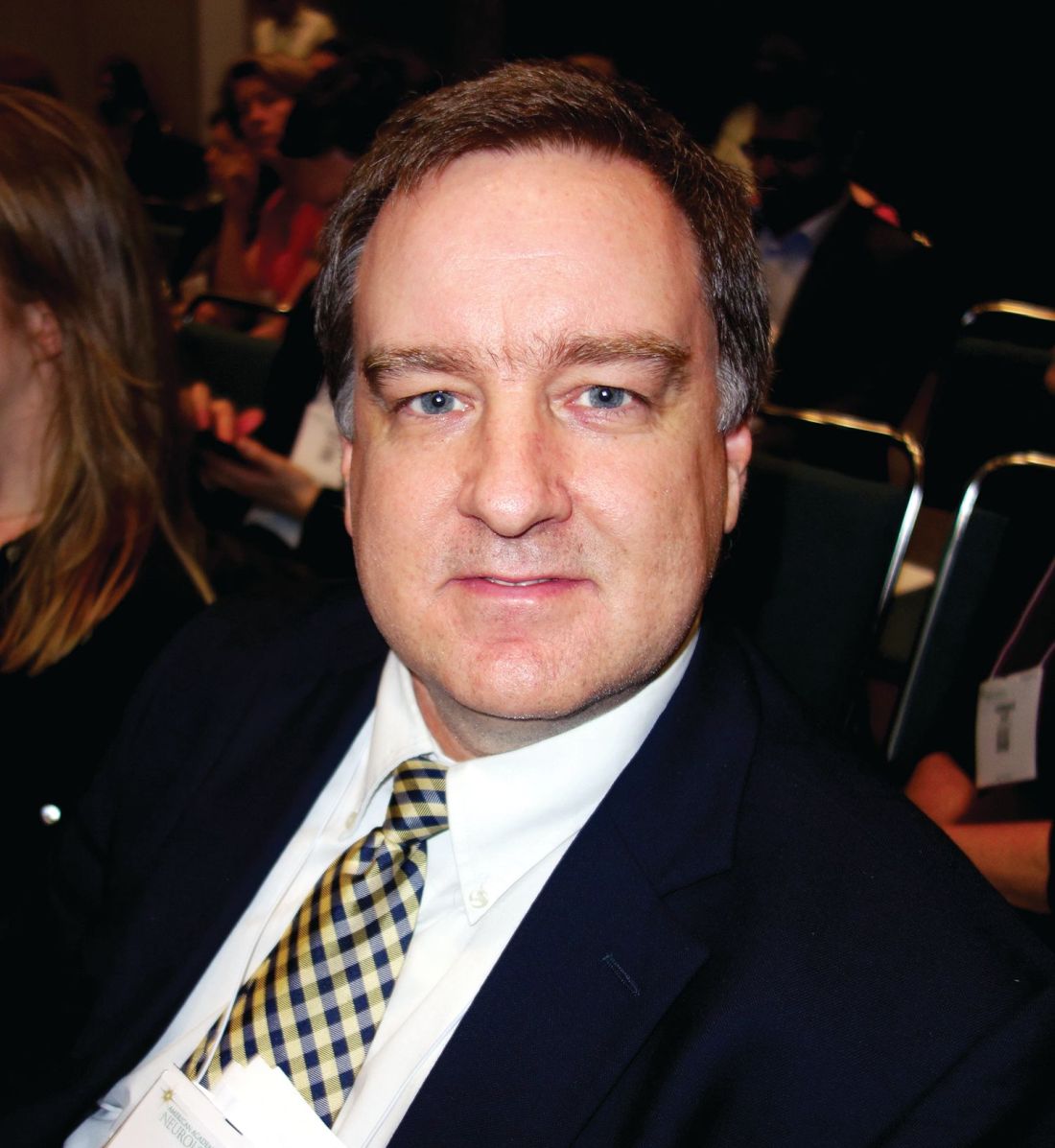
“I get emails all the time saying ‘how would you start treatment at this age?’ and we say there are no data,” he said at the American Academy of Neurology annual meeting.
Dr. Krueger presented a survey of 19 TSC centers to get the ball rolling. They reported on treating 45 children under 2 years old, at least one child at each center. The goal of the survey was to establish a baseline “to allow us to move forward with what proper studies would look like, and what needs to be done. Two-thirds of patients have seizures before their first birthday; we need to establish a safety [and efficacy] profile well before 2 years of age,” and evidence-based guidelines, he said.
On average, everolimus treatment started at 11.7 months, and continued for 27 months. Sirolimus treatment started at a mean of 16 months and continued for 16 months. Dosing ranged from once a week to daily. In contrast to case reports that skew heavily toward rhabdomyomas, most of the children were treated for epilepsy and subependymal giant cell astrocytoma (SEGA). Almost all the centers had participated in a clinical evaluation of everolimus for TSC epilepsy and SEGAs in older children, “so there was familiarity and a comfort level” for those indications, Dr. Krueger said.
Everolimus was used much more often than was sirolimus, which probably reflects ongoing clinical development of the drug. A handful of children switched between the two, probably because of side effects. Most centers reported a favorable response. The average initial daily dose of everolimus was 1.05 mg/m2; the average initial daily dose of sirolimus was 0.42 mg/m2.
Thirty-five children (78%) had an adverse event (AE), usually infections or lipid problems. Most were mild or moderate and didn’t affect treatment. Seven children (16%) had severe grade 3 events.
“The frequency of these AEs and the severity and type were not different from the earlier trials,” but almost 40% of the children discontinued treatment due to AEs, which was “much higher” than in past trials. “I think this represents the lack of real data with which to guide clinical judgment. There was a high tendency with any AE for discontinuation,” Dr. Krueger said.
There were more boys than girls among the 45 children. The majority were from the United States, and predominantly Cincinnati Children’s Hospital. Most had TSC2 mutations, which are associated with worse disease than TSC1 mutations.
Dr. Krueger reported research funding and personal compensation from Novartis, maker of everolimus. Almost all of his coinvestigators reported ties to the company. The work was supported by the Tuberous Sclerosis Alliance, which is funded in part by Novartis.
SOURCE: Krueger D et al. Neurology. 2018 Apr 90(15 Suppl.):S35.003.
REPORTING FROM AAN 2018
Key clinical point: A survey of treatment centers has begun to plug the evidence gap for using mTOR inhibitors in infants with tuberous sclerosis.
Major finding: Adverse events don’t seem more common than in trials of adults and older children, but the discontinuation rate is much higher, at about 40%.
Study details: Review of 45 children under 2 years old.
Disclosures: The lead investigator reported research funding and personal compensation from Novartis, maker of everolimus.
Source: Krueger D et al. Neurology. 2018 Apr 90(15 Suppl.):S35.003.
Parkinson’s disease patients have impaired insulin secretion
LOS ANGELES – Parkinson’s disease patients with long-standing disease should be screened for glucose dysregulation and treated with insulin releasing drugs, instead of insulin sensitizers, when necessary, according to a French investigation that compared 50 patients with 50 healthy controls matched for age, sex, and body mass index.
The subjects underwent a 75-g oral glucose tolerance test. Glucose levels were higher in the Parkinson’s disease (PD) group from about 60 minutes through the end of the test at 180 minutes; the differences were statistically significant at 90 and 150 minutes. The total blood glucose area under the time curve (AUC) was significantly higher in the PD group (1,187 vs. 1,101 mmol.min l-1; P = .05).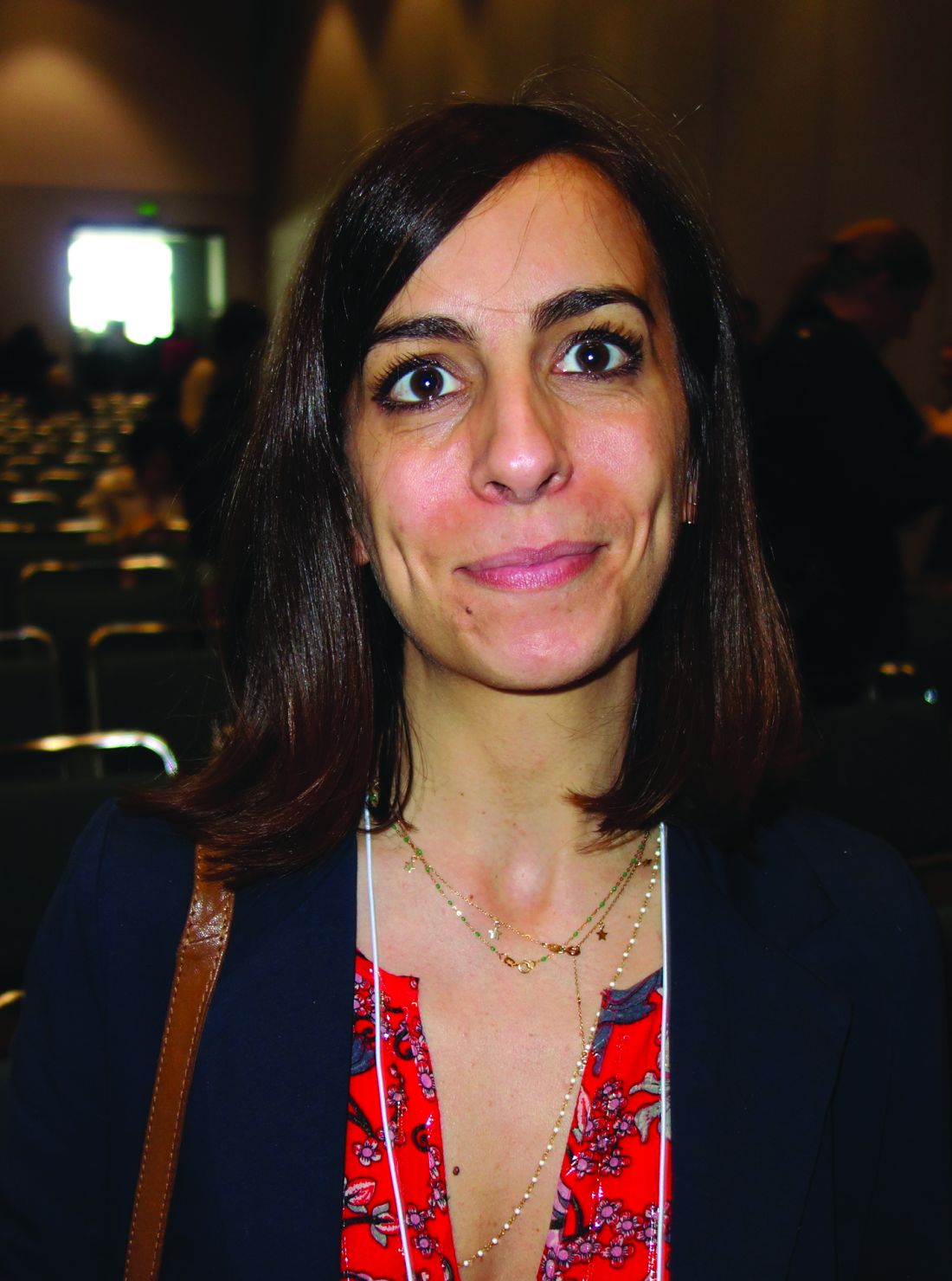
In short, “PD patients had higher blood glucose following oral glucose intake without … the expected concomitant rise in insulin levels, suggesting an under-active insulin response. PD patients with advanced disease” – all the patients had had PD for more than 5 years – “have impaired blood glucose levels in response to oral glucose intake,” said lead investigator Ana Marques, MD, of the Université Clermont Auvergne in Clermont-Ferrand, France.
“Blood glucose dysregulation should be screened in PD patients with moderate to advanced disease. Insulin releasing drugs should possibly be preferred [over] insulin sensitizer drugs in PD patients with diabetes,” she said at the annual meeting of the American Academy of Neurology.
Higher blood glucose levels were also associated with longer PD duration, lower dopaminergic therapy doses, and higher degrees of dysautonomia. Mean PD duration in the study was about 8 years, and mean levodopa-equivalent dose 884 mg/d.
The findings add weight to the proposed and still somewhat controversial link between PD and diabetes. “Dysglycemia appears to be another nonmotor consequence of PD,” Dr. Marques said. Because insulin production “is modulated by the autonomic nervous system, the severity of dysautonomia in PD could be linked with blood glucose dysregulation. Sympathetic denervation might lead to beta-cell dysfunction.”
Subjects were 61 years old, on average; two-thirds were men. The mean BMI was about 25 kg/m2. Patients were excluded if they had a change in dopaminergic therapy in the previous month, previous deep brain stimulation, medications that would interfere with glucose metabolism, or diabetes, among other things.
PD patients had slightly higher fasting urine glucose (P = .02). They also had lower fasting plasma insulin, but the difference wasn’t statistically significant (P = .5). Dysglycemia was also associated with higher BMI. Male gender and higher levodopa-equivalent doses were protective.
“The association between dysglycemia and PD is bilateral. In many studies, PD enhances the risk, but dysglycemia and particularly diabetes have been reported to increase the risk of PD. It goes both ways. There’s a lot that remains to be understood,” Dr. Marques said.
There was no external funding, and the investigators had nothing to disclose.
SOURCE: Marques A et al. Neurology. 2018 Apr 90(15 Suppl.):S3.008
REPORTING FROM AAN 2018
Key clinical point: instead of insulin sensitizers, when necessary.
Major finding: The total blood glucose area under the time curve was significantly higher in the PD group after oral glucose challenge.
Study details: A study of 50 PD patients and 50 controls.
Disclosures: There was no external funding, and the investigators had nothing to disclose.
Source: Marques A et al. Neurology. 2018 Apr 90(15 Suppl.):S3.008