User login
European ANCA-associated vasculitis guidance gets first makeover since 2009
LONDON – Updated management recommendations for patients with antineutrophil cytoplasmic antibody (ANCA)–associated vasculitis from the European League Against Rheumatism and the European Renal Association-European Dialysis and Transplant Association aim to provide clinicians with reliable guidance on the best approach to treatment.
The update, presented at the European Congress of Rheumatology and recently published online in Annals of the Rheumatic Diseases (Ann Rheum Dis. 2016 Jun 23. doi:10.1136/annrheumdis-2016-209133), reassessed items in the 2009 recommendations for the management of primary systemic vasculitis and focused only on the management of ANCA-associated vasculitis (AAV), according to recommendations task force member Dr. Max Yates.

“In the past 5 years, 1,691 papers have been published on primary systemic vasculitis in internal medicine, rheumatology, and nephrology journals. Together with the licensing of rituximab for AAV, it was an opportune time to update the recommendations with an AAV focus,” Dr. Yates explained. The revised guidance is based on a systematic literature review from January 2007 to February 2015, focusing in particular on specific items that needed updating, such as the importance of ANCA testing and biopsy in diagnosis and follow-up, disease staging at diagnosis, the choices for remission-induction and remission-maintenance therapies, and the drug choices for relapsing and refractory disease. The task force considered for the first time the choice of immunosuppressive drugs and biologic agents (principally rituximab) and immunologic monitoring. They identified patient education as another priority.
“These updated recommendations provide a framework of practice and should apply to the majority of patients with AAV,” added Dr. Yates, who is a clinical fellow at Norwich Medical School at the University of East Anglia and works in the department of rheumatology at the Norfolk and Norwich (England) University Hospital.
The 22-member task force included rheumatologists, internists, nephrologists, a clinical immunologist, an otorhinolaryngologist, a chest physician, an ophthalmologist, a vasculitis nurse, and a patient with vasculitis from 11 countries in Europe and the United States. The task force was convened by rheumatologist Dr. Chetan Mukhtyar of the Norfolk and Norwich University Hospital on behalf of EULAR and by vasculitis and renal specialist Dr. David Jayne of Addenbrooke’s Hospital in Cambridge (England) on behalf of the European Renal Association-European Dialysis and Transplant Association.
The recommendations now contain one single, simple overarching principle, Dr. Mukhtyar said at the congress. That is, the need for shared decision making between the patient and the clinician. This principle is also included as the first point in many of the other recently updated EULAR recommendations on the management of rheumatic diseases.
Both previous and updated versions of the vasculitis recommendations contain 15 recommendations, with some changed and others combined. One key recommendation is about who should treat patients with AAV; it states that patients “should be managed in close collaboration with, or at, centers of expertise,” Dr. Mukhtyar said.“Patients with ANCA-associated vasculitis have often very complex presentations that involve several different specialties, and it is always worthwhile that these patients are looked after by people who commonly see them, because these are rare conditions,” he observed.
Deciding when to perform a biopsy is also covered, with the recommendation being that it can be used to establish a new diagnosis and to further evaluate cases of suspected relapsing vasculitis. “When do you do a biopsy?” Dr. Mukhtyar asked. “Well, every time you can, every time it is clinically feasible,” he suggested.
As for treatment, there are different recommendations depending on whether the aim is to induce or maintain remission and whether there has been a major relapse. In patients with organ- or life-threatening disease, for example, the advice is to use glucocorticoids and either cyclophosphamide or rituximab to induce remission, Dr. Mukhtyar said. The specific dosing or administration of glucocorticoids is not specified as this will depend on the clinical situation, but the advice is to taper down when possible, somewhere between month 3 and 5.
For remission induction in less severe (non–organ threatening) disease, the recommendation is to use glucocorticoids plus either methotrexate or mycophenolate mofetil. Situations when methotrexate or mycophenolate mofetil should and should not be used are specified, notably when cyclophosphamide or rituximab are not available or are contraindicated.
For maintenance of remission, the task force advised using low-dose glucocorticoids plus azathioprine, rituximab, methotrexate, or mycophenolate mofetil.
Guidance on when to use plasma exchange is given for patients with severe disease and options following failure of remission-induction therapy, and when to switch therapy is also covered.
There are also several follow-up recommendations, such as the periodic assessment of cardiovascular risk, and patient-focused recommendations on awareness of the nature, benefits, and risks of therapy.
The recommendations should provide clinicians with reliable guidance on the best approach to treating AAV, according to Dr. Yates. “From the patients’ point of view, these recommendations should provide useful insight into which treatments they are likely to be offered and when. They also emphasize that as a patient, you should have a voice in your treatment and if you have any questions or concerns, be sure to speak with your specialist.”
Dr. Yates and Dr. Mukhtyar did not report having any relevant disclosures.
The prior 2009 EULAR recommendations were very much in need of updating given the plethora of studies in the past 7 years addressing ANCA-associated vasculitis (AAV). The emergence of rituximab as an effective therapy in AAV had to be considered and included in these newer guidelines. Its potential role in both remission induction, as well as remission maintenance of AAV, is addressed.
The recommendations are somewhat complicated, particularly as eosinophilic granulomatosis with polyangiitis (EGPA, previously referred to as Churg-Strauss syndrome) has been included, but most of the well-done prospective clinical trials addressing remission induction and remission maintenance in AAV were limited to patients with granulomatosis with polyangiitis or microscopic polyangiitis and did not include patients with EGPA. The role of plasma exchange is also discussed, but the results of the PEXIVAS trial, which will address that more definitively, are not yet forthcoming. Those results are anticipated in the not too distant future and will much better define that component of management in those most severely ill patients with AAV.
 |
| Dr. Robert Spiera |
These recommendations serve as a framework for helping clinicians understand what is widely accepted as standard of care for these diseases but in no way can define individual treatment decisions as the authors acknowledge. Such decisions must become very personalized in relation to details of the patient’s individual comorbidities and other features of their medical and even socioeconomic status. For example, when choosing between rituximab and cyclophosphamide for remission induction in a young woman (or man, for that matter), future fertility concerns (which cyclophosphamide could potentially compromise) are very relevant. Moreover, the costs of rituximab are substantial, and the lack of superiority of rituximab over cyclophosphamide in many situations, particularly in patients with new severe disease, could be an important factor to consider when choosing which immunosuppressive will be used.
Many of the unanswered questions await results of ongoing or upcoming trials, including some addressing the relative efficacy of various remission maintenance regimens (rituximab vs. azathioprine) or the role of plasmapheresis. Many questions in AAV are not easily addressable in clinical trials, such as whether there are some groups of patients in whom remission maintenance therapy should never be withdrawn. However, such questions may be addressed through observational studies of the well-defined patient cohorts and registries that have been developed in the United States and Europe.
Robert F. Spiera, MD, is director of the Scleroderma, Vasculitis, & Myositis Center at the Hospital for Special Surgery, N.Y. He is also professor of clinical medicine at Cornell University, N.Y. He has received research funding and consulting fees from Roche/Genentech, which markets rituximab.
The prior 2009 EULAR recommendations were very much in need of updating given the plethora of studies in the past 7 years addressing ANCA-associated vasculitis (AAV). The emergence of rituximab as an effective therapy in AAV had to be considered and included in these newer guidelines. Its potential role in both remission induction, as well as remission maintenance of AAV, is addressed.
The recommendations are somewhat complicated, particularly as eosinophilic granulomatosis with polyangiitis (EGPA, previously referred to as Churg-Strauss syndrome) has been included, but most of the well-done prospective clinical trials addressing remission induction and remission maintenance in AAV were limited to patients with granulomatosis with polyangiitis or microscopic polyangiitis and did not include patients with EGPA. The role of plasma exchange is also discussed, but the results of the PEXIVAS trial, which will address that more definitively, are not yet forthcoming. Those results are anticipated in the not too distant future and will much better define that component of management in those most severely ill patients with AAV.
|
| Dr. Robert Spiera |
These recommendations serve as a framework for helping clinicians understand what is widely accepted as standard of care for these diseases but in no way can define individual treatment decisions as the authors acknowledge. Such decisions must become very personalized in relation to details of the patient’s individual comorbidities and other features of their medical and even socioeconomic status. For example, when choosing between rituximab and cyclophosphamide for remission induction in a young woman (or man, for that matter), future fertility concerns (which cyclophosphamide could potentially compromise) are very relevant. Moreover, the costs of rituximab are substantial, and the lack of superiority of rituximab over cyclophosphamide in many situations, particularly in patients with new severe disease, could be an important factor to consider when choosing which immunosuppressive will be used.
Many of the unanswered questions await results of ongoing or upcoming trials, including some addressing the relative efficacy of various remission maintenance regimens (rituximab vs. azathioprine) or the role of plasmapheresis. Many questions in AAV are not easily addressable in clinical trials, such as whether there are some groups of patients in whom remission maintenance therapy should never be withdrawn. However, such questions may be addressed through observational studies of the well-defined patient cohorts and registries that have been developed in the United States and Europe.
Robert F. Spiera, MD, is director of the Scleroderma, Vasculitis, & Myositis Center at the Hospital for Special Surgery, N.Y. He is also professor of clinical medicine at Cornell University, N.Y. He has received research funding and consulting fees from Roche/Genentech, which markets rituximab.
LONDON – Updated management recommendations for patients with antineutrophil cytoplasmic antibody (ANCA)–associated vasculitis from the European League Against Rheumatism and the European Renal Association-European Dialysis and Transplant Association aim to provide clinicians with reliable guidance on the best approach to treatment.
The update, presented at the European Congress of Rheumatology and recently published online in Annals of the Rheumatic Diseases (Ann Rheum Dis. 2016 Jun 23. doi:10.1136/annrheumdis-2016-209133), reassessed items in the 2009 recommendations for the management of primary systemic vasculitis and focused only on the management of ANCA-associated vasculitis (AAV), according to recommendations task force member Dr. Max Yates.
“In the past 5 years, 1,691 papers have been published on primary systemic vasculitis in internal medicine, rheumatology, and nephrology journals. Together with the licensing of rituximab for AAV, it was an opportune time to update the recommendations with an AAV focus,” Dr. Yates explained. The revised guidance is based on a systematic literature review from January 2007 to February 2015, focusing in particular on specific items that needed updating, such as the importance of ANCA testing and biopsy in diagnosis and follow-up, disease staging at diagnosis, the choices for remission-induction and remission-maintenance therapies, and the drug choices for relapsing and refractory disease. The task force considered for the first time the choice of immunosuppressive drugs and biologic agents (principally rituximab) and immunologic monitoring. They identified patient education as another priority.
“These updated recommendations provide a framework of practice and should apply to the majority of patients with AAV,” added Dr. Yates, who is a clinical fellow at Norwich Medical School at the University of East Anglia and works in the department of rheumatology at the Norfolk and Norwich (England) University Hospital.
The 22-member task force included rheumatologists, internists, nephrologists, a clinical immunologist, an otorhinolaryngologist, a chest physician, an ophthalmologist, a vasculitis nurse, and a patient with vasculitis from 11 countries in Europe and the United States. The task force was convened by rheumatologist Dr. Chetan Mukhtyar of the Norfolk and Norwich University Hospital on behalf of EULAR and by vasculitis and renal specialist Dr. David Jayne of Addenbrooke’s Hospital in Cambridge (England) on behalf of the European Renal Association-European Dialysis and Transplant Association.
The recommendations now contain one single, simple overarching principle, Dr. Mukhtyar said at the congress. That is, the need for shared decision making between the patient and the clinician. This principle is also included as the first point in many of the other recently updated EULAR recommendations on the management of rheumatic diseases.
Both previous and updated versions of the vasculitis recommendations contain 15 recommendations, with some changed and others combined. One key recommendation is about who should treat patients with AAV; it states that patients “should be managed in close collaboration with, or at, centers of expertise,” Dr. Mukhtyar said.“Patients with ANCA-associated vasculitis have often very complex presentations that involve several different specialties, and it is always worthwhile that these patients are looked after by people who commonly see them, because these are rare conditions,” he observed.
Deciding when to perform a biopsy is also covered, with the recommendation being that it can be used to establish a new diagnosis and to further evaluate cases of suspected relapsing vasculitis. “When do you do a biopsy?” Dr. Mukhtyar asked. “Well, every time you can, every time it is clinically feasible,” he suggested.
As for treatment, there are different recommendations depending on whether the aim is to induce or maintain remission and whether there has been a major relapse. In patients with organ- or life-threatening disease, for example, the advice is to use glucocorticoids and either cyclophosphamide or rituximab to induce remission, Dr. Mukhtyar said. The specific dosing or administration of glucocorticoids is not specified as this will depend on the clinical situation, but the advice is to taper down when possible, somewhere between month 3 and 5.
For remission induction in less severe (non–organ threatening) disease, the recommendation is to use glucocorticoids plus either methotrexate or mycophenolate mofetil. Situations when methotrexate or mycophenolate mofetil should and should not be used are specified, notably when cyclophosphamide or rituximab are not available or are contraindicated.
For maintenance of remission, the task force advised using low-dose glucocorticoids plus azathioprine, rituximab, methotrexate, or mycophenolate mofetil.
Guidance on when to use plasma exchange is given for patients with severe disease and options following failure of remission-induction therapy, and when to switch therapy is also covered.
There are also several follow-up recommendations, such as the periodic assessment of cardiovascular risk, and patient-focused recommendations on awareness of the nature, benefits, and risks of therapy.
The recommendations should provide clinicians with reliable guidance on the best approach to treating AAV, according to Dr. Yates. “From the patients’ point of view, these recommendations should provide useful insight into which treatments they are likely to be offered and when. They also emphasize that as a patient, you should have a voice in your treatment and if you have any questions or concerns, be sure to speak with your specialist.”
Dr. Yates and Dr. Mukhtyar did not report having any relevant disclosures.
AT THE EULAR 2016 CONGRESS

Primary care gout patients often discontinue allopurinol
LONDON – Many patients with newly diagnosed gout who are prescribed allopurinol to reduce their uric acid level and prevent recurrent episodes fail to stick with their treatment, according to an analysis of more than 47,000 U.K. gout patients who received prescriptions for allopurinol during the 28-year period of 1987-2014.
One possible contributing factor to this pattern may be physicians who inadequately stress to patients the importance of sticking with allopurinol treatment to improve their long-term health, Lieke E.J.M. Scheepers said at the European Congress of Rheumatology.

“We think that physicians underestimate” the low level of gout patient adherence to allopurinol, said Ms. Scheepers, a PhD student in the department of rheumatology at Maastricht (the Netherlands) University.
“We view gout as a chronic disease, but many physicians and patients believe gout can occur as a single episode, and then it’s over,” explained Dr. Annelies Boonen, the senior investigator on the study, in an interview. “Gout patients often don’t appreciate that they will need to take their medication for many years. We need to convince primary care physicians to follow gout patients closely and not wait [to resume treatment] until the patient has a new episode,” said Dr. Boonen, a professor of rheumatology at Maastricht University.
“Some physicians are not convinced that it harms a patient to have two or three acute gout attacks a year, but there is a subgroup that will have joint damage” from this pattern of recurrence, she noted. However, Dr. Boonen acknowledged that gout patients usually seen in primary care practice often don’t have the same level of disease severity and recurrence as the patients she sees in her referral clinic. “We don’t know which gout patients will develop joint damage,” she admitted.
Another barrier to good adherence with long-term uric acid–lowering treatment is that “patients who don’t have daily symptoms often question why they should continue to take their medication,” added Ms. Scheepers. “Many patients fear the possible adverse effects of their treatment” more than they fear a possible gout recurrence.
Ms. Scheepers and her associates analyzed data from 47,774 patients with newly diagnosed gout receiving treatment exclusively with allopurinol from about 680 primary care U.K. physicians and archived in the Clinical Practice Research Datalink maintained by the U.K. government. The patients averaged 64 years old, and three-quarters were men.
During their first year on treatment, 57% of the patients had at least one 30-day gap in their use of allopurinol, and 38% had at least one 90-day gap in their allopurinol treatment, Ms. Scheepers reported. During an average follow-up of nearly 6 years, 77% of patients had at least one 30-day gap in treatment and 54% had at least one 90-day gap. The median time to a 90-day gap in allopurinol treatment was just under 3 years (1,059 days).
The researchers also assessed patient compliance and adherence to therapy by analyzing the percentage of days during follow-up that they took allopurinol. The overall average percentage of days on treatment was 57%, and 39% of patients received allopurinol on at least 80% of the days when they were followed.
Another analysis focused specifically on 14,808 patients who restarted on allopurinol after they had stopped their use of the drug for at least 90 days. Among these patients, the rate of a new 30-day gap during their first year back on treatment was 72%, with 48% having a new gap of 90 days or more during their first year back on treatment. During total follow-up of this group of patients with an established history of stopping allopurinol, 82% had a new gap in treatment of at least 30 days and 63% had a gap of 90 days or more.
The researchers also examined demographic and clinical variables that significantly linked with either greater or lesser adherence to allopurinol treatment. Two subgroups – women and smokers – showed significantly worse adherence, while older patients, patients who also took other drugs (antihypertensive medications, colchicine, or statins), and patients with various comorbidities (dementia, diabetes, depression, or impaired renal function) all had significantly better adherence. One possible explanation for this pattern is that patients who are older, have comorbidities, or already take other drugs may have a better-established routine and mindset for adhering to medication regimens that helps them remain adherent to allopurinol, Ms. Scheepers said.
Dr. Scheepers and Dr. Boonen had no disclosures.
On Twitter @mitchelzoler
LONDON – Many patients with newly diagnosed gout who are prescribed allopurinol to reduce their uric acid level and prevent recurrent episodes fail to stick with their treatment, according to an analysis of more than 47,000 U.K. gout patients who received prescriptions for allopurinol during the 28-year period of 1987-2014.
One possible contributing factor to this pattern may be physicians who inadequately stress to patients the importance of sticking with allopurinol treatment to improve their long-term health, Lieke E.J.M. Scheepers said at the European Congress of Rheumatology.
“We think that physicians underestimate” the low level of gout patient adherence to allopurinol, said Ms. Scheepers, a PhD student in the department of rheumatology at Maastricht (the Netherlands) University.
“We view gout as a chronic disease, but many physicians and patients believe gout can occur as a single episode, and then it’s over,” explained Dr. Annelies Boonen, the senior investigator on the study, in an interview. “Gout patients often don’t appreciate that they will need to take their medication for many years. We need to convince primary care physicians to follow gout patients closely and not wait [to resume treatment] until the patient has a new episode,” said Dr. Boonen, a professor of rheumatology at Maastricht University.
“Some physicians are not convinced that it harms a patient to have two or three acute gout attacks a year, but there is a subgroup that will have joint damage” from this pattern of recurrence, she noted. However, Dr. Boonen acknowledged that gout patients usually seen in primary care practice often don’t have the same level of disease severity and recurrence as the patients she sees in her referral clinic. “We don’t know which gout patients will develop joint damage,” she admitted.
Another barrier to good adherence with long-term uric acid–lowering treatment is that “patients who don’t have daily symptoms often question why they should continue to take their medication,” added Ms. Scheepers. “Many patients fear the possible adverse effects of their treatment” more than they fear a possible gout recurrence.
Ms. Scheepers and her associates analyzed data from 47,774 patients with newly diagnosed gout receiving treatment exclusively with allopurinol from about 680 primary care U.K. physicians and archived in the Clinical Practice Research Datalink maintained by the U.K. government. The patients averaged 64 years old, and three-quarters were men.
During their first year on treatment, 57% of the patients had at least one 30-day gap in their use of allopurinol, and 38% had at least one 90-day gap in their allopurinol treatment, Ms. Scheepers reported. During an average follow-up of nearly 6 years, 77% of patients had at least one 30-day gap in treatment and 54% had at least one 90-day gap. The median time to a 90-day gap in allopurinol treatment was just under 3 years (1,059 days).
The researchers also assessed patient compliance and adherence to therapy by analyzing the percentage of days during follow-up that they took allopurinol. The overall average percentage of days on treatment was 57%, and 39% of patients received allopurinol on at least 80% of the days when they were followed.
Another analysis focused specifically on 14,808 patients who restarted on allopurinol after they had stopped their use of the drug for at least 90 days. Among these patients, the rate of a new 30-day gap during their first year back on treatment was 72%, with 48% having a new gap of 90 days or more during their first year back on treatment. During total follow-up of this group of patients with an established history of stopping allopurinol, 82% had a new gap in treatment of at least 30 days and 63% had a gap of 90 days or more.
The researchers also examined demographic and clinical variables that significantly linked with either greater or lesser adherence to allopurinol treatment. Two subgroups – women and smokers – showed significantly worse adherence, while older patients, patients who also took other drugs (antihypertensive medications, colchicine, or statins), and patients with various comorbidities (dementia, diabetes, depression, or impaired renal function) all had significantly better adherence. One possible explanation for this pattern is that patients who are older, have comorbidities, or already take other drugs may have a better-established routine and mindset for adhering to medication regimens that helps them remain adherent to allopurinol, Ms. Scheepers said.
Dr. Scheepers and Dr. Boonen had no disclosures.
On Twitter @mitchelzoler
AT THE EULAR 2016 CONGRESS
Key clinical point: A majority of newly diagnosed gout patients in the U.K. have significant gaps in treatment and only a minority show good treatment adherence.
Major finding: During their first year of allopurinol treatment, 57% of gout patients had a treatment gap of 30 days or longer.
Data source: A database of about 680 U.K. general practice physicians maintained by the Clinical Practice Research Datalink that included 47,774 patients with incident gout treated exclusively with allopurinol during 1987-2014.
Disclosures: Dr. Scheepers and Dr. Boonen had no disclosures.

VIDEO: No major malformations ascribed to bisphosphonate use in pregnancy
LONDON – One of the largest studies of pregnancy outcomes after bisphosphonate exposure has found no evidence for major teratogenic effects in women with inflammatory diseases and glucocorticoid-induced osteoporosis and women with bone diseases.
However, the investigators for the French case-control study did find higher rates of neonatal complications and spontaneous abortion among infants of mothers with systemic inflammatory diseases and bisphosphonate use, but the results could be the result of confounding because of the severity of underlying disease and exposure to other medications.
“I think if a women is worried about bisphosphonate exposure during pregnancy, this study can bring her some reassuring news,” although it does not necessarily mean that bisphosphonates are safe during pregnancy, first author Aurélien Sokal said in an interview at the European Congress of Rheumatology. He is a medical student at Beaujon Hospital, Clichy, France, but conducted the study with colleagues during his time in training in the rheumatology department at Paris-Sud University.
“Very little is known about the effect of bisphosphonates on pregnancy outcomes and fetal development,” Mr. Sokal said, and they are feared for possible teratogenic effects in pregnancy because of their long half-life in bone – where they can be released even 1 year after their administration – as well as their ability to cross the placenta and high affinity for high-turnover bones, such as those in a growing fetus. He also noted that abnormalities in bone length, low birth weights, and bone diseases have been observed in rats exposed to bisphosphonates during gestation.
The study compared 23 patients with inflammatory diseases and bisphosphonate exposure during pregnancy against 92 controls with inflammatory diseases but no exposure, and 16 with bone diseases and exposure to bisphosphonates against 64 healthy controls with no underlying disease or bisphosphonate use. The patients came from a database assembled by the French Reference Center of Teratogenic Agents (CRAT) in Paris that has collected information since 1975 on patients referred for any drug exposure during pregnancy and followed their care through the end of pregnancy. The 39 patients who were exposed to bisphosphonates took the drugs during 1987-2014 within the 6 weeks preceding (n = 6) or during pregnancy (n = 33). They had a mean age of 33 years.
Systemic inflammatory diseases
The systemic inflammatory diseases found in women in the study included systemic lupus erythematosus (SLE), rheumatoid arthritis, antiphospholipid syndrome, systemic vasculitis, and other diseases. Of the 23 cases with systemic inflammatory diseases, 16 took risedronate, 5 took alendronate, 1 took etidronate, and the bisphosphonate was unknown in 1. Bisphosphonate exposure occurred before pregnancy in 2, during the first trimester in 21, second trimester in 4, third trimester in 4, and in all trimesters in 1.
Other types of medications were used significantly more often by patients with systemic inflammatory diseases than by controls: steroids (78% vs. 47%), methotrexate (26% vs. 5%), colchicine (17% vs. 2%), proton pump inhibitors (22% vs. 5%), and reproductive hormones (17% vs. 2%). Controls took antimalarials significantly more often (50% vs. 22%).
Voluntary abortions occurred at a similar rate in both exposed and unexposed women (12% vs. 9%), whereas significantly more therapeutic pregnancy terminations occurred among women exposed to bisphosphonates (17% vs. 1%). Live births occurred in 94% of the remaining exposed pregnant women, compared with 80% of controls.
Newborns were delivered at a mean of 38 weeks in both cases and controls, and there were no differences in birth weight, length, or rate of congenital malformation (9% vs. 2%).
The two malformations in neonates from exposed women had an uncertain link to bisphosphonates. One involved a neonate with severe malformative syndrome and advanced bone maturation who had a mother with SLE and was exposed to multiple drugs, including mycophenolate mofetil. The other neonate had ductus arteriosus, inguinal hernia, and negative otoacoustic emission; the baby’s mother had Crohn’s disease but had not taken known teratogenic drugs.
Two neonatal malformations among control women involved one neonate with severe malformative syndrome who had a mother with SLE but who was without exposure to known teratogenic drugs, and another with convulsant encephalopathy whose mother had systemic sclerosis and took pentoxifylline, cisapride, dihydroergocryptine, and colchicine.
However, cases had a 25% rate of neonatal complications, compared with a significantly lower 5% in controls. No infants had hypocalcemia.
Bone diseases
The 16 women with bone diseases included 9 with osteoporosis, 3 with malignancy, and 4 with miscellaneous bone conditions. A total of 5 received intravenous bisphosphonates and 11 received oral drugs (9 alendronate, 2 other). Most received a bisphosphonate in the first trimester (9 patients), but also 4 received it before pregnancy and 3 in the second trimester. More pregnancy terminations (voluntary or therapeutic) occurred among women with bone disease when compared with controls (19% vs. 3%), but the difference was not statistically significant. However in the remaining patients, live births occurred significantly less often in cases than in controls (69% vs. 100%). Birth weight, length, gestational age at birth, and the rates of congenital malformation and neonatal complications were otherwise similar.
The results of the study fall in line with those from the two major previous controlled studies on 24 women (Reprod Toxicol. 2006 Nov;22:578-9) and 21 women (Bone. 2009 Mar;44:428-30). Another series of 10 bisphosphonate-exposed pregnancies described 2 malformations, including 1 ventricular septal defect and 1 kidney and cardiac malformation (Autoimmun Rev. 2010 Jun;9:547-52). Another single case report described a neonate with bilateral talipes equinovarus (J Bone Miner Res. 2004 Oct;19:1742-5). A literature review of 78 cases of bisphosphonate exposure during or prior to pregnancy reported three malformations (Hormones [Athens]. 2011 Oct-Dec;10:280-91).
The study is ongoing and continues to collect data on the follow-up of children, Mr. Sokal said.
The study had no specific funding, and none of the investigators had disclosures to report.
The video associated with this article is no longer available on this site. Please view all of our videos on the MDedge YouTube channel
AT THE EULAR 2016 CONGRESS
Key clinical point: One of the largest controlled studies on pregnancy outcomes following bisphosphonate use found no increase in major malformations.
Major finding: Congenital malformations occurred at similar rates for both cases and controls with systemic inflammatory disease and also for women with bone diseases and bisphosphonate exposure in comparison with healthy control women.
Data source: A French case-control study involving 23 patients with systemic inflammatory diseases and bisphosphonate exposure during pregnancy, 92 controls with systemic inflammatory diseases but no exposure, 16 with bone diseases and exposure to bisphosphonates, and 64 healthy controls with no underlying disease or bisphosphonate exposure.
Disclosures: The study had no specific funding and none of the investigators had disclosures to report.

Single rituximab dose slows rheumatoid arthritis development
LONDON – A single, intravenous infusion of 1,000 mg of rituximab to people with arthralgia and a high risk for developing rheumatoid arthritis cut the subsequent rate of rheumatoid arthritis development roughly in half during more than 18 months of follow-up in a proof-of-concept, placebo-controlled study that randomized 81 people.
“This is the first study to evaluate the effects of a biopharmaceutical in subjects at risk of developing RA [rheumatoid arthritis],” Dr. Daniëlle M. Gerlag said at the European Congress of Rheumatology. “These results strongly support the rationale for future clinical trials aimed at prevention of RA by a targeted intervention,” added Dr. Gerlag, a rheumatologist at the Academic Medical Center in Amsterdam.

Additional studies are needed to confirm this effect and to examine whether the period of protection against RA development can be extended by administration of additional rituximab (Rituxan) doses. In the current study, the protective effect from the single dose administered appeared to wane over time, she noted.
The idea behind this strategy is that a window of opportunity exists in people at high risk for developing RA to prevent the disease by blocking production of the autoantibodies that trigger the development of a subclinical synovitis that eventually leads to RA. Rituximab is a cytolytic antibody directed against the CD20 antigen on B cells that already has regulatory approval for treating moderately to severely active RA as well as certain other diseases.
Dr. Gerlag and her associates recently published an analysis that detailed their rationale for hypothesizing that prophylactic treatment with rituximab might prove effective at delaying or preventing the development of RA in susceptible people (Rheumatology [Oxford]. 2016 April;55[4]:607-14).
The Prevention of RA by Rituximab (PRAIRI) study ran at three Dutch centers. The investigators enrolled people with arthralgia who had never been diagnosed with arthritis, had never used a disease-modifying antirheumatic drug, and had at least one of these two risk factors: a serum level of IgM rheumatoid factor of more than 12.5 IU/mL and a serum level of anticitrullinated peptide antibodies of more than 25 IU/mL. Enrolled participants also needed to have at least one of the following: a serum level of C-reactive protein greater than 3 mg/L, an erythrocyte sedimentation rate of greater than 28 mm/hr, and evidence of subclinical synovitis identified by either ultrasound or MRI.
The researchers found these participants largely through screening sessions run at health fairs and by publicizing the study during television appearances, Dr. Gerlag said. About three-quarters of the participants were first-degree relatives of patients already diagnosed with RA, but this was not a criterion for enrollment. The participants averaged about 53 years old, and nearly two-thirds were women.
Among the 81 people who underwent treatment, 41 received a single, 1,000-mg infusion of rituximab, and 40 received a placebo infusion. The researchers then followed the participants with scheduled, periodic examinations during a median of 29 months.
During follow-up, 16 of the 40 people in the placebo group (40%) developed RA after a median of 12 months, and 14 of the 41 in the treated arm (34%) developed RA after a median of 17 months.
The researchers performed two different statistical analyses on these outcomes. They used a Kaplan-Meier survival analysis to determine the time until 25% of people in each arm developed RA. Among the placebo patients, this occurred after 12 months, while in the intervention arm, it did not occur until 24 months, a statistically significant doubling of the time to this outcome with rituximab treatment, Dr. Gerlag reported.
The second analysis calculated a Cox proportional hazard ratio based on the time to development of rheumatoid arthritis among those in each of the treatment groups. This determined a 55% reduced hazard ratio after 12 months among people treated with rituximab, compared with the placebo-treated controls, and a 53% reduced hazard after 18 months, both statistically significant differences.
A safety analysis showed that some people treated with rituximab had mild infusion-related symptoms, but no participants had serious infections. Serious adverse events occurred in 11 people in the rituximab group and in 3 in the placebo arm, but none of these serious adverse events was judged to be related to treatment by the study’s data safety monitoring board, said Dr. Gerlag, who is also on the staff of GlaxoSmithKline in Cambridge, England.
The PRAIRI study received no commercial funding. Dr. Gerlag is also a shareholder in GlaxoSmithKline, but the company played no role in the study.
On Twitter@mitchelzoler
LONDON – A single, intravenous infusion of 1,000 mg of rituximab to people with arthralgia and a high risk for developing rheumatoid arthritis cut the subsequent rate of rheumatoid arthritis development roughly in half during more than 18 months of follow-up in a proof-of-concept, placebo-controlled study that randomized 81 people.
“This is the first study to evaluate the effects of a biopharmaceutical in subjects at risk of developing RA [rheumatoid arthritis],” Dr. Daniëlle M. Gerlag said at the European Congress of Rheumatology. “These results strongly support the rationale for future clinical trials aimed at prevention of RA by a targeted intervention,” added Dr. Gerlag, a rheumatologist at the Academic Medical Center in Amsterdam.
Additional studies are needed to confirm this effect and to examine whether the period of protection against RA development can be extended by administration of additional rituximab (Rituxan) doses. In the current study, the protective effect from the single dose administered appeared to wane over time, she noted.
The idea behind this strategy is that a window of opportunity exists in people at high risk for developing RA to prevent the disease by blocking production of the autoantibodies that trigger the development of a subclinical synovitis that eventually leads to RA. Rituximab is a cytolytic antibody directed against the CD20 antigen on B cells that already has regulatory approval for treating moderately to severely active RA as well as certain other diseases.
Dr. Gerlag and her associates recently published an analysis that detailed their rationale for hypothesizing that prophylactic treatment with rituximab might prove effective at delaying or preventing the development of RA in susceptible people (Rheumatology [Oxford]. 2016 April;55[4]:607-14).
The Prevention of RA by Rituximab (PRAIRI) study ran at three Dutch centers. The investigators enrolled people with arthralgia who had never been diagnosed with arthritis, had never used a disease-modifying antirheumatic drug, and had at least one of these two risk factors: a serum level of IgM rheumatoid factor of more than 12.5 IU/mL and a serum level of anticitrullinated peptide antibodies of more than 25 IU/mL. Enrolled participants also needed to have at least one of the following: a serum level of C-reactive protein greater than 3 mg/L, an erythrocyte sedimentation rate of greater than 28 mm/hr, and evidence of subclinical synovitis identified by either ultrasound or MRI.
The researchers found these participants largely through screening sessions run at health fairs and by publicizing the study during television appearances, Dr. Gerlag said. About three-quarters of the participants were first-degree relatives of patients already diagnosed with RA, but this was not a criterion for enrollment. The participants averaged about 53 years old, and nearly two-thirds were women.
Among the 81 people who underwent treatment, 41 received a single, 1,000-mg infusion of rituximab, and 40 received a placebo infusion. The researchers then followed the participants with scheduled, periodic examinations during a median of 29 months.
During follow-up, 16 of the 40 people in the placebo group (40%) developed RA after a median of 12 months, and 14 of the 41 in the treated arm (34%) developed RA after a median of 17 months.
The researchers performed two different statistical analyses on these outcomes. They used a Kaplan-Meier survival analysis to determine the time until 25% of people in each arm developed RA. Among the placebo patients, this occurred after 12 months, while in the intervention arm, it did not occur until 24 months, a statistically significant doubling of the time to this outcome with rituximab treatment, Dr. Gerlag reported.
The second analysis calculated a Cox proportional hazard ratio based on the time to development of rheumatoid arthritis among those in each of the treatment groups. This determined a 55% reduced hazard ratio after 12 months among people treated with rituximab, compared with the placebo-treated controls, and a 53% reduced hazard after 18 months, both statistically significant differences.
A safety analysis showed that some people treated with rituximab had mild infusion-related symptoms, but no participants had serious infections. Serious adverse events occurred in 11 people in the rituximab group and in 3 in the placebo arm, but none of these serious adverse events was judged to be related to treatment by the study’s data safety monitoring board, said Dr. Gerlag, who is also on the staff of GlaxoSmithKline in Cambridge, England.
The PRAIRI study received no commercial funding. Dr. Gerlag is also a shareholder in GlaxoSmithKline, but the company played no role in the study.
On Twitter@mitchelzoler
AT THE EULAR 2016 CONGRESS
Key clinical point: A single, intravenous dose of 1,000 mg rituximab to people with arthralgia and a high risk for developing rheumatoid arthritis halved the incidence of rheumatoid arthritis during the 18 months after treatment in a placebo-controlled study.
Major finding: Rituximab cut the rheumatoid arthritis incidence, compared with placebo, by 55% after 12 months and 53% after 18 months.
Data source: PRAIRI, a multicenter, placebo-controlled, randomized trial with 81 people at high risk for developing rheumatoid arthritis.
Disclosures: PRAIRI received no commercial funding. Dr. Gerlag is an employee of and shareholder in GlaxoSmithKline, but the company played no role in the study.

Investigational Wnt inhibitor shows promise in knee osteoarthritis
LONDON – Early clinical data show that a novel injectable drug holds promise for becoming the first disease-modifying osteoarthritis drug.
The results of a randomized, placebo-controlled, double-blind phase I trial involving 61 patients showed that a single intra-articular injection of SM04690 was associated with improved Western Ontario and McMaster Universities Arthritis Index (WOMAC) function and pain scores. The investigational drug also seemed to slow joint-space narrowing, compared with baseline values, with the suggestion that it may even increase joint space width.
However, those were exploratory efficacy analyses because the primary objective of the trial was to examine the safety of SM04690, a small molecule that inhibits the Wnt signaling pathway.

“The Wnt pathway has been implicated in the development of osteoarthritis [OA],” said Dr. Yusuf Yazici during a poster presentation at the European Congress of Rheumatology.
“Overactivity of Wnt signaling leads to stem cells constantly differentiating into osteoblasts, leading to osteophyte formation,” he explained, noting that Wnt signaling also stimulates the secretion of cartilage-destroying metalloproteases (Osteoarthritis Cartilage. 2012;20:162-71). “It has been very well established in the literature that if you could somehow turn that off you, could maybe improve some of the things that are happening in osteoarthritis.”
SM04690 works by “pushing the lineage fate of progenitor stem cells in the knee towards chondrocyte formation and away from osteoblast formation,” said Dr. Yazici of New York University Langone Medical Center, New York, and the chief medical officer of Samumed, the San Diego–based company developing the novel Wnt inhibitor.
He noted preclinical data had been presented orally at the EULAR congress showing that there was cartilage growth, suppressed protease production, and reduced proinflammatory cytokine (interleukin-6 and tumor necrosis factor–alpha) production.
The phase I data represent the first in-human results, with three doses of SM04690 evaluated (0.03 mg, 0.07 mg, and 0.23 mg) versus placebo in patients with moderate to severe symptomatic OA. For inclusion, patients had to have a WOMAC total score of between 36 and 72 and Kellgren-Lawrence (KL) grade 2 or 3 knee OA, and be willing to forgo pain medication for 24 hours prior to pain assessments being performed.
At baseline, the mean age of patients ranged from 60 to 64 years, their body mass index ranged from 28.7 kg/m2 to 31.4 kg/m2, and 41%-69% had KL grade 3 knee OA.
In terms of safety, the primary objective of the trial, there were no reports of serious adverse events related to the study drug. One patient who had reported increased knee pain and paroxysmal tachycardia 2 months after the injection was found to have a history of the condition, and after unblinding, none of the patients had detectable drug levels outside of the knee.
Overall, the number of adverse events was low and no different from placebo, Dr. Yazici said. The percentage of patients reporting an adverse event with the three rising doses of SM04690 were 53%, 35%, and 44%, respectively, compared with 55% of those given placebo.
WOMAC function scores for the 0.03-mg dose declined by a mean of –18.4 at week 12 and by –20.1 at week 24 from a baseline of 39.1; for 0.07 mg, by –19.5 at week 12 and by –18.9 at 24 weeks from 37.5; for 0.23 mg, by –17.8 at week 12 and by –12.4 at week 24 from 40.4; and for placebo, by –14.9 at week 12 and by –16.0 at week 24 from 34.4.
WOMAC pain scores at baseline were a respective 10.8, 10.8, 11.4, and 9.9, and the mean changes at week 12 were –4.4, –5.8, –5.7, and –4.2. At week 24, the mean declines were –5.6, –5.3, –4.3, and –4.8.
Medial joint space width was a mean of 4.5, 3.72, 3.62, and 3.74 mm at baseline in the four treatment groups, with mean changes from baseline to 24 weeks of 0.00, 0.49, –0.15, and –0.33 for the 0.03-mg, 0.07-mg, and 0.23-mg SM04690 and placebo groups, respectively.
Although the trial was not powered to detect any statistically significant differences between the active treatment dose and placebo, there was an indication that more patients treated with SM04690 than with placebo were likely to achieve an OMERACT-OARSI strict response.
These data support the ongoing phase II trial that is being conducted in 455 patients, Dr. Yazici said. The results of that trial are expected around October 2016, which should be in time for their presentation at the annual meeting of the American College of Rheumatology.
LONDON – Early clinical data show that a novel injectable drug holds promise for becoming the first disease-modifying osteoarthritis drug.
The results of a randomized, placebo-controlled, double-blind phase I trial involving 61 patients showed that a single intra-articular injection of SM04690 was associated with improved Western Ontario and McMaster Universities Arthritis Index (WOMAC) function and pain scores. The investigational drug also seemed to slow joint-space narrowing, compared with baseline values, with the suggestion that it may even increase joint space width.
However, those were exploratory efficacy analyses because the primary objective of the trial was to examine the safety of SM04690, a small molecule that inhibits the Wnt signaling pathway.
“The Wnt pathway has been implicated in the development of osteoarthritis [OA],” said Dr. Yusuf Yazici during a poster presentation at the European Congress of Rheumatology.
“Overactivity of Wnt signaling leads to stem cells constantly differentiating into osteoblasts, leading to osteophyte formation,” he explained, noting that Wnt signaling also stimulates the secretion of cartilage-destroying metalloproteases (Osteoarthritis Cartilage. 2012;20:162-71). “It has been very well established in the literature that if you could somehow turn that off you, could maybe improve some of the things that are happening in osteoarthritis.”
SM04690 works by “pushing the lineage fate of progenitor stem cells in the knee towards chondrocyte formation and away from osteoblast formation,” said Dr. Yazici of New York University Langone Medical Center, New York, and the chief medical officer of Samumed, the San Diego–based company developing the novel Wnt inhibitor.
He noted preclinical data had been presented orally at the EULAR congress showing that there was cartilage growth, suppressed protease production, and reduced proinflammatory cytokine (interleukin-6 and tumor necrosis factor–alpha) production.
The phase I data represent the first in-human results, with three doses of SM04690 evaluated (0.03 mg, 0.07 mg, and 0.23 mg) versus placebo in patients with moderate to severe symptomatic OA. For inclusion, patients had to have a WOMAC total score of between 36 and 72 and Kellgren-Lawrence (KL) grade 2 or 3 knee OA, and be willing to forgo pain medication for 24 hours prior to pain assessments being performed.
At baseline, the mean age of patients ranged from 60 to 64 years, their body mass index ranged from 28.7 kg/m2 to 31.4 kg/m2, and 41%-69% had KL grade 3 knee OA.
In terms of safety, the primary objective of the trial, there were no reports of serious adverse events related to the study drug. One patient who had reported increased knee pain and paroxysmal tachycardia 2 months after the injection was found to have a history of the condition, and after unblinding, none of the patients had detectable drug levels outside of the knee.
Overall, the number of adverse events was low and no different from placebo, Dr. Yazici said. The percentage of patients reporting an adverse event with the three rising doses of SM04690 were 53%, 35%, and 44%, respectively, compared with 55% of those given placebo.
WOMAC function scores for the 0.03-mg dose declined by a mean of –18.4 at week 12 and by –20.1 at week 24 from a baseline of 39.1; for 0.07 mg, by –19.5 at week 12 and by –18.9 at 24 weeks from 37.5; for 0.23 mg, by –17.8 at week 12 and by –12.4 at week 24 from 40.4; and for placebo, by –14.9 at week 12 and by –16.0 at week 24 from 34.4.
WOMAC pain scores at baseline were a respective 10.8, 10.8, 11.4, and 9.9, and the mean changes at week 12 were –4.4, –5.8, –5.7, and –4.2. At week 24, the mean declines were –5.6, –5.3, –4.3, and –4.8.
Medial joint space width was a mean of 4.5, 3.72, 3.62, and 3.74 mm at baseline in the four treatment groups, with mean changes from baseline to 24 weeks of 0.00, 0.49, –0.15, and –0.33 for the 0.03-mg, 0.07-mg, and 0.23-mg SM04690 and placebo groups, respectively.
Although the trial was not powered to detect any statistically significant differences between the active treatment dose and placebo, there was an indication that more patients treated with SM04690 than with placebo were likely to achieve an OMERACT-OARSI strict response.
These data support the ongoing phase II trial that is being conducted in 455 patients, Dr. Yazici said. The results of that trial are expected around October 2016, which should be in time for their presentation at the annual meeting of the American College of Rheumatology.
AT THE EULAR 2016 CONGRESS
Key clinical point: Early clinical data show that a novel injectable drug holds promise for becoming the first disease-modifying osteoarthritis drug.
Major finding: SM04690 was well tolerated, and exploratory efficacy analyses showed improved function, pain, and joint space width.
Data source: A multicenter, randomized, placebo-controlled, double-blind phase I trial involving 61 patients with knee osteoarthritis.
Disclosures: Dr. Yazici is chief medical officer of Samumed, the company that funded the study.

VIDEO: Depression worsens newly diagnosed juvenile idiopathic arthritis
LONDON – Depression is relatively common among teenagers newly diagnosed with juvenile idiopathic arthritis, and adolescents with both disorders appeared to have a less complete response to their treatment in a study of 102 patients.
Juvenile idiopathic arthritis (JIA) that first manifests when a patient is a teenager comes at a “vulnerable time” that can drive the development and worsening of depression, and depression can potentially exacerbate inflammation and also interfere with treatment compliance, Dr. John Ioannou said at the European Congress of Rheumatology,
Depression and JIA can produce a “vicious cycle in which depression exacerbates the disease and the disease exacerbates depression,” explained Dr. Ioannou, a rheumatologist at University College Hospital in London.
Although no study results have yet identified an effective intervention for depression identified in teenagers with newly diagnosed JIA, the immediate message from these new findings is that clinicians must assess the psychological health of adolescents with JIA both when they are first diagnosed as well as at subsequent visits, and if depression is found it requires some sort of intervention, Dr. Ioannou said in an interview.
He and his associates studied 102 patients from the United Kingdom, who were newly diagnosed with JIA and were 11-16 years old at baseline and enrolled in the Childhood Arthritis Prospective Study (CAPS), a nationwide cohort of patients with childhood-onset arthritis of various types. The average age of the group they studied was just under 13 years old, 57% were girls, 52% had persistent oligoarticular arthritis, 30% had polyarticular arthritis, and 18% had enthesitis-related arthritis. All patients underwent assessment at baseline for depression using the Mood and Feelings Questionnaire and 15 (15%) had a score that flagged them as having “probable” depression.
This depression prevalence is about three- to fourfold higher than for an otherwise healthy group of similarly aged adolescents, Dr. Ioannou said.
At baseline, the subgroup of teens with depression had a significantly higher number of inflamed joints, restricted joints, and also more overall pain and disability as measured on the Childhood Health Assessment Questionnaire.
The 102 teens with JIA underwent follow-up assessment 1-3 years later, after they had received ongoing treatment for their JIA. At follow-up, standard JIA treatment had largely resulted in resolution of joint inflammation and movement restriction among all patients, including those with depression at baseline. However the adolescents who had both JIA and depression at entry continued to have significantly more pain and disability at follow-up than did the nondepressed JIA patients, suggesting a link between depression and refractory pain and disability in JIA patients, the researchers reported.
“We need to ensure that psychological assessments and support are available to all young people diagnosed with JIA, and that this is fully integrated into routine care” for newly diagnosed JIA patients, Dr. Ioannou said. He had no disclosures.
The video associated with this article is no longer available on this site. Please view all of our videos on the MDedge YouTube channel
On Twitter @mitchelzoler
Prior study results showed that about a third of adult patients with rheumatoid arthritis are depressed. There seems to be a vulnerability to depression among patients diagnosed with inflammatory arthritis. It makes sense that chronic arthritis can cause depression as it is a painful, debilitating, and long-term disease that is often nonremitting.
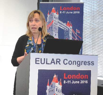 |
| Mitchel L. Zoler/Frontline Medical News Susan Barlett, Ph.D. |
Good evidence also suggests that people who are depressed are more vulnerable to develop rheumatoid arthritis or other autoimmune diseases.
The important new findings reported by Dr. Ioannou and his associates underscore the importance of providing psychological support to adolescents newly diagnosed with juvenile idiopathic arthritis. Among its many effects, depression is one of the few robust predictors of nonadherence to medical treatments by patients. Patients who are depressed are less likely to take their medications as prescribed. Depressed patients are also more likely to smoke because tobacco smoking can produce some depression relief. But smoking also contributes to the development and worsening of rheumatoid arthritis and likely other forms of inflammatory arthritis.
Susan Bartlett, Ph.D., is a psychologist and clinical epidemiologist at McGill University, Montreal, who specializes in chronic diseases including arthritis. She had no disclosures. She made these comments during a press conference.
Prior study results showed that about a third of adult patients with rheumatoid arthritis are depressed. There seems to be a vulnerability to depression among patients diagnosed with inflammatory arthritis. It makes sense that chronic arthritis can cause depression as it is a painful, debilitating, and long-term disease that is often nonremitting.
|
| Mitchel L. Zoler/Frontline Medical News Susan Barlett, Ph.D. |
Good evidence also suggests that people who are depressed are more vulnerable to develop rheumatoid arthritis or other autoimmune diseases.
The important new findings reported by Dr. Ioannou and his associates underscore the importance of providing psychological support to adolescents newly diagnosed with juvenile idiopathic arthritis. Among its many effects, depression is one of the few robust predictors of nonadherence to medical treatments by patients. Patients who are depressed are less likely to take their medications as prescribed. Depressed patients are also more likely to smoke because tobacco smoking can produce some depression relief. But smoking also contributes to the development and worsening of rheumatoid arthritis and likely other forms of inflammatory arthritis.
Susan Bartlett, Ph.D., is a psychologist and clinical epidemiologist at McGill University, Montreal, who specializes in chronic diseases including arthritis. She had no disclosures. She made these comments during a press conference.
LONDON – Depression is relatively common among teenagers newly diagnosed with juvenile idiopathic arthritis, and adolescents with both disorders appeared to have a less complete response to their treatment in a study of 102 patients.
Juvenile idiopathic arthritis (JIA) that first manifests when a patient is a teenager comes at a “vulnerable time” that can drive the development and worsening of depression, and depression can potentially exacerbate inflammation and also interfere with treatment compliance, Dr. John Ioannou said at the European Congress of Rheumatology,
Depression and JIA can produce a “vicious cycle in which depression exacerbates the disease and the disease exacerbates depression,” explained Dr. Ioannou, a rheumatologist at University College Hospital in London.
Although no study results have yet identified an effective intervention for depression identified in teenagers with newly diagnosed JIA, the immediate message from these new findings is that clinicians must assess the psychological health of adolescents with JIA both when they are first diagnosed as well as at subsequent visits, and if depression is found it requires some sort of intervention, Dr. Ioannou said in an interview.
He and his associates studied 102 patients from the United Kingdom, who were newly diagnosed with JIA and were 11-16 years old at baseline and enrolled in the Childhood Arthritis Prospective Study (CAPS), a nationwide cohort of patients with childhood-onset arthritis of various types. The average age of the group they studied was just under 13 years old, 57% were girls, 52% had persistent oligoarticular arthritis, 30% had polyarticular arthritis, and 18% had enthesitis-related arthritis. All patients underwent assessment at baseline for depression using the Mood and Feelings Questionnaire and 15 (15%) had a score that flagged them as having “probable” depression.
This depression prevalence is about three- to fourfold higher than for an otherwise healthy group of similarly aged adolescents, Dr. Ioannou said.
At baseline, the subgroup of teens with depression had a significantly higher number of inflamed joints, restricted joints, and also more overall pain and disability as measured on the Childhood Health Assessment Questionnaire.
The 102 teens with JIA underwent follow-up assessment 1-3 years later, after they had received ongoing treatment for their JIA. At follow-up, standard JIA treatment had largely resulted in resolution of joint inflammation and movement restriction among all patients, including those with depression at baseline. However the adolescents who had both JIA and depression at entry continued to have significantly more pain and disability at follow-up than did the nondepressed JIA patients, suggesting a link between depression and refractory pain and disability in JIA patients, the researchers reported.
“We need to ensure that psychological assessments and support are available to all young people diagnosed with JIA, and that this is fully integrated into routine care” for newly diagnosed JIA patients, Dr. Ioannou said. He had no disclosures.
The video associated with this article is no longer available on this site. Please view all of our videos on the MDedge YouTube channel
On Twitter @mitchelzoler
AT THE EULAR 2016 CONGRESS
Key clinical point: Pain and disability often persist despite effective antirheumatic treatment in depressed adolescents with juvenile idiopathic arthritis.
Major finding: Disability and pain levels remained significantly elevated among JIA teens with depression, compared with JIA teens without baseline depression.
Data source: The 102 adolescents enrolled in the Childhood Arthritis Prospective Study with juvenile idiopathic arthritis, including 15 patients with depression at baseline.
Disclosures: Dr. Ioannou had no disclosures.

VIDEO: RA patients on subcutaneous methotrexate avoid biologics
LONDON – Subcutaneous methotrexate monotherapy may be more effective at helping recently diagnosed patients with rheumatoid arthritis avoid biologic therapy, compared with similar patients on oral methotrexate, based on an analysis of data collected from 483 Canadian patients in routine care and enrolled in a national registry.
“This is a signal for improved efficacy with subcutaneous methotrexate, compared with oral methotrexate,” said Dr. Stephanie Gottheil, who reported these results at the European Congress of Rheumatology.
“In general, as long as patients with rheumatoid arthritis are under good control without a biologic drug, that is preferable” to initiating biologic treatment, said Dr. Gottheil, a researcher at Western University in London, Ont. Delaying the start of biologic treatment saves money, avoids the increased risk of infection that comes with biologic treatment, and defers a patient’s immune response to a biologic drug that can eventually compromise the biologic’s efficacy, she said in an interview.
“These data did not come from a randomized trial and so are by no means conclusive, but this is a signal that supports other data that subcutaneous methotrexate potentially puts patients into remission faster, and we know that earlier remission predicts more sustained remission,” she said.
“The biggest barrier to subcutaneous administration of methotrexate is patient preference to not inject themselves, but results from some studies have also shown that subcutaneous methotrexate is better tolerated,” compared with oral dosing, she added.
The study used data collected in the Canadian Early Arthritis Cohort (CATCH), which enrolls patients at several centers throughout Canada diagnosed with rheumatoid arthritis for less than 12 months. Dr. Gottheil and her associates particularly focused on 1,189 early RA patients with moderate to severe disease activity enrolled in CATCH during 2007-2012 who received methotrexate and had never previously received a biologic drug. The study’s primary endpoint was time to first treatment with a biologic during 3 years of follow-up after entry into the registry.
The patients’ average age at enrollment was 56 years, more than two-thirds were women, and their average methotrexate dosage was 20 mg/week. The cohort included 483 patients on methotrexate monotherapy – with virtually equal numbers on oral methotrexate and subcutaneous methotrexate – and 706 on a regimen that combined methotrexate with one or more additional (nonbiologic) drugs at baseline. The patients in each of the methotrexate monotherapy subgroups, those on oral or subcutaneous therapy, were very similar in their demographic and clinical profiles.
The analysis showed no statistically significant difference in time to first biologic use between the patients on a combination regimen and those on oral methotrexate monotherapy.
But when the researchers compared the time to first biologic among those on subcutaneous methotrexate monotherapy with those on oral methotrexate monotherapy, the subcutaneous patients showed a statistically significant, 47% reduced rate of starting any biologic drug during follow-up in an analysis that controlled for age, sex, education, comorbidities, disease duration, baseline disease activity, baseline corticosteroid use, joint erosions at baseline, and score on the health-assessment questionnaire at baseline, Dr. Gottheil reported.
The analysis also revealed three other variables that significantly linked with a slower progression to biologic treatment: older age, no use of corticosteroid treatment at baseline, and lower disease activity at baseline.
The CATCH registry research program is sponsored by AbbVie, Amgen, Bristol-Myers Squibb, Hoffmann-La Roche, Janssen, Pfizer, and UCB. Dr. Gottheil had no relevant disclosures.
The video associated with this article is no longer available on this site. Please view all of our videos on the MDedge YouTube channel
On Twitter @mitchelzoler
AT THE EULAR 2016 CONGRESS
Key clinical point: Among patients with recently diagnosed, moderate to severe rheumatoid arthritis, subcutaneous methotrexate monotherapy postponed the need for biologic therapy longer than did oral methotrexate monotherapy.
Major finding: Progression to biologic therapy was 47% less common among RA patients on subcutaneous methotrexate monotherapy, compared with oral methotrexate.
Data source: Three-year follow-up of 483 Canadian patients with recently diagnosed rheumatoid arthritis enrolled in CATCH, a national, real-world registry.
Disclosures: The CATCH registry research program is sponsored by AbbVie, Amgen, Bristol-Myers Squibb, Hoffmann-La Roche, Janssen, Pfizer, and UCB. Dr. Gottheil had no relevant disclosures.

Updated Behçet’s disease recommendations expand biologic treatment
LONDON – A EULAR task force issued the first update to recommendations for managing Behçet’s disease since 2008, with revised recommendations that reflect expanded use of biologic agents, and increased evidence to guide management of gastrointestinal involvement, use of anticoagulants in patients with venous involvement, and use of surgical and interventional treatments, Dr. Gülen Hatemi said while presenting the update at the European Congress of Rheumatology.
The task force, which included more than 20 members, identified 304 articles to apply to the update, and produced five overarching principal and 18 specific recommendations divided among six categories of clinical manifestations of Behçet’s disease, said Dr. Hatemi, convenor of the task force and a rheumatologist at Istanbul University.

For mucocutaneous involvement, the update included five items that all received a “strong” recommendation from the task force: For an oral or genital ulcer, use a topical agent, such as a local steroid. Try colchicine first to prevent recurrent mucocutaneous lesions, especially when the dominant lesion is erythema nodosum or a genital ulcer. Treat papulopustular or acnelike lesions with topical or systemic agents, as when treating acne vulgaris. Coordinate treatment of leg ulcers, which can be caused by venous stasis or obliterative vasculitis, with a dermatologist and vascular surgeon. And azathioprine, thalidomide, interferon-alpha, a tumor necrosis factor (TNF)–alpha antagonist, or apremilast (Otezla) may be necessary for selected patients.
The task force issued two strong recommendations for managing eye involvement along with one conditional recommendation. The first strong recommendation was that managing uveitis requires close collaboration with an ophthalmologist, with the goal of inducing and maintaining remission. Patients with an inflammatory eye disease affecting the posterior segment should receive treatment with azathioprine, cyclosporine, interferon-alpha, or a monoclonal TNF-alpha antagonist. Treatment with a systemic corticosteroid should occur only when combined with azathioprine or another systemic immunosuppressant.
The second strong recommendation was that patients who present with an initial or recurrent acute episode of sight-threatening uveitis should receive treatment with a high-dose glucocorticoid, infliximab, or interferon-alpha. Intravitreal injection with a glucocorticoid as an adjunct to systemic therapy is an option for patients with a unilateral exacerbation. The conditional recommendation was for patients with isolated anterior uveitis. When these patients have markers of a poor prognosis – such as young age, male sex, or early disease onset – systemic treatment with an immunosuppressant is a possible option.
The panel issued three strong recommendations along with one conditional recommendation for managing vascular involvement. One of the strong recommendations called for treating acute deep vein thrombosis with a glucocorticoid as well as an immunosuppresant such as azathioprine, cyclophosphamide, or cyclosporine. A conditional recommendation said patients with refractory venous thrombosis could be considered for treatment with a monoclonal TNF-alpha antagonist, along with an anticoagulant if the patient’s risk for bleeding was generally low and a coexistent pulmonary artery aneurysm was ruled out.
Management of arterial aneurysms received the other two strong recommendations. The panel recommended high-dose glucocorticoid plus cyclophosphamide for a pulmonary artery aneurysm, followed by a monoclonal TNF-alpha antagonist for refractory cases. Patients with these aneurysms who are at high risk for major bleeding should undergo embolization in preference to open surgery. When patients have aortic or peripheral artery aneurysms, treatment should start with cyclophosphamide and a corticosteroid before an aneurysm repair is attempted. But surgery or stenting of the aneurysm should not be delayed when patients are symptomatic.
Gastrointestinal involvement received one strong and two conditional recommendations. The panel strongly recommended confirming gastrointestinal involvement using endoscopy, imaging, or both, while also ruling out treatment with a nonsteroidal anti-inflammatory drug, inflammatory bowel disease, or an infection such as tuberculosis as the cause of gastrointestinal symptoms. One of the conditional recommendations called for an urgent surgical consult when patients have perforation, major bleeding, or obstruction. The second conditional recommendation called for considering glucocorticoid treatment to treat an acute exacerbation of gastrointestinal involvement. Additional treatment options to pair with a glucocorticoid include a disease-modifying drug such as 5-aminosalicylic acid or azathioprine. For patients with severe or refractory gastrointestinal symptoms or both, a monoclonal TNF-alpha antagonist or thalidomide is another potential option.
The panel issued two strong recommendations for managing nervous system involvement. The top treatment option for parenchymal involvement is a high-dose glucocorticoid followed by slow tapering while also treating with an immunosuppressant such as azathioprine. Treatment with cyclosporine should be avoided, the panel said. Treatment with a monoclonal TNF-alpha antagonist is an option to consider as first-line treatment for patients with severe nervous system involvement or for those with refractory disease. The second strong recommendation was to treat a cerebral venous thrombus with a high-dose glucocorticoid followed by tapering, with short-term anticoagulant treatment as an option. Patients also need screening for the presence of vascular disease at an extracranial location.
The panel’s final recommendation was a strong endorsement of colchicine as first-line treatment for arthritis in Behçet’s patients, although patients with acute monoarticular disease can be managed with an intra-articular injection of a glucocorticoid. For patients with recurrent or chronic arthritis, treatment options include azathioprine, interferon-alpha, or a TNF-alpha antagonist.
Dr. Hatemi has received research support from, received honoraria from, or has been a speaker for AbbVie, Celgene, Merck Sharp & Dohme, and Pfizer.
On Twitter @mitchelzoler
LONDON – A EULAR task force issued the first update to recommendations for managing Behçet’s disease since 2008, with revised recommendations that reflect expanded use of biologic agents, and increased evidence to guide management of gastrointestinal involvement, use of anticoagulants in patients with venous involvement, and use of surgical and interventional treatments, Dr. Gülen Hatemi said while presenting the update at the European Congress of Rheumatology.
The task force, which included more than 20 members, identified 304 articles to apply to the update, and produced five overarching principal and 18 specific recommendations divided among six categories of clinical manifestations of Behçet’s disease, said Dr. Hatemi, convenor of the task force and a rheumatologist at Istanbul University.
For mucocutaneous involvement, the update included five items that all received a “strong” recommendation from the task force: For an oral or genital ulcer, use a topical agent, such as a local steroid. Try colchicine first to prevent recurrent mucocutaneous lesions, especially when the dominant lesion is erythema nodosum or a genital ulcer. Treat papulopustular or acnelike lesions with topical or systemic agents, as when treating acne vulgaris. Coordinate treatment of leg ulcers, which can be caused by venous stasis or obliterative vasculitis, with a dermatologist and vascular surgeon. And azathioprine, thalidomide, interferon-alpha, a tumor necrosis factor (TNF)–alpha antagonist, or apremilast (Otezla) may be necessary for selected patients.
The task force issued two strong recommendations for managing eye involvement along with one conditional recommendation. The first strong recommendation was that managing uveitis requires close collaboration with an ophthalmologist, with the goal of inducing and maintaining remission. Patients with an inflammatory eye disease affecting the posterior segment should receive treatment with azathioprine, cyclosporine, interferon-alpha, or a monoclonal TNF-alpha antagonist. Treatment with a systemic corticosteroid should occur only when combined with azathioprine or another systemic immunosuppressant.
The second strong recommendation was that patients who present with an initial or recurrent acute episode of sight-threatening uveitis should receive treatment with a high-dose glucocorticoid, infliximab, or interferon-alpha. Intravitreal injection with a glucocorticoid as an adjunct to systemic therapy is an option for patients with a unilateral exacerbation. The conditional recommendation was for patients with isolated anterior uveitis. When these patients have markers of a poor prognosis – such as young age, male sex, or early disease onset – systemic treatment with an immunosuppressant is a possible option.
The panel issued three strong recommendations along with one conditional recommendation for managing vascular involvement. One of the strong recommendations called for treating acute deep vein thrombosis with a glucocorticoid as well as an immunosuppresant such as azathioprine, cyclophosphamide, or cyclosporine. A conditional recommendation said patients with refractory venous thrombosis could be considered for treatment with a monoclonal TNF-alpha antagonist, along with an anticoagulant if the patient’s risk for bleeding was generally low and a coexistent pulmonary artery aneurysm was ruled out.
Management of arterial aneurysms received the other two strong recommendations. The panel recommended high-dose glucocorticoid plus cyclophosphamide for a pulmonary artery aneurysm, followed by a monoclonal TNF-alpha antagonist for refractory cases. Patients with these aneurysms who are at high risk for major bleeding should undergo embolization in preference to open surgery. When patients have aortic or peripheral artery aneurysms, treatment should start with cyclophosphamide and a corticosteroid before an aneurysm repair is attempted. But surgery or stenting of the aneurysm should not be delayed when patients are symptomatic.
Gastrointestinal involvement received one strong and two conditional recommendations. The panel strongly recommended confirming gastrointestinal involvement using endoscopy, imaging, or both, while also ruling out treatment with a nonsteroidal anti-inflammatory drug, inflammatory bowel disease, or an infection such as tuberculosis as the cause of gastrointestinal symptoms. One of the conditional recommendations called for an urgent surgical consult when patients have perforation, major bleeding, or obstruction. The second conditional recommendation called for considering glucocorticoid treatment to treat an acute exacerbation of gastrointestinal involvement. Additional treatment options to pair with a glucocorticoid include a disease-modifying drug such as 5-aminosalicylic acid or azathioprine. For patients with severe or refractory gastrointestinal symptoms or both, a monoclonal TNF-alpha antagonist or thalidomide is another potential option.
The panel issued two strong recommendations for managing nervous system involvement. The top treatment option for parenchymal involvement is a high-dose glucocorticoid followed by slow tapering while also treating with an immunosuppressant such as azathioprine. Treatment with cyclosporine should be avoided, the panel said. Treatment with a monoclonal TNF-alpha antagonist is an option to consider as first-line treatment for patients with severe nervous system involvement or for those with refractory disease. The second strong recommendation was to treat a cerebral venous thrombus with a high-dose glucocorticoid followed by tapering, with short-term anticoagulant treatment as an option. Patients also need screening for the presence of vascular disease at an extracranial location.
The panel’s final recommendation was a strong endorsement of colchicine as first-line treatment for arthritis in Behçet’s patients, although patients with acute monoarticular disease can be managed with an intra-articular injection of a glucocorticoid. For patients with recurrent or chronic arthritis, treatment options include azathioprine, interferon-alpha, or a TNF-alpha antagonist.
Dr. Hatemi has received research support from, received honoraria from, or has been a speaker for AbbVie, Celgene, Merck Sharp & Dohme, and Pfizer.
On Twitter @mitchelzoler
AT THE EULAR 2016 CONGRESS

European initiative unveils pediatric care recommendations
LONDON – Recommendations on managing juvenile idiopathic arthritis and connective tissue disorders in children and young people across Europe were unveiled at the European Congress of Rheumatology.
The recommendations, which come from the SHARE (Single Hub and Access Point for Paediatric Rheumatology in Europe) project, cover best practices and provide guidance based on current evidence and expert opinion for the optimal diagnosis and treatment of these rare rheumatic diseases that affect the pediatric population.
It is hoped that the recommendations will be used to improve access to treatment and care within individual countries such that a child in one country will be able to receive the same standard of care as a child in another, Dr. Nico Wulffraat of University Medical Center Utrecht (the Netherlands) said in an interview.
Dr. Wulffraat, one of the driving forces behind the project, noted that the SHARE project was set up to look at making the management of rare pediatric rheumatic diseases more uniform across Europe. It addressed conditions such as juvenile idiopathic arthritis (JIA), childhood-onset systemic lupus erythematosus (cSLE), childhood antiphospholipid syndrome (APS), childhood vasculitis, juvenile dermatomyositis, and pediatric scleroderma. In addition, recommendations on diagnosis and treatment of periodic fever syndromes have been developed in collaboration with experts from the Eurofever Project.

“Our evidence- and consensus-based recommendations will hopefully drive access to uniform and optimal care throughout Europe, including off-label therapy when appropriate according to international consensus–derived expert advice,” Dr. Sebastiaan Vastert, SHARE project co-coordinator, said in an interview. He added: “The SHARE network will be invaluable for further international collaboration, both for optimization of care and for international collaboration in research as well.”
Dr. Wulffraat observed that while the recommendations are primarily directed at health care professionals, they also are of use for other stakeholders such as health authorities and insurance companies, and of course patients themselves to ensure the best level of care is being achieved throughout Europe.
The process for developing the guidelines was perhaps as important as the recommendations themselves, said Dr. Vastert, also of University Medical Center Utrecht. The process helped to build a network of international experts who could work together to develop future recommendations for improving patient care.
The recommendations for JIA and other pediatric rheumatic diseases included 51 “cross-cutting” statements, Dr. Vastert said. One of these statements was that a pediatric rheumatologist should manage children with signs of rheumatic disease. Another highlighted the members of a multidisciplinary team who should be involved as appropriate, such as a nurse specializing in pediatric rheumatic disease, a physiotherapist or occupational therapist, and a psychologist or psychosocial worker. Dr. Vastert also noted that good communication between team members is essential. In addition, there needs to be clear guidance on when to refer to a pediatric rheumatologist.
The SHARE project JIA recommendations include 10 evidence-based statements on diagnosis, 31 evidence-based statements on treatment, and 17 general statements on specific care for JIA, Dr. Vastert said. A few examples of the latter are that new patients should be seen in a specialist center within 4 weeks of referral; new patients and those starting a new therapy should be reviewed within 2-3 months to check on adherence, tolerance, and disease progression; and monitoring response to ongoing treatment should be every 3-6 months, preferably using existing standardized disease activity tools.

EULAR standard operating procedures were followed when developing the various SHARE recommendations, said Dr. Michael Beresford of the University of Liverpool (England) and the lead for the recommendations on childhood connective tissue disorders. Dr. Beresford noted that the latter were a rare, and in some cases extremely rare, complex group of pediatric rheumatic diseases that could lead to significant morbidity and mortality.
“Evidence-based guidelines have been lacking, and management is based mainly on physician experience. Consequently, treatment regimens vary widely throughout Europe,” Dr. Beresford observed. “These [recommendations] provide evidence-based, internationally agreed-upon standards of optimal care for pediatric connective tissue disorders.”
Specifically, the connective tissue disorder recommendations cover when to refer and how to diagnose, treat, and monitor cSLE (including neuropsychiatric SLE), childhood APS, and juvenile vasculitides, including rare pediatric vasculitides such as Takayasu arteritis. The SHARE recommendations for the management of juvenile dermatomyositis are currently in press in Annals of the Rheumatic Diseases, Dr. Beresford said.
Giving a few examples of recommendations for cSLE, Dr. Beresford noted that one of the challenges is to try to prevent delay in diagnosis. The expert panel decided that the 2012 Systemic Lupus International Collaborating Clinics (SLICC) criteria could be used for diagnosis. A referral to a pediatric rheumatologist is warranted, they determined, when a child has a positive antinuclear antibody (ANA) test and meets two clinical SLICC criteria. Dr. Beresford conceded that antibody testing might not be available because of cost in all countries, but they “decided to draw a line in the sand” to say that it is important that it is routinely done in order to come closer to a definitive diagnosis.
The aim of treatment for cSLE, the recommendations advise, is to optimize control and prevent damage caused by both the disease and by its treatment. For example, all children should be on hydroxychloroquine, and if tapering of prednisone is not possible, a disease-modifying antirheumatic drug should be added. It’s also important to actively check compliance with therapy, Dr. Beresford said.
The SHARE project was initially funded by a grant from the European Agency for Health and Consumers between 2012 and 2015 and now continues under the auspices of the Paediatric Rheumatology European Society. All speakers reported having no relevant disclosures.
LONDON – Recommendations on managing juvenile idiopathic arthritis and connective tissue disorders in children and young people across Europe were unveiled at the European Congress of Rheumatology.
The recommendations, which come from the SHARE (Single Hub and Access Point for Paediatric Rheumatology in Europe) project, cover best practices and provide guidance based on current evidence and expert opinion for the optimal diagnosis and treatment of these rare rheumatic diseases that affect the pediatric population.
It is hoped that the recommendations will be used to improve access to treatment and care within individual countries such that a child in one country will be able to receive the same standard of care as a child in another, Dr. Nico Wulffraat of University Medical Center Utrecht (the Netherlands) said in an interview.
Dr. Wulffraat, one of the driving forces behind the project, noted that the SHARE project was set up to look at making the management of rare pediatric rheumatic diseases more uniform across Europe. It addressed conditions such as juvenile idiopathic arthritis (JIA), childhood-onset systemic lupus erythematosus (cSLE), childhood antiphospholipid syndrome (APS), childhood vasculitis, juvenile dermatomyositis, and pediatric scleroderma. In addition, recommendations on diagnosis and treatment of periodic fever syndromes have been developed in collaboration with experts from the Eurofever Project.
“Our evidence- and consensus-based recommendations will hopefully drive access to uniform and optimal care throughout Europe, including off-label therapy when appropriate according to international consensus–derived expert advice,” Dr. Sebastiaan Vastert, SHARE project co-coordinator, said in an interview. He added: “The SHARE network will be invaluable for further international collaboration, both for optimization of care and for international collaboration in research as well.”
Dr. Wulffraat observed that while the recommendations are primarily directed at health care professionals, they also are of use for other stakeholders such as health authorities and insurance companies, and of course patients themselves to ensure the best level of care is being achieved throughout Europe.
The process for developing the guidelines was perhaps as important as the recommendations themselves, said Dr. Vastert, also of University Medical Center Utrecht. The process helped to build a network of international experts who could work together to develop future recommendations for improving patient care.
The recommendations for JIA and other pediatric rheumatic diseases included 51 “cross-cutting” statements, Dr. Vastert said. One of these statements was that a pediatric rheumatologist should manage children with signs of rheumatic disease. Another highlighted the members of a multidisciplinary team who should be involved as appropriate, such as a nurse specializing in pediatric rheumatic disease, a physiotherapist or occupational therapist, and a psychologist or psychosocial worker. Dr. Vastert also noted that good communication between team members is essential. In addition, there needs to be clear guidance on when to refer to a pediatric rheumatologist.
The SHARE project JIA recommendations include 10 evidence-based statements on diagnosis, 31 evidence-based statements on treatment, and 17 general statements on specific care for JIA, Dr. Vastert said. A few examples of the latter are that new patients should be seen in a specialist center within 4 weeks of referral; new patients and those starting a new therapy should be reviewed within 2-3 months to check on adherence, tolerance, and disease progression; and monitoring response to ongoing treatment should be every 3-6 months, preferably using existing standardized disease activity tools.
EULAR standard operating procedures were followed when developing the various SHARE recommendations, said Dr. Michael Beresford of the University of Liverpool (England) and the lead for the recommendations on childhood connective tissue disorders. Dr. Beresford noted that the latter were a rare, and in some cases extremely rare, complex group of pediatric rheumatic diseases that could lead to significant morbidity and mortality.
“Evidence-based guidelines have been lacking, and management is based mainly on physician experience. Consequently, treatment regimens vary widely throughout Europe,” Dr. Beresford observed. “These [recommendations] provide evidence-based, internationally agreed-upon standards of optimal care for pediatric connective tissue disorders.”
Specifically, the connective tissue disorder recommendations cover when to refer and how to diagnose, treat, and monitor cSLE (including neuropsychiatric SLE), childhood APS, and juvenile vasculitides, including rare pediatric vasculitides such as Takayasu arteritis. The SHARE recommendations for the management of juvenile dermatomyositis are currently in press in Annals of the Rheumatic Diseases, Dr. Beresford said.
Giving a few examples of recommendations for cSLE, Dr. Beresford noted that one of the challenges is to try to prevent delay in diagnosis. The expert panel decided that the 2012 Systemic Lupus International Collaborating Clinics (SLICC) criteria could be used for diagnosis. A referral to a pediatric rheumatologist is warranted, they determined, when a child has a positive antinuclear antibody (ANA) test and meets two clinical SLICC criteria. Dr. Beresford conceded that antibody testing might not be available because of cost in all countries, but they “decided to draw a line in the sand” to say that it is important that it is routinely done in order to come closer to a definitive diagnosis.
The aim of treatment for cSLE, the recommendations advise, is to optimize control and prevent damage caused by both the disease and by its treatment. For example, all children should be on hydroxychloroquine, and if tapering of prednisone is not possible, a disease-modifying antirheumatic drug should be added. It’s also important to actively check compliance with therapy, Dr. Beresford said.
The SHARE project was initially funded by a grant from the European Agency for Health and Consumers between 2012 and 2015 and now continues under the auspices of the Paediatric Rheumatology European Society. All speakers reported having no relevant disclosures.
EXPERT ANALYSIS FROM THE EULAR 2016 CONGRESS

VIDEO: FDG-PET/CT useful for fever, inflammation of unknown origin
LONDON – The use of combined modality imaging with 18F-fluorodeoxyglucose-PET/CT may provide enough information to make a definitive diagnosis in patients who present with fever or inflammation of unknown origin, particularly in those who are aged 50 years or older, have elevated C-reactive protein, and have no fever, according to findings from a single-center study of 240 cases.
The retrospective study of patients seen at the University Clinic of Erlangen (Germany) during 2007-2015 found that 18F-FDG-PET/CT was helpful in finding a diagnosis for a majority of patients with fever of unknown origin (FUO) and inflammation of unknown origin (IUO).
In an interview prior to his presentation at the European Congress of Rheumatology, the study’s senior investigator Dr. Georg Schett said that “By implementing a single 18F-FDG-PET/CT scan in a structured diagnostic approach for patients with FUO or IUO we were able to catch the underlying disease in the majority (79%) of the 240 patients studied. In the FUO group the leading diagnosis was adult-onset Still’s disease, [and] in the IUO group it was large-vessel vasculitis and polymyalgia rheumatica.”
FUO was defined about 50 years ago as several episodes of temperature exceeding 38.3° C that accompany an illness lasting more than 3 weeks, with no diagnosis after a week of testing following hospital admittance. If inflammation but no fever is involved, the condition is termed IUO.
FUO and IUO are severe, sometimes even life-threatening conditions, in which the cause of fever and inflammation, respectively, has not been defined using standard diagnostic approaches. This makes diagnosis challenging and requires a costly and complicated work-up. A delayed diagnosis can be serious, resulting in severe organ damage in patients with FUO and IUO due to the underlying, and uncontrolled, inflammatory disease.
The current diagnostic approaches for FUO and IUO include a thorough medical history, physical examination, laboratory testing, and imaging. 18F-FDG-PET/CT imaging could be potentially useful for the diagnosis of FUO/IUO because of its high-resolution detection of inflammation and malignancy. Dr. Schett and his colleagues explored this potential and examined clinical markers that would increase the likelihood of accurate 18F-FDG-PET/CT-based diagnosis in patients presenting with FUO or IUO.
The 240 patients in the study included 72 with FUO and 142 with IUO; the remaining 26 no longer fulfilled the criteria for either condition when they presented to the clinic (“ex-FUO/IUO” patients). The diagnostic work-up included 18F-FDG-PET/CT scans. Scans were considered to be positive when uptake of the tracer occurred at foci in addition to the other expected locations. The investigators explored whether the scans aided the final diagnosis, with multivariable regression analysis clarifying clinical parameters that aided the success of the scans in patients with and without FUO or IUO.
The mean age was 52 for FUO patients, 61 for IUO, and 51 for patients who no longer had IUO or FUO symptoms at presentation. These patients had mean C-reactive protein (CRP) levels of 95, 48, and 2 mg/L, respectively. Males comprised 64% of FUO, 40% of IUO, and 58% of ex-FUO/IUO patients.
18F-FDG-PET/CT was helpful in finding the diagnosis in 57% of all patients and 72% of the patients with a later diagnosis. A definitive diagnosis was not reached in 29% of patients with FUO and 17% of patients with IUO. Predictive markers for a diagnostic 18F-FDG-PET/CT for FUO and IUO were age over 50 years (P = .002 and P = .005, respectively), CRP level over 30 mg/L (P = .003 and P = .005, respectively), and the absence of fever (both P = .003). If all three parameters were fulfilled, 18F-FDG-PET/CT was diagnostic in nearly 80% of the cases, while it was successful in only 8% of cases where none of the three parameters was met.
The latter finding is particularly important, according to Dr. Schett, as it “indicates which patient subgroup is profiting the most from 18F-FDG-PET/CT.”
“FUO and IUO patients should be referred to specialized centers where 18F-FDG-PET/CT scanning is available to improve diagnosis. Simple clinical parameters such as age, CRP-level, and presence/absence of fever can guide targeted use of 18F-FDG-PET/CT,” said Dr. Schett, director of the department of internal medicine III and the Institute for Clinical Immunology at the University of Erlangen-Nuremberg (Germany).
False-positive results with 18F-FDG-PET/CT – when patients had tracer uptake that did not lead to diagnosis of the underlying diseases – are a challenge. “False-positives happen quite often due to activation of bone marrow and lymph node metabolism during inflammation, which does not support diagnosis,” Dr. Schett said. He added that, when tracer uptake associated with systemic inflammation was not considered, false positives were much less common. False-negative results – when 18F-FDG-PET/CT was negative but a diagnosis was made using other approaches – were rare, occurring in only 12 out of the 240 patients.
The research will support establishing recommendations for the use of 18F-FDG-PET/CT in FUO and IUO patients. Other patients could benefit as well. “It may be important to investigate also those patients who were referred for FUO or IUO but do not show fever or inflammation at time of admission,” Dr. Schett said. Of these ex-FUO/IUO patients, four were diagnosed with IgG4-related disease and three with familial Mediterranean syndrome by applying 18F-FDG-PET/CT.
Dr. Schett and the other authors had no disclosures.
The video associated with this article is no longer available on this site. Please view all of our videos on the MDedge YouTube channel
AT THE EULAR 2016 CONGRESS
Key clinical point: An 18F-FDG-PET/CT scan is most likely to aid diagnosis in patients who present with fever of unknown origin or inflammation of unknown origin if they are aged over 50 years, have elevated CRP level over 30 mg/L, and do not have fever.
Major finding: 18F-FDG-PET/CT was helpful in finding a diagnosis in 57% of all patients and 72% of the patients who eventually received a diagnosis.
Data source: A single-center study of 240 cases of fever of unknown origin or inflammation of unknown origin who underwent 18F-FDG-PET/CT scanning during 2007-2015.
Disclosures: Dr. Schett and the other authors had no disclosures.