User login
European Society of Cardiology (ESC): Annual Congress
Positive CvLPRIT Results Lead ACC to Change Guidelines
BARCELONA – Heart attack patients who had complete revascularization of all blocked arteries had better outcomes than those who had only the “culprit” artery unblocked, according to results from the CvLPRIT (Complete Versus Lesion-Only Primary PCI Trial) study.
The open label, randomized trial showed that among patients with acute ST-segment elevation myocardial infarction (STEMI), those who had stenting of significant coronary stenoses not responsible for the infarction as well as the infarct-producing lesion had a 55% reduction in major adverse cardiac events (MACE) at 1 year, compared with the group that had only the infarct-related artery treated. The results were presented at the annual congress of the European Society of Cardiology.
The positive results mirror the results of the PRAMI trial presented at last year’s ESC annual congress, and seem to be the tipping point for the American College of Cardiology to withdraw one of its Choosing Wisely recommendations, which had questioned any intervention beyond unblocking just the artery responsible for the heart attack.
“The newest findings regarding coronary revascularization are great examples of science on the move, and we are responding accordingly,” wrote ACC President Patrick T. O’Gara in a statement issued on Sept. 22, not too long after the results of CvLPRIT were presented.
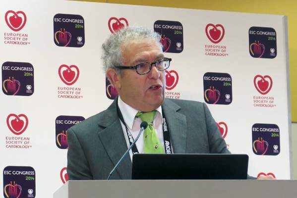
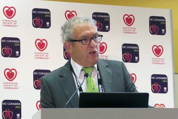
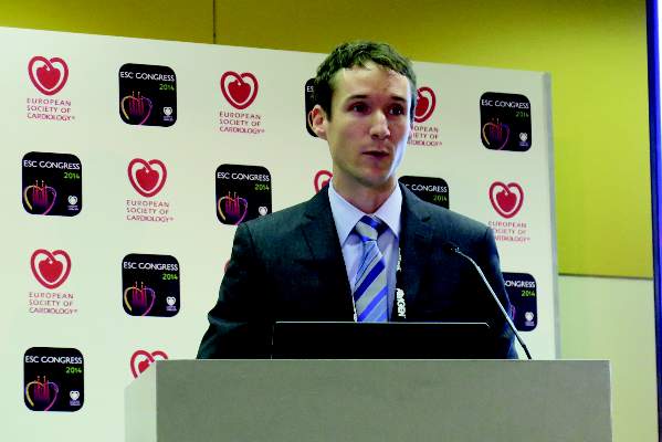
Dr. Anthony Gershlick, who presented the results of CvLPRIT at ESC, also concluded that “this strategy may be needed to be considered for future STEMI guidelines committees.”
But the topic remains controversial, and not all experts agree that it’s time for a change in clinical practice.
Dr. Shamir R. Mehta of McMaster University in Hamilton, Ont., said that both the CvLPRIT and PRAMI trials are still relatively small to measure up to the results of large meta-analyses, which show that revascularization of nonculprit arteries at the time of primary percutaneous coronary intervention (PCI) could be associated with higher mortality rates.
“The important question is, was there a significant hazard with doing revascularization at a later time point, and unfortunately this trial was too small to answer that question,” Dr. Mehta said at ESC. Dr. Gershlick, of University Hospitals of Leicester NHS Trust in England, disagreed.
“One question for me was, if a clinician is presented with angiographically significant stenoses in a non–infarct-related artery, should these be treated on that admission?” said Dr. Gershlick in a press conference. He said although retrospective registry data suggest otherwise, the results of PRAMI showed a 65% reduction in MACE with total revascularization at the time of primary PCI.
For CvLPRIT, he and his colleagues randomized 296 heart attack patients to receive either revascularization of only the infarct-related artery (146 patients), or have complete revascularization at the time of primary PCI.
The primary endpoint was MACE, which is a composite of total mortality, recurrent myocardial infarction (MI), heart failure, and ischemia-driven revascularization at 12 months.
Patients were on average 65 years old and mostly male. More than 80% had stenoses of a non–infarct-related artery, and more than 70% were treated via the radial approach.
In the complete revascularization group, the non–infarct-related arteries were treated after the infarct-related artery during the same sitting or during the same hospital admission.
At 12 months, there was a 55% reduction in MACE among patients who had complete revascularization. All components of the composite endpoint also showed a decrease, although they didn’t reach significance, compared with the group that received stenting of only the infarct-related artery.
There also was a reduction in all-cause mortality, recurrent MI, heart failure, and repeat revascularization in the complete revascularization group.
In addition, there were no safety signals, Dr. Gershlick said.
The study had several limitations, including its small size, combined endpoint, and loss to follow-up.
Experts agreed that there’s a need for larger randomized trials, such as the COMPLETE trial, which is currently enrolling patients.
Dr. Gershlick and Dr. Mehta had no disclosures.
AT THE ESC CONGRESS 2014
Positive CvLPRIT results lead ACC to change guidelines
BARCELONA – Heart attack patients who had complete revascularization of all blocked arteries had better outcomes than those who had only the “culprit” artery unblocked, according to results from the CvLPRIT (Complete Versus Lesion-Only Primary PCI Trial) study.
The open label, randomized trial showed that among patients with acute ST-segment elevation myocardial infarction (STEMI), those who had stenting of significant coronary stenoses not responsible for the infarction as well as the infarct-producing lesion had a 55% reduction in major adverse cardiac events (MACE) at 1 year, compared with the group that had only the infarct-related artery treated. The results were presented at the annual congress of the European Society of Cardiology.
The positive results mirror the results of the PRAMI trial presented at last year’s ESC annual congress, and seem to be the tipping point for the American College of Cardiology to withdraw one of its Choosing Wisely recommendations, which had questioned any intervention beyond unblocking just the artery responsible for the heart attack.
“The newest findings regarding coronary revascularization are great examples of science on the move, and we are responding accordingly,” wrote ACC President Patrick T. O’Gara in a statement issued on Sept. 22, not too long after the results of CvLPRIT were presented.
Dr. Anthony Gershlick, who presented the results of CvLPRIT at ESC, also concluded that “this strategy may be needed to be considered for future STEMI guidelines committees.”
But the topic remains controversial, and not all experts agree that it’s time for a change in clinical practice.
Dr. Shamir R. Mehta of McMaster University in Hamilton, Ont., said that both the CvLPRIT and PRAMI trials are still relatively small to measure up to the results of large meta-analyses, which show that revascularization of nonculprit arteries at the time of primary percutaneous coronary intervention (PCI) could be associated with higher mortality rates.
“The important question is, was there a significant hazard with doing revascularization at a later time point, and unfortunately this trial was too small to answer that question,” Dr. Mehta said at ESC. Dr. Gershlick, of University Hospitals of Leicester NHS Trust in England, disagreed.
“One question for me was, if a clinician is presented with angiographically significant stenoses in a non–infarct-related artery, should these be treated on that admission?” said Dr. Gershlick in a press conference. He said although retrospective registry data suggest otherwise, the results of PRAMI showed a 65% reduction in MACE with total revascularization at the time of primary PCI.
For CvLPRIT, he and his colleagues randomized 296 heart attack patients to receive either revascularization of only the infarct-related artery (146 patients), or have complete revascularization at the time of primary PCI.
The primary endpoint was MACE, which is a composite of total mortality, recurrent myocardial infarction (MI), heart failure, and ischemia-driven revascularization at 12 months.
Patients were on average 65 years old and mostly male. More than 80% had stenoses of a non–infarct-related artery, and more than 70% were treated via the radial approach.
In the complete revascularization group, the non–infarct-related arteries were treated after the infarct-related artery during the same sitting or during the same hospital admission.
At 12 months, there was a 55% reduction in MACE among patients who had complete revascularization. All components of the composite endpoint also showed a decrease, although they didn’t reach significance, compared with the group that received stenting of only the infarct-related artery.
There also was a reduction in all-cause mortality, recurrent MI, heart failure, and repeat revascularization in the complete revascularization group.
In addition, there were no safety signals, Dr. Gershlick said.
The study had several limitations, including its small size, combined endpoint, and loss to follow-up.
Experts agreed that there’s a need for larger randomized trials, such as the COMPLETE trial, which is currently enrolling patients.
Dr. Gershlick and Dr. Mehta had no disclosures.
On Twitter @naseemmiller
AT THE ESC CONGRESS 2014
Key clinical point: Complete revascularization at the time of primary PCI may be considered by future STEMI guidelines committees.
Major finding: There was a 55% reduction in MACE among heart attack patients who received complete revascularization at the time of primary PCI.
Data source: An open-label, randomized trial of 296 heart attack patients.
Disclosures: Dr. Gershlick and Dr. Mehta had no disclosures.
Lower-dose perindopril/amlodipine combo advances as novel antihypertensive therapy
BARCELONA – A low- and fixed-dose combination of perindopril 3.5 mg/amlodipine 2.5 mg once daily showed considerable promise as a potential first-step treatment in mild to moderate hypertension in a large, international, randomized trial.
The 8-week, double-blind, placebo- and active-controlled trial involved 1,297 patients, of whom 1,073 completed the study. The fixed-dose combo showed reductions in mean 24-hour blood pressure that were clinically meaningful and significantly greater than in patients randomized to perindopril monotherapy at 5 mg/day or placebo, Dr. Gianfranco Parati reported at the annual congress of the European Society of Cardiology.
Twenty-four–hour BP reductions in the lower-dose perindopril/amlodipine combination group were virtually identical to those seen in patients assigned to amlodipine monotherapy at 5 mg/day. However, the fixed-dose combo was better tolerated, with an incidence of peripheral edema of 1.6%, compared with 4.9% in patients on amlodipine at 5 mg/day.
Moreover, the rate of treatment withdrawal due to adverse effects was 1.2% with combination therapy compared to 2.6% with perindopril at 5 mg/day and 3.4% with amlodipine at 5 mg/day, added Dr. Parati of the University of Milan.
The decrease in mean 24-hour systolic BP in the lower-dose combo group was 3.8 mm Hg greater than with higher-dose perindopril monotherapy and 6.2 mm Hg greater than with placebo. The reduction in mean 24-hour diastolic BP with the fixed-dose combo was 2.4 mm Hg greater than with perindopril at 5 mg/day and 4.0 mm Hg more than with placebo.
The lower-dose combination also performed well in terms of the secondary endpoints of diurnal and nocturnal blood pressure lowering, as well as BP lowering during the previous 6 hours and in the morning.
Dr. Parati noted that current guidelines, as well as guidance from regulatory agencies, emphasize the high rate of uncontrolled hypertension and take a favorable view of the early use of fixed-dose combinations in patients with uncomplicated hypertension as a means of addressing the problem.
He received a research grant from Servier, which funded the multicenter study.
AT THE ESC CONGRESS 2014
Key clinical point: A fixed combination of perindopril and amlodipine, in lower doses than customary when either drug is used as monotherapy, provides a favorable combination of efficacy and safety in treating mild to moderate hypertension.
Major finding: Fixed-dose perindopril 3.5 mg/amlodipine 2.5 mg once daily achieved reduction in mean 24-hour systolic and diastolic blood pressure that were 3.8 and 2.4 mm Hg greater, respectively, than with perindopril at 5 mg/day.
Data source: An 8-week, double-blind, multicenter, international clinical trial of 1,297 patients with grade-1 or -2 uncomplicated hypertension.
Disclosures: The presenter received a research grant from Servier, which funded the study.
‘Healthy immigrant effect’ persists even after a decade
BARCELONA – In what is being called “the healthy immigrant effect,” Canadian investigators have found that adult diabetic immigrants from low-income countries with high cardiovascular mortality rates are relatively protected against cardiovascular events, compared with matched long-time or lifetime Canadian residents. This healthy immigrant effect appears to last for at least a decade following the immigrants’ arrival in their new land, and perhaps longer, Dr. Karen Okrainec reported at the annual congress of the European Society of Cardiology. Ontario is fertile territory in which to study the health of immigrants. Fully 43% of Ontario residents are foreign born, the highest proportion in all of Canada’s provinces, noted Dr. Okrainec of the University of Toronto.
She presented a population-based cohort study involving 87,707 adult diabetic subjects who immigrated to Ontario during 1965-2005 and an equal number of long-term or lifetime diabetic Ontario residents matched for age, sex, and neighborhood. Most of the immigrants came from South Asia and East Asia, although there were also significant numbers from the Caribbean, Sub-Saharan Africa, North Africa, and the Middle East, and Eastern and Western Europe. The immigrants had been in Canada for a mean of 11.6 years at the time of the analysis.
The primary outcome in the study was the composite endpoint of all-cause mortality or one or more hospitalizations or emergency department visits for acute MI, heart failure, unstable angina, stroke, or TIA between April 2005 and February 2012. There were 13,685 of these events among the immigrants, for an event rate of 2.4 cases/100 person-years.
This was fully 32% lower than the unadjusted event rate among the control group.
After researchers adjusted for years since diagnosis of diabetes, education level, income, the presence of hypertension, and other comorbid conditions, language barriers, marital status, and other potential confounders, they found that the risk of the composite endpoint remained 24% lower in immigrants than controls. Thus, it is clear there is not an accelerated risk of cardiovascular events among diabetic immigrants, despite their change in lifestyle in moving to a highly developed country where they may encounter cultural or language barriers to health care access, according to Dr. Okrainec.
Not all immigrants benefited from the healthy immigrant effect, however. Refugees did not. Neither did those who were single, nor did immigrants from Eastern and Central Europe or Latin America. Also, the healthy immigrant effect – the diabetic immigrants’ health advantage over longer-term Ontario residents – appeared to grow stronger with time; in other words, the healthy immigrant effect was weaker in those who arrived in Ontario less than 10 years earlier.
Dr. Okrainec’s study was funded by the Ontario Ministry of Health and Long-Term Care. She reported having no financial conflicts.
AT THE ESC CONGRESS 2014
Key clinical point: Diabetic immigrants to Ontario from low-income countries appear to have a lower risk of cardiovascular events and all-cause mortality than do long-term or lifetime residents with diabetes.
Major finding: Diabetic immigrants had a rate of major cardiovascular events or all-cause mortality of 2.4 events/100 person-years during a mean follow-up of 11.6 years since arrival in Ontario, a rate 24% lower than in matched controls.
Data source: A population-based cohort study in nearly 88,000 adult diabetic immigrants to Ontario – the majority from South or East Asia – and an equal number of matched long-term or lifetime Ontario residents with diabetes who served as controls.
Disclosures: The study was funded by the Ontario Ministry of Health and Long-Term Care. The presenter reported having no financial conflicts.
Meta-analysis: Transapical TAVR associated with higher mortality
BARCELONA – The transfemoral approach to aortic valve replacement is associated with lower risk of death, stroke, and bleeding, compared with the more invasive transapical approach, according to a meta-analysis of more than a dozen studies.
After adjustment for baseline characteristics, Italian researchers found that, compared with patients who underwent transapical transcatheter aortic valve replacement (TAVR), the transfemoral group was nearly 20% less likely to die within 30 days and during the first year after the procedure. The transfemoral group also was significantly less likely to have periprocedural bleeding or stroke.
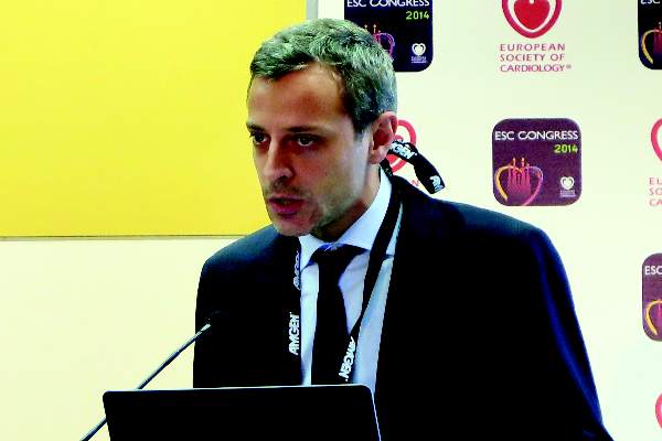
Although the findings should be viewed as hypothesis generating, they suggest that “transapical access should be reserved as a last option in TAVR patients. This choice may guarantee less mortality, less stroke, and less bleeding in this frail and elderly population,” Dr. Federico Conrotto said at the annual congress of the European Society of Cardiology.
Although the transfemoral approach for aortic valve replacement is completely percutaneous, the transapical approach is more invasive and involves the direct puncture of the left ventricle. Also, the transapical approach is mostly reserved for patients in whom femoral vessels are unapproachable. These patients also turn out to be sicker, hence making it difficult to compare the two approaches, and there are no randomized trials comparing the two, said Dr. Conrotto of Città della Salute e della Scienza Hospital in Turin, Italy.
In the absence of randomized trials, Dr. Conrotto and his colleagues selected 13 studies that reported the adjustments for clinical baseline characteristics and reported the baseline, periprocedural, and midterm (median, 365 days) outcomes for patients who underwent transfemoral and transapical TAVR.
Their primary endpoint was all-cause mortality at 1-year follow-up. The secondary endpoint was 30-day mortality and in-hospital complications, including bleeding and stroke.
In total, the studies included 10,468 patients who underwent TAVR between 2005 and 2012. The patients’ median age was 82 years and half were men: one-fourth had diabetes, 18% had renal dysfunction, 61% had coronary artery disease, and the average ejection fraction was 50%. Almost 70% of the TAVR procedures were performed transfemorally.
Results showed that patients who underwent the transfemoral approach were 15% less likely to die at 365 days following TAVR and 20% less likely to die at 30 days, compared with those who underwent the transapical approach, Dr. Conrotto said. Both differences were statistically significant.
Those who underwent the transfemoral approach were almost 30% less likely to have periprocedural bleeding and 10% less likely to have a periprocedural stroke. Again, both differences were statistically significant, he noted.
“Not only does the transfemoral approach lead to longer life, but it’s safer,” said Dr. Conrotto.
But Dr. Michael A. Borger, director of the cardiovascular institute at Columbia University Medical Center, New York, had his reservations about drawing any practice-changing conclusion from the study. In many centers, the transfemoral approach is the preferred method, so naturally only sicker patients undergo transapical TAVR, “and those patients are going to have worse outcomes. So no matter how much they try with statistics, they will not be able to account for that selection bias. And really, the only way to know is with a randomized trial, but there’s no desire within the cardiology community for a randomized trial,” he said, citing the current widespread preference for the transfemoral approach.
Dr. Conrotto and Dr. Borger had no disclosures.
On Twitter @naseemmiller
AT THE ESC CONGRESS 2014
Key clinical finding:The improved outcomes with transfemoral TAVR may be a result of patient selection because transapical TAVR is used primarily in patients unsuitable for the transfemoral procedure.
Major finding:Compared with transapical approach, patients who underwent transfemoral TAVR were nearly 20% less likely to die within 30 days and during the first year after the procedure.
Data source: Meta-analysis of 13 studies involving almost 10,500 patients.
Disclosures:Dr. Conrotto and Dr. Borger had no disclosures.
Beating heart could power batteryless pacemaker
BARCELONA – Swiss researchers have modified an automatic wristwatch, attached it to a pig’s heart, and successfully harvested more than enough energy from this constantly beating muscle to create the first batteryless pacemaker of its kind.
The feasibility study was presented at the annual congress of the European Society of Cardiology, and Dr. Sylvia G. Priori of NYU Langone Medical Center, New York, said that it “could revolutionize how the devices are made.”
Batteries take up almost half of the space in today’s pacemakers, said Adrian Zurbuchen, an engineer at the University of Bern, Switzerland, who presented the study. In addition, they have to be replaced, which increases costs and the risk of complications.
Meanwhile, automatic watches, which have a history trailing back to the 18th century, have proven effective in harvesting the power of motion to keep the clock running. So what happens if this motion comes from the beating heart to power a pacemaker?
To find the answer, the research team modified a commercially available automatic wristwatch, and after performing a sternotomy in domestic pigs, they sutured it to the myocardium on the anteroapical part of the left ventricle.
The system includes an electric circuit that transforms the energy and powers a custom-made pacemaker, and epicardial leads that deliver the electrical stimuli to the heart.
The group successfully tested the system in vivo and performed batteryless pacing at 130 beats per minute, Mr. Zurbuchen reported. The device generated a mean output power of 52 microwatts, which is nearly five times more than the power required by a modern pacemaker, he said.
Researchers are now looking into less invasive ways of implanting the device, which has not been commercialized.
Dr. Priori and Mr. Zurbuchen had no disclosures.
On Twitter @naseemmiller
AT THE ESC CONGRESS 2014
Key clinical point: The heartbeat can be harvested to power a medical device such as a pacemaker.
Major finding: A modified automatic wristwatch successfully powered a batteryless pacemaker in pigs.
Data source: In vivo study in pigs.
Disclosures: Dr. Priori and Mr. Zurbuchen had no disclosures.
ESC issues cascade of new cardiology practice guidelines
BARCELONA – Routine use of a new risk calculator tool to estimate the 5-year risk of sudden cardiac death is recommended in all patients with hypertrophic cardiomyopathy in updated guidelines launched at the annual congress of the European Society of Cardiology.
“Based on that estimate, you can now have an intelligent conversation with the patient about what the threshold is for implantable cardioverter-defibrillator placement given an individual’s risk/benefit ratio,” explained Dr. Perry M. Elliott, chair of the guidelines task force and professor of inherited cardiovascular disease at The Heart Hospital, London.
The hypertrophic cardiomyopathy guidelines were among five new practice guidelines introduced at the annual congress. Others addressed acute pulmonary embolism, noncardiac surgery, myocardial revascularization, and aortic diseases.
Here are selected highlights of the new guidelines:
• Hypertrophic cardiomyopathy. The novel sudden cardiac death risk prognostication tool, known as HCM Risk-SCD, is a major innovation in the new guidelines. The risk calculator grew out of a recent multicenter study of nearly 3,700 patients conducted by Dr. Elliott and his coinvestigators (Eur. Heart J. 2014;35:2010-20).
Now physicians can plug a series of validated risk factors into an online calculator and receive an estimated 5-year risk figure. The input factors include maximal wall thickness, left atrial diameter, family history of sudden cardiac death, unexplained syncope, and maximal left ventricular outflow gradient. If the estimated 5-year risk is 6% or more and the patient has a life expectancy of more than a year, implantation of a cardioverter-defibrillator should be seriously considered, according to the guidelines.
“What we’re trying to do in these guidelines is to change a mind-set, because hypertrophic cardiomyopathy is not really a diagnosis; it actually represents a family of diseases. Running throughout this entire document is an emphasis on individualization, from diagnosis all the way through treatment,” Dr. Elliott said. “There’s a strong emphasis on making a specific diagnosis if you possibly can, because these subtypes of cardiomyopathy have totally different natural histories and in the future will have very different treatments.”
Other highlights of the new guidelines include a stepwise approach to management of left ventricular outflow tract obstruction and heart failure, advice on reproduction, suggestions regarding simple laboratory tests with diagnostic utility, and guidance on the effective utilization of ECG, echocardiography, and cardiac magnetic resonance imaging.
“We show a number of echocardiographic red flags, some of which are perhaps not readily appreciated in everyday practice,” Dr. Elliott said. “For example, the presence of hypertrophy with impaired systolic function immediately narrows down your diagnosis to one of five or six different conditions.”
• Pulmonary embolism. The new guidelines place the novel oral anticoagulants rivaroxaban (Xarelto), apixaban (Eliquis), dabigatran (Pradaxa), and edoxaban (Savaysa) on equal footing with once-standard warfarin.
Task force chair Dr. Stavros Konstantinides highlighted as new in the 2014 guidelines a strong recommendation that patients who do not have a high-risk pulmonary embolism – that is, those who are not in shock or hypotensive – should be scored using the Pulmonary Embolism Severity Index or another validated clinical risk prediction score. The purpose is to distinguish between those at low versus intermediate risk.
Those at intermediate risk should undergo right ventricular imaging via CT or echocardiography, along with a biomarker test for myocardial injury. If both the imaging study and biomarker are positive, the patient is classified as being at intermediate-high risk; if not, intermediate-low. This has treatment implications, since it’s recommended that systemic thrombolysis be considered only in those with intermediate-high-risk pulmonary embolism and clinical signs of hemodynamic decompensation, explained Dr. Konstantinides of University Medical Center in Mainz (Germany).
• Noncardiac surgery. Perioperative initiation of beta-blocker therapy is no longer recommended routinely, but may be considered in patients scheduled for high-risk noncardiac surgery who also have two or more cardiovascular risk factors or known ischemic heart disease. Atenolol and bisoprolol are recommended as the perioperative beta-blockers of choice. If a patient is already on a beta-blocker prior to surgery, the drug can be continued perioperatively, according to task force cochair Dr. Juhani Knuuti of the University of Turku (Finland).
The guidelines emphasize that prophylactic coronary revascularization is seldom indicated in patients undergoing noncardiac surgery that has a low or intermediate cardiovascular risk.
• Myocardial revascularization. These guidelines, with more than 300 recommendations, are the lengthiest of the five new sets of guidelines. The myocardial revascularization guidelines rely heavily upon the findings of a recent meta-analysis conducted by task force cochairs Dr. Stephan Windecker of Bern (Switzerland) University and Dr. Philippe Kohl of the University of Liege (Belgium) and their coinvestigators. The meta-analysis encompassed 100 randomized controlled trials with nearly 94,000 randomized patients and more than 262,000 patient-years of follow-u p (BMJ 2014 June 23 [doi: 10.1136.bmj.g3859]).
The guidelines introduce substantial changes in the recommended method of revascularization in various situations. There is a strong emphasis on risk stratification using the SYNTAX score; in fact, the guidelines include a primer on how to calculate it.
Based largely on the results of the meta-analysis as well as 5-year follow-up in the SYNTAX trial (Lancet 2013;381:629-38), the guidelines now regard percutaneous coronary intervention as equivalent to coronary artery bypass graft surgery in several patient subsets where CABG was previously preferred. These include patients with left main or triple-vessel disease and a SYNTAX score below 22. In patients with one- or two-vessel disease and proximal left anterior descending coronary artery stenosis, both PCI and CABG get a Class I recommendation.
The new guidelines emphasize the value of intracoronary fractional flow reserve measurement to identify hemodynamically relevant lesions warranting revascularization in patients with stable coronary artery disease lacking noninvasive evidence of ischemia.
• Aortic diseases. Routine screening for abdominal aortic aneurysm via ultrasound is recommended in all men over age 65 and “may be considered” in women over 65 with a history of smoking, according to the new guidelines. That’s a much stronger proscreening stance than has been taken by the U.S. Preventive Services Task Force.
Former ESC guidelines were confined to aortic dissection and focused on the thoracic aorta. The new guidelines expand in scope to include intramural hematomas, thoracic and abdominal aneurysms, aortic valve lesions, and penetrating ulcers.
“We are not only dealing with the thoracic aorta, but are taking the holistic view of the aorta as one organ,” according to guidelines task force cochair Dr. Raimund Erbel, professor of medicine at the University of Essen (Germany).
“When an aortic aneurysm is identified at any location, assessment of the entire aorta and aortic valve is recommended at baseline and during follow-up,” he added.
All of the new guidelines can be downloaded in their entirely at the ESC website (escardio.org/guidelines).
AT THE ESC CONGRESS 2014
Diabetes Increases Risk for Atrial Fibrillation
BARCELONA – Adults with diabetes mellitus are at increased risk of subsequent new-onset atrial fibrillation – and the younger the age at diabetes onset, the greater the likelihood of developing the arrhythmia.
That’s the key finding from a Danish national registry study in which all 5,168,416 Danish adults without atrial fibrillation in 1996 were followed through 2012 for development of atrial fibrillation (AF). The study population included 75,197 Danes with diabetes at baseline and another 235,327 who developed the disease during follow-up, Dr. Jannik L. Pallisgaard explained at the annual congress of the European Society of Cardiology.
During follow-up, 5.6% of those with diabetes and 3.3% of those without diabetes developed AF. The mean time from diabetes onset to AF onset was 5 years, reported Dr. Pallisgaard of the University of Copenhagen.
"What was particularly interesting, I think, is that we found the youngest patients were the group at highest risk" of developing AF, he said. "We suggest that starting at the onset of diabetes, routine pulse palpation, ECGs, and focused patient interviews asking about any signs of atrial fibrillation could prove beneficial in detecting the arrhythmia."
The incidence rate ratio for developing AF per 1,000 person-years of follow-up was roughly 2.5-fold greater in 18- to 39-year-olds with diabetes than in their nondiabetic peers. From this peak rate in young adults, the magnitude of relative risk dropped in stepwise fashion with age: The variability in risk was lower in 40- to 60-year-old diabetics than in the 18- to 39-year olds and lower still in 65- to 74-year olds. Variability in the incidence rate ratio finally bottomed out at a still statistically significant 1.3-fold increased risk of developing AF in diabetic individuals ages 75 and older compared to their nondiabetic peers.
Dr. Pallisgaard noted that while the relative risk of developing AF was greatest in the 18- to 39-year-olds, the absolute number of new cases of AF was far greater in older patients because there were so many more of them with diabetes. He cautioned that as the obesity epidemic leads to more and more patients developing type 2 diabetes at younger ages, more cases of AF can be expected in young adults.
Dr. Pallisgaard cited two likely mechanisms underlying the observed increased risk of AF in diabetic patients: left ventricular hypertrophy and vascular inflammation, which are both often present in the diabetic population.
He reported having no financial conflicts regarding this study, conducted with Danish institutional research funds.
AT THE ESC CONGRESS 2014
Wearable defibrillator impresses as bridge to ICD decision
BARCELONA – Use of the LifeVest wearable cardioverter defibrillator for 3 months is a safe and effective means of buying time to decide whether to place a permanent implantable cardioverter defibrillator for primary prevention of sudden cardiac death in potential candidates, according to the first report from a large prospective U.S. registry.
The wearable cardioverter defibrillator (WCD) simultaneously provides patient monitoring and reliable protection against fatal arrhythmias while physicians wait to see if, for example, a patient’s left ventricular ejection fraction, depressed to less than 35% immediately post-MI, will recover over the first several months or if life-threatening arrhythmias will arise warranting implantable cardioverter defibrillator (ICD) implantation, Dr. Valentina Kutyifa explained while presenting the results of the WEARIT-II registry at the annual congress of the European Society of Cardiology.
"The WCD seems to be a powerful risk-assessment tool to identify patients at a high risk for sudden cardiac death who need subsequent ICD implantation. The WCD can be used as a bridge to a decision for appropriate ICD implantation in patients with a low ejection fraction who are immediately post-MI, or following coronary revascularization, or with new-onset dilated cardiomyopathy who are at high risk for sudden cardiac death until stabilization, or who have an inherited arrhythmia or congenital disorder," said Dr. Kutyifa of the University of Rochester (N.Y.).
She reported on 2,000 U.S. patients in those categories who participated in the prospective WEARIT-II (Prospective Registry and Follow-Up of Patients Using the Wearable Defibrillator) registry. All received from their physicians a 3-month prescription for the LifeVest WCD, which is covered by Medicare and by most health plans. Of the 2,000 patients, 927 had a left ventricular ejection fraction (LVEF) of 35% or below owing to nonischemic cardiomyopathy, 805 had ischemic cardiomyopathy, and 268 had an inherited or congenital arrhythmogenic disorder, such as long QT syndrome or arrhythmogenic right ventricular dysplasia.
Participants’ median ejection fraction at enrollment was 25%, and 52% of subjects had heart failure symptoms at that time. Patients wore the WCD for a median of 22.5 hours per day, with no difference in wear time by disease etiology.
In terms of WCD safety, the inappropriate shock rate was just 0.5% in 2,000 patients during 3 months. No study deaths occurred related to unsuccessful attempts at termination of ventricular tachycardia (VT) or ventricular fibrillation (VF) by the WCD. Three patients died while wearing the WCD, and in all three cases the WCD detected asystole at the time of death.
During 90 days of WCD wear, 2.1% of patients experienced VT or VF, for an event rate of 22 per 100 person-years. The WCD administered a shock for VT/VF in 1.1% of patients. Sustained VT or VF occurred in 1.4% of patients, while 3.6% of participants experienced atrial arrhythmias or supraventricular tachycardia, with an event rate of 121 per 100 person-years.
At the end of the 3-month period of WCD use, 42% of patients with ischemic cardiomyopathy got an ICD, as did 36% of those with nonischemic cardiomyopathy and 46% who had a congenital or inherited condition. The main reason for not implanting an ICD was a boost in ejection fraction.
The WCD frequently detected arrhythmias which facilitated the decision whether to implant an ICD or not. Eighty-five percent of patients with VT/VF treated with a WCD shock got an ICD, as did 65% of those with sustained VT that terminated spontaneously during the wearable device’s detection time. Of patients with WCD-detected atrial arrhythmias, 48% received an ICD, as did 39% of those with no arrhythmias during the 3-month period.
Dr. Kutyifa characterized the WCD as helping to fill an unmet need for improved selection of patients for primary ICD therapy. In the landmark MADIT-II trial, for example, only 4% of patients received an appropriate ICD shock during 4 years of follow-up (N. Engl. J. Med. 2002;346:877-82).
Asked about the cost-effectiveness of WCD as a bridge to the ICD decision, she replied that such data weren’t included in the WEARIT-II registry.
"The up-front cost of the device may seem rather high, but if you think about the fact that if we use the WCD and are then able to make the decision to not implant an ICD in patients who don’t need it, we really need to look at the long-term savings: the cost of the ICD, battery changes, and issues of possible lead extraction. So I think long-term this management strategy would be cost-effective," the electrophysiologist said, adding that the WCD is also rentable.
At the congress-closing conference highlights overview session, Dr. Sylvia G. Priori singled out the WEARIT-II registry as one of the top developments in the field of arrhythmias presented at this year’s meeting. She noted that the WCD has been available for several years – Medicare covers it, as do most U.S. health plans – yet until now many physicians have wanted to see stronger evidence of safety and effectiveness before incorporating the WCD into their own practices. WEARIT-II, she said, provides that supporting evidence.
"This was quite an important study. It showed the device is able to recognize life-threatening arrhythmias, so it is safe, and it does not deliver an excessive number of inappropriate shocks. And there were no deaths related to the device," commented Dr. Priori of the University of Pavia (Italy).
"The take-home message is that, yes, we know there are patients where we are reluctant to immediately say that an ICD should be implanted forever, and now we should consider testing with this WCD, which has a low risk of inappropriate shocks and a good record in shocking patients out of the arrhythmia," she added.
The WEARIT-II registry is now expanding via enrollment of patients in Europe and Israel. The registry is funded by Zoll, which markets the WCD. Dr. Kutyifa reported having received research grants from the company as well as from other device manufacturers.
AT THE ESC CONGRESS 2014
Key clinical point: The temporary use of a commercially available wearable cardioverter defibrillator buys physicians time to decide whether or not to take the big step of permanent implantable cardioverter defibrillator placement in potential candidates for whom questions exist.
Major finding: During 3 months use of the LifeVest wearable cardioverter defibrillator, the vest detected VT/VF and administered an arrhythmia-ending shock in 1.1% of device wearers with an LVEF of 35% or less.
Data source: The prospective WEARIT-II registry includes to date 2,000 patients who are potential candidates for prophylactic implantation of a permanent cardioverter defibrillator for primary prevention of sudden cardiac arrest.
Disclosures: The presenter reported receiving research grant support from Zoll, which funded the WEARIT-II registry.
Pulmonary vein isolation alone may be best ablative procedure for persistent atrial fibrillation
BARCELONA – More extensive catheter ablation procedures offered no benefit over pulmonary vein isolation alone for persistent atrial fibrillation in the largest-ever randomized trial examining outcomes of the three most popular ablation strategies.
"This study, the STAR AF 2 trial, will force a change in thinking both in the guidelines as well as in clinical practice," Dr. Atul Verma predicted, in presenting the study findings at the annual congress of the European Society of Cardiology.
Because of a widespread belief that catheter ablation success rates are probably lower in persistent AF than in paroxysmal AF, guidelines suggest "operators should consider more ablation based on linear lesions or complex fractionated electrograms," in addition to pulmonary vein isolation, in treating patients with persistent AF (Heart Rhythm 2012;9:632-96). The guidelines noted, however, that there is little evidence to support this recommendation.
The STAR AF 2 trial was conducted to learn if more complex ablation procedures really do provide greater efficacy than pulmonary vein isolation (PVI) alone. The study included 589 patients at 48 centers in 12 countries. All patients had persistent AF refractory to at least one antiarrhythmic drug and were about to undergo their first-ever catheter ablation.
Participants were randomized 1:4:4 to PVI alone with the procedural endpoint of entrance and exit block by circular mapping catheter, or PVI plus mapping and ablation of complex fractionated electrograms during AF identified using a validated 3-D mapping system, or PVI plus a left atrial roof line and another line along the mitral valve isthmus with the endpoint of bidirectional block confirmed by prespecified pacing maneuvers.
Patients remained blinded as to which of the three treatments they received. They were prospectively followed with 24-hour Holter monitoring at 3, 6, 9, 12, and 18 months along with weekly transtelephonic monitoring transmissions or at any time they felt symptoms.
Successful PVI was achieved in 97% of patients, complex fractionated electrograms were eliminated in 80% of patients assigned to that strategy, and both target lines were blocked in 74% of patients who underwent linear ablation.
The primary outcome was freedom from a documented episode of AF lasting more than 30 seconds after one procedure with or without antiarrhythmic medication through 18 months. The rates were 59% with PVI only, 48% with PVI plus complex fractionated electrograms, and 44% with PVI and linear ablation. These rates weren’t significantly different.
There were downsides to the two more elaborate ablation strategies. Procedural times were roughly 1 hour longer. Moreover, mean fluoroscopy time was 29 minutes in the PVI-only group, compared with 41 and 42 minutes with the more complex procedures. That translates to 44% more radiation exposure for both operators and patients, with absolutely no resultant added benefit over PVI alone, noted Dr. Verma, an electrophysiologist at Southlake Regional Health Center in Newmarket, Ont.
Complication rates across the board in STAR AF 2 were among the lowest ever reported in a multicenter clinical trial of catheter ablation. Of note, however, the sole fatal complication was the result of an atrial esophageal fistula in a patient assigned to PVI plus electrogram ablation.
Discussant Dr. Jagmeet P. Singh, director of the cardiac resynchronization therapy program at Massachusetts General Hospital, Boston, called STAR AF 2 "a fantastic trial."
"This study surely advocates that less ablation is more – and less works quite well," he said, noting that the roughly 50% success rate at 18 months with PVI alone is comparable to prior published success rates in paroxysmal AF.
Discussant Dr. Paulus Kirchhof said his own recent informal survey of high-volume catheter ablation centers in the United States and Europe indicated roughly one-third do PVI alone for patients with persistent AF, one-third do PVI plus ablation of complex fractionated electrograms, and one-third do PVI plus linear ablation.
"So I would say this was a question at equipoise," added Dr. Kirchhof, professor of cardiovascular sciences at the University of Birmingham (England).
Zeroing in on the added fluoroscopy time associated with the more complex ablation procedures, he noted that observational data suggest lengthier fluoroscopy may be associated with silent, subclinical brain lesions. Based upon the STAR 2 AF results, therefore, a reasonable strategy now for persistent AF is to do PVI alone, then wait and see what happens before considering additional ablation procedures later, he said.
"More importantly, I think this study shows we have to go back to the drawing board. The time pattern of AF – its duration, whether it’s paroxysmal or persistent, the left atrial size – all these things we believe identify patients who need more therapy, they may not actually help us. We just have to accept that not all patients with AF are the same, and that the pattern of AF does not discriminate so well. I think what we can really learn from this trial moving forward is that we need a clinical classification of AF patients. We have to define the patient who would benefit before we continue to develop ever-more intensive interventional strategies," he commented.
Factors worthy of further study as potential tools for separating AF patients into subgroups for treatment purposes include markers of atrial fibrosis, whether by imaging, blood, or ECG patterns; markers of parasympathetic/sympathetic imbalance; clinical markers of abnormal calcium metabolism; or blood markers, Dr. Kirchhof added.
The STAR AF 2 trial was funded by St. Jude Medical. Dr. Verma, Dr. Singh, and Dr. Kirchhof reported receiving grant support from St. Jude Medical as well as other pharmaceutical and medical device companies. In addition, Dr. Verma and Dr. Singh have served on advisory boards for St. Jude.
AT THE ESC CONGRESS 2014
Key clinical point: Pulmonary vein isolation alone may offer advantages over more elaborate procedures for persistent atrial fibrillation.
Major finding: The rates of freedom from a documented episode of AF lasting more than 30 seconds were 59% with pulmonary vein isolation only, 48% with PVI plus complex fractionated electrograms, and 44% with PVI and linear ablation.
Data source: The STAR AF 2 trial was a randomized, multicenter prospective study in which 589 patients with persistent AF were randomized to one of three popular catheter ablation strategies.
Disclosures: The study was funded by St. Jude Medical. The presenter has received research grants from and served on advisory boards for St. Jude and other medical device and pharmaceutical companies.
