User login
American Academy of Neurology (AAN): Annual Meeting 2019
Insomnia symptoms correlate with seizure frequency
PHILADELPHIA – . Insomnia symptoms are not associated with epilepsy type, number of antiepileptic drugs (AEDs), or AED standardized dose, however.

“Given the potential benefits of sleep therapies on epilepsy outcomes, routine screening of insomnia symptoms is warranted,” said lead study author Thapanee Somboon, MD, a researcher at the sleep disorders center at Cleveland Clinic Neurological Institute in Ohio and at Prasat Neurological Institute in Bangkok.
Insomnia is common and associated with depression in patients with epilepsy, but prior studies that looked at the relationship between insomnia and epilepsy-related characteristics yielded limited and conflicting results, according to Dr. Somboon.
To evaluate potential associations between insomnia and epilepsy, Dr. Somboon and colleagues conducted a prospective analysis of data from 270 patients with epilepsy who presented to the Cleveland Clinic Epilepsy Center for an initial evaluation between January and August 2018. The patients completed the Insomnia Severity Index (ISI). An ISI score of 8 or greater indicated clinical insomnia symptoms, and an ISI score of 15 or greater indicated moderate or severe insomnia symptoms.
The researchers used Spearman’s correlation and the Kruskal-Wallis test to evaluate associations among insomnia symptoms and AED standardized dose, monthly seizure frequency, Patient Health Questionnaire (PHQ-9), Generalized Anxiety Disorder Questionnaire (GAD-7), and Quality of Life in Epilepsy-10 (QOLIE10).
Among the 270 patients, the average age was 43.5 years, 58% were female, 74% had focal epilepsy, and 26% had one or more seizures per month. The population’s median ISI score was 7. Nearly half had an ISI score of 8 or greater, and 23% had an ISI score of 15 or greater.
“A positive correlation was found between ISI and PHQ-9 (r = 0.64, P less than .001), GAD-7 (r = 0.68, P less than .001), QOLIE (r = 0.55, P less than .001), and monthly seizure frequency (r = 0.31, P less than .001),” the researchers reported. Insomnia symptoms had a significantly stronger correlation with PHQ-9 and GAD-7 than with seizure frequency.
Dr. Somboon had no disclosures. A coinvestigator has received research support from Jazz Pharmaceuticals.
SOURCE: Somboon T et al. AAN 2019, Abstract P3.6-026.
PHILADELPHIA – . Insomnia symptoms are not associated with epilepsy type, number of antiepileptic drugs (AEDs), or AED standardized dose, however.
“Given the potential benefits of sleep therapies on epilepsy outcomes, routine screening of insomnia symptoms is warranted,” said lead study author Thapanee Somboon, MD, a researcher at the sleep disorders center at Cleveland Clinic Neurological Institute in Ohio and at Prasat Neurological Institute in Bangkok.
Insomnia is common and associated with depression in patients with epilepsy, but prior studies that looked at the relationship between insomnia and epilepsy-related characteristics yielded limited and conflicting results, according to Dr. Somboon.
To evaluate potential associations between insomnia and epilepsy, Dr. Somboon and colleagues conducted a prospective analysis of data from 270 patients with epilepsy who presented to the Cleveland Clinic Epilepsy Center for an initial evaluation between January and August 2018. The patients completed the Insomnia Severity Index (ISI). An ISI score of 8 or greater indicated clinical insomnia symptoms, and an ISI score of 15 or greater indicated moderate or severe insomnia symptoms.
The researchers used Spearman’s correlation and the Kruskal-Wallis test to evaluate associations among insomnia symptoms and AED standardized dose, monthly seizure frequency, Patient Health Questionnaire (PHQ-9), Generalized Anxiety Disorder Questionnaire (GAD-7), and Quality of Life in Epilepsy-10 (QOLIE10).
Among the 270 patients, the average age was 43.5 years, 58% were female, 74% had focal epilepsy, and 26% had one or more seizures per month. The population’s median ISI score was 7. Nearly half had an ISI score of 8 or greater, and 23% had an ISI score of 15 or greater.
“A positive correlation was found between ISI and PHQ-9 (r = 0.64, P less than .001), GAD-7 (r = 0.68, P less than .001), QOLIE (r = 0.55, P less than .001), and monthly seizure frequency (r = 0.31, P less than .001),” the researchers reported. Insomnia symptoms had a significantly stronger correlation with PHQ-9 and GAD-7 than with seizure frequency.
Dr. Somboon had no disclosures. A coinvestigator has received research support from Jazz Pharmaceuticals.
SOURCE: Somboon T et al. AAN 2019, Abstract P3.6-026.
REPORTING FROM AAN 2019
Inebilizumab reduces the risk of NMOSD attacks
PHILADELPHIA – , according to results presented at the annual meeting of the American Academy of Neurology. The antibody also reduces the risks of disability worsening, MRI lesion activity, and hospitalization.
No approved therapies are available for NMOSD, which is thought to be a B-cell-mediated disorder. Research suggests that CD19 is a potential therapeutic target in NMOSD. Inebilizumab has a high affinity for CD19 and reliably depleted B cells in preclinical and clinical studies.
Participants did not receive concomitant immunosuppressants
Bruce Cree, MD, PhD, clinical research director at the University of California, San Francisco, Multiple Sclerosis Center, and colleagues conducted a phase 3 study to evaluate the safety and efficacy of inebilizumab in NMOSD. Eligible participants were older than 18 years, had NMOSD, and were seropositive for aquaporin-4 (AQP4) antibodies. Seronegative patients were allowed to participate, provided that they met the 2006 Wingerchuk criteria for neuromyelitis optica. Participants were required to have had one attack in the previous year or two attacks in the 2 previous years. Participants had a wide range of disability. No background use of immune suppressants was permitted.
The trial was conducted at 99 centers in 24 countries. After a 4-week screening period, investigators randomized patients in a 3:1 ratio to inebilizumab or placebo. Treatment was administered on days 1 and 15 by IV infusion. The randomized, controlled period lasted for 197 days. If a participant did not have a relapse by the end of this period, he or she was given the choice of participating in a yearlong open-label study, in which he or she would receive 300 mg of inebilizumab every 6 months. Participants who had an attack were treated for the attack.
An independent eligibility committee reviewed and confirmed the diagnostic criteria for AQP4 seronegative participants. A masked attack-adjudication committee provided real-time assessment of NMOSD attacks. An independent data monitoring committee reviewed patient safety and ensured the ethical conduct of the study.
The trial’s primary endpoint was the time from randomization to first attack. Secondary endpoints included a measure of disability, a measure of visual acuity, cumulative lesions on MRI, and NMOSD-related inpatient hospitalizations.
Treatment reduced the risk of hospitalization
Dr. Cree and colleagues screened 467 subjects and randomized 231. Among those patients, 175 received inebilizumab, and 56 received placebo. The population’s mean age was 43 years. About 90% were women, and 72% were white or Asian. The median baseline disability score was 3.5, indicating moderate disability. More than 90% of patients were AQP4 seropositive. The mean number of attacks at entry was 4.3. About 60% of participants had had prior exposure to immune suppressants.
Enrollment into the randomized, controlled period was stopped at 231 patients after 43 attacks had occurred, based on the recommendation of the independent data monitoring committee, which concluded that further recruitment into the placebo arm would not be ethical. The committee also recommended that all participants be shifted into the open-label period.
Among AQP4 seropositive patients, 88% of the inebilizumab group did not have an attack, compared with 57% of the control group. This result represents a 77% reduction in the risk of a relapse following inebilizumab treatment. The number needed to treat was 3.23. In the population overall, 87% of patients treated with inebilizumab did not have an attack, compared with 60% of controls, which corresponded to a 73% reduction in risk of an attack and a number needed to treat of 3.7.
In addition, Dr. Cree and colleagues found that inebilizumab was associated with a 63% reduction in risk of disability, a 43% reduction in risk of developing new lesions, and a 71% reduction in NMOSD-related hospitalizations. These results were statistically significant and clinically meaningful, he said. The only secondary endpoint that was not met was the difference in binocular low-contrast visual acuity scores. “This [result] may be due to the low frequency of optic neuritis events, a floor effect within the placebo arm (many of those patients had profound visual loss at baseline), or selection of a binocular acuity test that diluted the impact of monocular events,” said Dr. Cree.
Adverse events and serious adverse events were well balanced between the two treatment arms. The rate of infusion-related reactions was low in both arms and was “perhaps slightly lower in the inebilizumab treatment arm,” said Dr. Cree. No deaths occurred during the randomized, controlled period, and the researchers found no pattern of serious adverse events. Two deaths occurred in the open-label period: one related to a severe attack, and the other related to a brain event of unclear etiology without definite diagnosis.
B cells were depleted within approximately 4 weeks of treatment, and this depletion was sustained throughout the randomized, controlled period.
This study was funded by Viela Bio and MedImmune. Dr. Cree has received personal compensation for consulting, serving on a scientific advisory board, speaking, or other activities from Abbvie, Akili, Biogen, EMD Serono, GeNeuro and Novartis.
SOURCE: Cree B et al. AAN 2019. Abstract Plen02.001.
REPORTING FROM AAN 2019
Perplexing text messages may be the sole sign of stroke
PHILADELPHIA – described at the annual meeting of the American Academy of Neurology. The phenomenon, dubbed “dystextia” and characterized by confusing interpersonal electronic communications, is not new but is increasingly relevant to clinical practice now that smartphones are essentially ubiquitous, said the case report authors.
In fact, time to intervention may be positively impacted if access to a patient’s texts and emails can be obtained, said lead author Taylor R. Anderson, a medical student at Wayne State University, Detroit.
The findings sparked interest in further research into the underlying causes and implications of dystextia: “It will be interesting to see if there are specific regions of the brain that are responsible for texting, and how they relate to other forms of communication such as handwriting and typing,” the authors said in their poster presentation.
Mr. Anderson and colleagues described two patients evaluated at Ascension St. John Hospital and Medical Center in Detroit who had stroke presenting as difficulty in typing text messages.
One case was a 43-year-old woman who experienced headache consistent with her usual migraine and spelling errors in her texts and posts on Facebook. She had visuospatial anomalies and left facial droop on evaluation. A brain MRI revealed acute embolic infarcts in the parietal and right frontal lobes, according to Mr. Anderson and coauthors.
The second case was a 66-year-old woman who had difficulty writing texts and typed notes. A head CT done in an urgent care facility showed a left frontal subacute infarct, which, according to the authors, was likely related to risk factors including hypertension, diabetes, and dyslipidemia.
These cases show that dystextia can arise after lesions in either hemisphere, the authors said. However, they emphasized that both cases involved the dominant cerebral hemisphere, while by contrast, there have not been previous reports of dystextia due to nondominant hemispheric infarct.
It stands to reason that stroke would affect the ability to text, a multipurpose task that involves use of motor, language, and vision skills, the authors said. Strokes could affect not only skills needed to type, read, and express thoughts, but also visuospatial memory mapping to letters on the device’s keyboard, they noted.
The left frontal and superior parietal regions have been implicated in handwriting in neuroimaging experiments, the authors said, while the operculum and left second frontal convolution are involved in typing.
The authors had nothing to disclose.
SOURCE: Anderson T et al. AAN 2019. Abstract P5.3-062.
REPORTING FROM AAN 2019
More than one in six patients with status epilepticus are readmitted after hospital discharge
PHILADELPHIA – according to research presented at the annual meeting of the American Academy of Neurology. It is possible to identify patients at high risk of readmission, which could allow neurologists to reduce their clinical and economic burden, said the investigators.
Status epilepticus is a major neurologic emergency. Patients often have significant disability and may represent a burden on their families and on the health care system. To identify independent predictors of 30-day hospital readmission among patients discharged after generalized convulsive status epilepticus, Mohamad Rahwan, MD, a neurologist at the Medical University of South Carolina, Charleston, and colleagues examined data from the 2014 Nationwide Readmission Database.
The investigators included adults with a primary discharge diagnosis of generalized convulsive status epilepticus, identified by the ICD-9-CM code 345.3, in their study. Patients who died during hospitalization, had missing information on the length of stay, or were discharged in December 2014 were excluded from analysis. Dr. Rahwan and colleagues calculated the overall 30-day readmission rate for the sample and compared prespecified groups by their 30-day readmission status. They performed multiple logistic regression analysis to identify independent predictors of 30-day readmission, adjusting for potential confounders.
In all, 14,562 adults were discharged with a diagnosis of generalized convulsive status epilepticus. Of this population, 2,520 patients (17.3%) were readmitted within 30 days. Multivariate logistic regression analysis indicated that patients discharged against medical advice (odds ratio, 1.45), those discharged to short-term hospital (OR, 1.39), those with comorbid conditions (OR for Charlson Comorbidity Index of 1, 1.12; OR for Charlson Comorbidity Index of 2 or greater, 1.32), and those with a length of stay exceeding 6 days (OR, 1.42) had a greater risk of 30-day readmission. The researchers observed an inverse association for patients aged 45 years or older and for those in high-income households. “Greater attention to high-risk subgroups may identify opportunities to ameliorate the clinical and economic burden of early readmissions after generalized convulsive status epilepticus,” said the researchers.
The researchers had no disclosures.
SOURCE: Rahwan M et al. AAN 2019, Abstract S36.006.
REPORTING FROM AAN 2019
Changes in brain networks may predict MS worsening
PHILADELPHIA – (MS), according to a study described at the annual meeting of the American Academy of Neurology.
Neurologists do not have reliable biomarkers to predict disease evolution in the medium or long term for patients with MS. The ability to predict disease evolution accurately could aid in the choice of treatment.
Maria Assunta Rocca, MD, head of the Neuroimaging of CNS White Matter Unit, Department of Neurology, Institute of Experimental Neurology, Scientific Institute Ospedale, San Raffaele, Milan, Italy, and colleagues sought to evaluate structural and functional network MRI measures as predictors of clinical deterioration over 6.5 years. They obtained conventional, 3D, T1-weighted, diffusion-weighted MRI, and resting-state functional MRI images at baseline from 233 patients with MS and 77 healthy controls. Patients underwent a neurologic examination at baseline and after a median follow-up period of 6.5 years. At follow-up, the researchers classified patients as clinically stable or worsened, according to their change in Expanded Disability Status Scale (EDSS) score. They also evaluated conversion to secondary progressive MS among patients with relapsing remitting MS at baseline.
To identify the main large-scale resting state functional connectivity networks, Dr. Rocca and colleagues applied spatial independent component analysis to resting state functional MRI data. They applied the same technique to gray matter probability maps and fractional anisotropy maps to identify the corresponding structural gray matter and white matter networks.
At follow-up, 105 patients with MS (45%) had significant EDSS worsening. Of 157 patients with relapsing remitting MS, 26 (16%) converted to secondary progressive MS. The multivariable model, after adjustment for follow-up duration, identified baseline EDSS score (odds ratio, 1.59), normalized gray matter volume (OR, 0.99), and abnormally high baseline resting state functional connectivity of the left precentral gyrus in the sensorimotor network (OR, 1.67) as predictors of EDSS worsening. These variables remained significant after the researchers adjusted for treatment effect. Independent variables associated with conversion to secondary progressive MS included baseline EDSS score (OR, 2.8) and atrophy of gray matter networks associated with sensory (OR, 0.5) and motor (OR, 0.4) functions.
Dr. Rocca received personal compensation from Biogen Idec, Novartis, Genzyme, Sanofi-Aventis, Teva, Merck Serono, and Roche. Coauthors reported research support from Biogen, Merck Serono, Novartis, Teva, and Roche..
SOURCE: Filippi M et al. AAN 2019, Abstract S49.004.
REPORTING FROM AAN 2019
Key clinical point: Structural and functional network MRI measures predict long-term worsening in multiple sclerosis.
Major finding: The odds ratio of worsening for patients with abnormally high baseline resting state functional connectivity is 1.67.
Study details: A prospective imaging study of 233 patients with multiple sclerosis and 77 healthy controls.
Disclosures: Dr. Rocca received personal compensation from Biogen Idec, Novartis, Genzyme, Sanofi-Aventis, Teva, Merck Serono, and Roche. Coauthors reported research support from Biogen, Merck Serono, Novartis, Teva, and Roche.
Source: Filippi M et al. AAN 2019, Abstract S49.004.
Ibudilast’s efficacy differs in primary and secondary progressive MS
PHILADELPHIA – researchers reported at the annual meeting of the American Academy of Neurology.
The difference may be related to faster atrophy rates among patients with primary progressive MS who received placebo, compared with those with secondary progressive MS who received placebo.
The finding was surprising, said Andrew Goodman, MD, professor of neurology, chief of the neuroimmunology unit, and director of the multiple sclerosis center at the University of Rochester (N.Y.). “Going into the trial, it was my bias and expectation that both primary and secondary progressive MS would behave more similarly than different.”
The trial, SPRINT-MS, included more than 250 patients with progressive MS at 28 sites. Patients were aged 18-65 years and were followed for 96 weeks. Patients had primary progressive MS (n = 134) or secondary progressive MS (n = 121) and were randomized 1:1 to ibudilast or placebo.
Ibudilast is an orally administered small molecule that has been used in Japan for approximately 30 years for asthma and other indications, Dr. Goodman said. Preclinical models suggested that the drug may have neuroprotective effects.
The trial’s primary result – a 48% slowing in the rate of whole brain atrophy as measured by brain parenchymal fraction with ibudilast – was reported last year (N Engl J Med. 2018 Aug 30;379[9]:846-55).
The present study examined whether the treatment effect of ibudilast was similar by progressive disease type using a linear mixed model analytic approach.
The group with primary progressive MS included a smaller percentage of women. Patients with secondary progressive MS had longer disease duration and more brain atrophy at baseline.
“The overall benefit which we previously reported appears to be driven by subjects with primary progressive rather than secondary progressive MS,” Dr. Goodman said. Accounting for baseline covariates did not affect this result.
Among patients who received placebo, brain atrophy in those with secondary progressive MS was 57% slower than in those with primary progressive MS. The rate of atrophy for untreated patients with primary progressive MS “was roughly twice as fast as that in the secondary progressive MS group, which we think may explain in part the differential in efficacy,” Dr. Goodman said. “These findings may impact future trial design for progressive MS.”
The SPRINT-MS trial was funded by the National Institute of Neurological Disorders and Stroke. The National Multiple Sclerosis Society and MediciNova also supported the study. Dr. Goodman reported receiving research support from pharmaceutical companies, as well as personal compensation from companies for consulting, serving on a scientific advisory board, and speaking.
SOURCE: Goodman A et al. AAN 2019, Abstract S12.007.
REPORTING FROM AAN 2019
Rimegepant dissolving tablets treat acute migraine in phase 3 trial
PHILADELPHIA – according to phase 3 trial results presented at the annual meeting of the American Academy of Neurology. The treatment’s efficacy is sustained for 2-48 hours, researchers reported.

Rimegepant is a small molecule calcitonin gene–related peptide receptor antagonist. A 75-mg oral tablet formulation was effective in phase 3 trials. The present study evaluated a novel, orally dissolving tablet formulation that is intended to speed the drug’s onset. The tablet’s time to peak concentration is 1.50 hours, compared with 1.99 hours for the oral tablet.
Formulation preferences
“People with migraine prefer orally dissolving tablets to oral tablets, mainly for their convenience, onset of action, and ability to be taken without drinking liquids,” said first author Richard B. Lipton, MD, of Albert Einstein College of Medicine, New York, and colleagues.
To assess the formulation’s efficacy and safety, the investigators conducted a double-blind, randomized, placebo-controlled, multicenter trial. Participants were aged at least 18 years and had migraine for at least 1 year. They had 2-8 moderate or severe migraine attacks and fewer than 15 headache days per month during the 3 months before the trial. Their preventive migraine medication doses, if any, had been stable for at least 3 months.
Coprimary efficacy endpoints were pain freedom 2 hours post dose and freedom from the most bothersome symptom at 2 hours post dose. The efficacy analyses included randomized subjects who had a qualifying migraine attack, took the study medication, and provided at least one postbaseline efficacy data point.
The investigators included 1,351 patients in their efficacy analysis – 669 who received rimegepant and 682 who received placebo. About 85% were female, and patients’ mean age was 40.2 years. They averaged 4.6 migraine attacks per month, and their most bothersome symptoms included photophobia (57%), nausea (23.5%), and phonophobia (19.3%). About 14% used preventive treatment.
Within 45 days of randomization, patients treated a migraine attack of moderate to severe intensity and completed an electronic diary predose to 48 hours post dose.
Less use of rescue medication
At 2 hours post dose, patients who received 75 mg rimegepant were more likely than patients who received placebo to achieve pain freedom (21.2% vs. 10.9%) and freedom from the most bothersome symptom (35.1% vs. 26.8%).
Numerical differences in the likelihood of pain relief between group began 15 minutes post dose, and the difference was statistically significant at 60 minutes (36.8% vs. 31.2%).
Various secondary endpoints, including ability to function normally at 2 hours post dose (38.1% vs. 25.8%), sustained pain relief from 2-48 hours (42.2% vs. 25.2%), and use of rescue medications within 24 hours (14.2% vs. 29.2%), also were statistically significant.
In the safety analysis, the most common adverse events were nausea (1.6% in the rimegepant group and 0.4% in the placebo group) and urinary tract infection (1.5% in the rimegepant group and 0.6% in the placebo group). There were no serious adverse events. “Safety and tolerability were similar to placebo,” Dr. Lipton and colleagues said.
Biohaven Pharmaceuticals, the developer of the drug, sponsored the study. Dr. Lipton has received honoraria and research support from Biohaven and holds stock in the company. Coauthors are employees of Biohaven.
PHILADELPHIA – according to phase 3 trial results presented at the annual meeting of the American Academy of Neurology. The treatment’s efficacy is sustained for 2-48 hours, researchers reported.
Rimegepant is a small molecule calcitonin gene–related peptide receptor antagonist. A 75-mg oral tablet formulation was effective in phase 3 trials. The present study evaluated a novel, orally dissolving tablet formulation that is intended to speed the drug’s onset. The tablet’s time to peak concentration is 1.50 hours, compared with 1.99 hours for the oral tablet.
Formulation preferences
“People with migraine prefer orally dissolving tablets to oral tablets, mainly for their convenience, onset of action, and ability to be taken without drinking liquids,” said first author Richard B. Lipton, MD, of Albert Einstein College of Medicine, New York, and colleagues.
To assess the formulation’s efficacy and safety, the investigators conducted a double-blind, randomized, placebo-controlled, multicenter trial. Participants were aged at least 18 years and had migraine for at least 1 year. They had 2-8 moderate or severe migraine attacks and fewer than 15 headache days per month during the 3 months before the trial. Their preventive migraine medication doses, if any, had been stable for at least 3 months.
Coprimary efficacy endpoints were pain freedom 2 hours post dose and freedom from the most bothersome symptom at 2 hours post dose. The efficacy analyses included randomized subjects who had a qualifying migraine attack, took the study medication, and provided at least one postbaseline efficacy data point.
The investigators included 1,351 patients in their efficacy analysis – 669 who received rimegepant and 682 who received placebo. About 85% were female, and patients’ mean age was 40.2 years. They averaged 4.6 migraine attacks per month, and their most bothersome symptoms included photophobia (57%), nausea (23.5%), and phonophobia (19.3%). About 14% used preventive treatment.
Within 45 days of randomization, patients treated a migraine attack of moderate to severe intensity and completed an electronic diary predose to 48 hours post dose.
Less use of rescue medication
At 2 hours post dose, patients who received 75 mg rimegepant were more likely than patients who received placebo to achieve pain freedom (21.2% vs. 10.9%) and freedom from the most bothersome symptom (35.1% vs. 26.8%).
Numerical differences in the likelihood of pain relief between group began 15 minutes post dose, and the difference was statistically significant at 60 minutes (36.8% vs. 31.2%).
Various secondary endpoints, including ability to function normally at 2 hours post dose (38.1% vs. 25.8%), sustained pain relief from 2-48 hours (42.2% vs. 25.2%), and use of rescue medications within 24 hours (14.2% vs. 29.2%), also were statistically significant.
In the safety analysis, the most common adverse events were nausea (1.6% in the rimegepant group and 0.4% in the placebo group) and urinary tract infection (1.5% in the rimegepant group and 0.6% in the placebo group). There were no serious adverse events. “Safety and tolerability were similar to placebo,” Dr. Lipton and colleagues said.
Biohaven Pharmaceuticals, the developer of the drug, sponsored the study. Dr. Lipton has received honoraria and research support from Biohaven and holds stock in the company. Coauthors are employees of Biohaven.
REPORTING FROM AAN 2019
Can an antisense oligonucleotide benefit patients with SOD1-ALS?
PHILADELPHIA – In patients with amyotrophic lateral sclerosis (ALS) caused by gain-of-toxic function mutations in the SOD1 gene, according to phase 1/2 trial results presented at the annual meeting of the American Academy of Neurology. Exploratory analyses suggest that the drug, known as tofersen (also known as BIIB067), may lessen declines in function, respiratory function, and strength. The treatment generally was safe and well tolerated, researchers said.
Most ALS cases are sporadic, but about 10% are genetic, of which approximately 20% are caused by SOD1 mutations. “Although SOD1-ALS disease progression is heterogeneous, the underlying pathophysiology, attributable to mutant SOD1 toxicity, is thought to be consistent across SOD1 mutation types,” said Timothy M. Miller, MD, PhD, of Washington University, St. Louis, and colleagues. “As such, effective reduction of SOD1 protein, irrespective of mutation, has the potential to alter the disease course of people with SOD1-ALS.”
Tofersen is an antisense oligonucleotide ribonuclease H1-mediated inhibitor of SOD1 messenger RNA. To study its safety, tolerability, pharmacokinetics, pharmacodynamics, and exploratory efficacy in patients with SOD1-ALS, investigators conducted a double-blind study. The investigators randomized 50 patients with ALS with a SOD1 mutation to 20 mg (n = 10), 40 mg (n = 9), 60 mg (n = 9), or 100 mg (n = 10) of tofersen or placebo (n = 12).
Patients received tofersen by intrathecal bolus over 1-3 minutes. They received a loading regimen on days 1, 15, and 29 and maintenance dosing on days 57 and 85. After the dosing period, patients completed a 12-week follow-up period.
Adverse events
All patients received at least one dose of the study treatment, and 48 received all treatments. Three patients died during the study. One patient in the 20-mg group died of pulmonary embolism, and one patient in both the 60-mg group and placebo group died of respiratory failure. Investigators considered the deaths secondary to ALS or other conditions and not drug related.
Most adverse events were mild or moderate. The most common adverse events among tofersen-treated patients were headache (n = 16), procedural pain (n = 14), and post–lumbar puncture syndrome (n = 13). Five patients who received tofersen and two who received placebo experienced serious adverse events; no serious adverse events occurred in the 100-mg dose group.
“A reduction from baseline in CSF SOD1 concentrations was observed in the tofersen 40-, 60-, and 100-mg cohorts with the maximal reduction observed in the 100 mg–treated group [37% vs. no reduction in the placebo group; P less than 0.002] at day 85,” the investigators reported.
Possible efficacy
In exploratory analyses, the 100-mg dose slowed decline on the Amyotrophic Lateral Sclerosis Functional Rating Scale–Revised (ASLFRS-R), compared with placebo. Mean change in ASLFRS-R from baseline to day 85 was –1.1 in patients who received 100 mg of tofersen, compared with –5.3 for patients who received placebo. Declines on measures of respiratory function and muscle strength also slowed. “Across clinical measures, separation from placebo was most apparent in participants with fast progressing disease,” the researchers said.
“Lower concentrations of the protein in the spinal fluid suggest that there were also lower concentrations in the brain and spinal cord,” Dr. Miller said in a news release. “Such reductions could lead to preservation of motor neurons and slow progression of the disease, but more study is needed to examine this further.”
The study was sponsored by Biogen, which is developing tofersen. Some of the study authors are Biogen employees. Dr. Miller is on Biogen’s medical advisory board and receives clinical research support from Biogen. In addition, he consults, has licensing agreements with, and is a principal investigator for other companies.
REPORTING FROM AAN 2019
Experts discuss what’s new in migraine treatment
PHILADELPHIA – At the annual meeting of the American Academy of Neurology, Alan M. Rapoport, MD, clinical professor of neurology at the University of California, Los Angeles, sat down with Stewart J. Tepper, MD, professor of neurology at the Geisel School of Medicine at Dartmouth, Hanover, N.H., to discuss in a video some of the new data presented at the meeting regarding the CGRP monoclonal antibodies, the small molecule receptor antagonists (gepants), and what Dr. Tepper described as “a real shift in paradigm and a watershed moment in migraine.”
The three gepants that are farthest along in clinical trials are ubrogepant, rimegepant, and atogepant. “Reassuring data” was presented, Dr. Tepper said, regarding liver toxicity, which has been a concern with earlier generations of the gepants. The Food and Drug Administration had mandated a close look at liver function with the use of these drugs, which are metabolized in the liver, and, to date, no safety signals have emerged.
The three CGRP monoclonal antibodies that are currently on the market are erenumab (Aimovig), fremanezumab (Ajovy), and galcanezumab (Emgality). Data from numerous open-label extension studies were presented. In general, it seems that “the monoclonal antibodies accumulate greater efficacy over time,” Dr. Tepper said. No safety concerns have emerged from 5 years of clinical trial data. With 250,000 patients on these drugs worldwide, that is “very reassuring,” Dr. Tepper said.
New data also show that the majority of patients with chronic migraine who are taking monoclonal antibodies convert from chronic migraine to episodic migraine. Additionally, new data show that use of monoclonal antibodies “dramatically reduce all migraine medication use,” Dr. Tepper said.
EXPERT ANALYSIS FROM AAN 2019
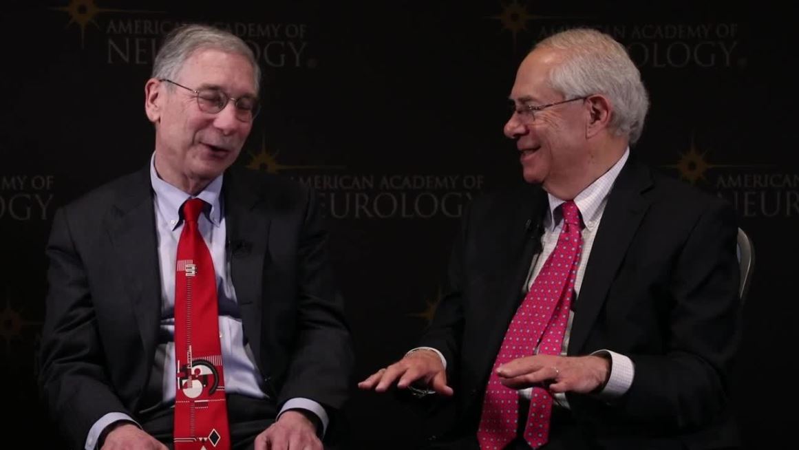
Galcanezumab reduces cluster headache attack frequency
PHILADELPHIA – according to study results presented at the annual meeting of the American Academy of Neurology. The treatment has a similar safety profile in this patient population as it has among people with episodic or chronic migraine, said the researchers.

Calcitonin gene-related peptide (CGRP) has an important role in the pathogenesis of cluster headache. Galcanezumab (Emgality)is a humanized monoclonal antibody that binds to CGRP. Eli Lilly & Co. developed the molecule as a treatment for migraine. Cluster headache is characterized by recurrent unilateral headache attacks accompanied by autonomic symptoms. The most common acute treatments for cluster headache are sumatriptan (Imitrex) and high-flow oxygen, but some patients do not respond to these therapies.
David W. Dodick, MD, director of the headache, sports neurology, and concussion programs at Mayo Clinic in Phoenix, and colleagues conducted a trial to evaluate the efficacy and safety of galcanezumab in patients with episodic cluster headache.
After screening, participants underwent a prospective baseline diary phase for 7 consecutive days. The investigators subsequently randomized patients in equal groups to galcanezumab (300 mg subcutaneously) or placebo. Treatment was administered subcutaneously once monthly. The double-blind treatment period lasted for 8 weeks, and a washout period followed. The trial’s primary endpoint was the overall mean change from baseline in weekly cluster headache attack frequency during weeks 1-3, as recorded in patient diaries. The main secondary endpoint was the proportion of patients who had a reduction in weekly cluster headache attack frequency of 50% or more at week 3.
In all, 49 patients were randomized to galcanezumab, and 57 were randomized to placebo. Mean age was 45-47 years. Between 82% and 84% of patients were male. The mean number of weekly cluster headache attacks at baseline was approximately 17.5 in both groups.
During weeks 1-3, the mean change in weekly attack frequency was −8.7 in the galcanezumab group and −5.2 for controls. The difference between groups was statistically significant. The percentage of participants with a reduction in weekly attack frequency of at least 50% at week 3 was 76% for galcanezumab versus 57% for placebo. The between-group differences in these endpoints were statistically significant.
The discontinuation rate was 8% (4 participants) in the galcanezumab group and 21% (12 participants) in the placebo group. Eight participants (14%) in the placebo group discontinued treatment because of lack of efficacy, compared with one participant (2%) in the galcanezumab group. The researchers observed no clinically meaningful differences between treatment groups on tolerability or safety parameters except for a greater incidence of injection-site pain with galcanezumab versus placebo (8.2% vs. 0%).
Eli Lilly and Co. sponsored the study. Dr. Dodick has a consulting relationship with the company.
SOURCE: Bardos JN et al. AAN 2019, Abstract 02.004.
PHILADELPHIA – according to study results presented at the annual meeting of the American Academy of Neurology. The treatment has a similar safety profile in this patient population as it has among people with episodic or chronic migraine, said the researchers.
Calcitonin gene-related peptide (CGRP) has an important role in the pathogenesis of cluster headache. Galcanezumab (Emgality)is a humanized monoclonal antibody that binds to CGRP. Eli Lilly & Co. developed the molecule as a treatment for migraine. Cluster headache is characterized by recurrent unilateral headache attacks accompanied by autonomic symptoms. The most common acute treatments for cluster headache are sumatriptan (Imitrex) and high-flow oxygen, but some patients do not respond to these therapies.
David W. Dodick, MD, director of the headache, sports neurology, and concussion programs at Mayo Clinic in Phoenix, and colleagues conducted a trial to evaluate the efficacy and safety of galcanezumab in patients with episodic cluster headache.
After screening, participants underwent a prospective baseline diary phase for 7 consecutive days. The investigators subsequently randomized patients in equal groups to galcanezumab (300 mg subcutaneously) or placebo. Treatment was administered subcutaneously once monthly. The double-blind treatment period lasted for 8 weeks, and a washout period followed. The trial’s primary endpoint was the overall mean change from baseline in weekly cluster headache attack frequency during weeks 1-3, as recorded in patient diaries. The main secondary endpoint was the proportion of patients who had a reduction in weekly cluster headache attack frequency of 50% or more at week 3.
In all, 49 patients were randomized to galcanezumab, and 57 were randomized to placebo. Mean age was 45-47 years. Between 82% and 84% of patients were male. The mean number of weekly cluster headache attacks at baseline was approximately 17.5 in both groups.
During weeks 1-3, the mean change in weekly attack frequency was −8.7 in the galcanezumab group and −5.2 for controls. The difference between groups was statistically significant. The percentage of participants with a reduction in weekly attack frequency of at least 50% at week 3 was 76% for galcanezumab versus 57% for placebo. The between-group differences in these endpoints were statistically significant.
The discontinuation rate was 8% (4 participants) in the galcanezumab group and 21% (12 participants) in the placebo group. Eight participants (14%) in the placebo group discontinued treatment because of lack of efficacy, compared with one participant (2%) in the galcanezumab group. The researchers observed no clinically meaningful differences between treatment groups on tolerability or safety parameters except for a greater incidence of injection-site pain with galcanezumab versus placebo (8.2% vs. 0%).
Eli Lilly and Co. sponsored the study. Dr. Dodick has a consulting relationship with the company.
SOURCE: Bardos JN et al. AAN 2019, Abstract 02.004.
REPORTING FROM AAN 2019