User login
Experts discuss what’s new in migraine treatment
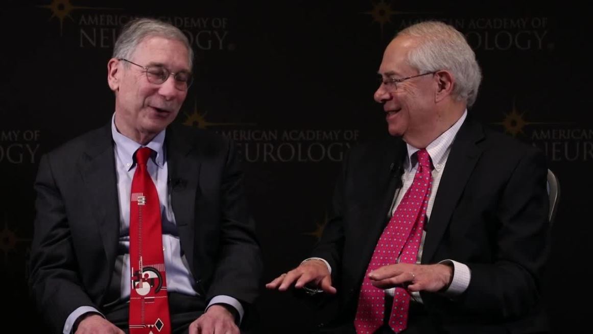
PHILADELPHIA – At the annual meeting of the American Academy of Neurology, Alan M. Rapoport, MD, clinical professor of neurology at the University of California, Los Angeles, sat down with Stewart J. Tepper, MD, professor of neurology at the Geisel School of Medicine at Dartmouth, Hanover, N.H., to discuss in a video some of the new data presented at the meeting regarding the CGRP monoclonal antibodies, the small molecule receptor antagonists (gepants), and what Dr. Tepper described as “a real shift in paradigm and a watershed moment in migraine.”
The three gepants that are farthest along in clinical trials are ubrogepant, rimegepant, and atogepant. “Reassuring data” was presented, Dr. Tepper said, regarding liver toxicity, which has been a concern with earlier generations of the gepants. The Food and Drug Administration had mandated a close look at liver function with the use of these drugs, which are metabolized in the liver, and, to date, no safety signals have emerged.
The three CGRP monoclonal antibodies that are currently on the market are erenumab (Aimovig), fremanezumab (Ajovy), and galcanezumab (Emgality). Data from numerous open-label extension studies were presented. In general, it seems that “the monoclonal antibodies accumulate greater efficacy over time,” Dr. Tepper said. No safety concerns have emerged from 5 years of clinical trial data. With 250,000 patients on these drugs worldwide, that is “very reassuring,” Dr. Tepper said.
New data also show that the majority of patients with chronic migraine who are taking monoclonal antibodies convert from chronic migraine to episodic migraine. Additionally, new data show that use of monoclonal antibodies “dramatically reduce all migraine medication use,” Dr. Tepper said.
EXPERT ANALYSIS FROM AAN 2019
Patients describe significant impact of epilepsy on their lives
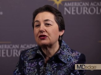
PHILADELPHIA – said Jacqueline French, MD, a professor at the Comprehensive Epilepsy Center at New York University.
“This underscores the need to consider these experiences, and potentially the stage of disease, when developing patient-reported outcome measures,” she said at the annual meeting of the American Academy of Neurology.
To describe the patient’s experience of living with epilepsy, including the occurrence of disease-related signs and symptoms and impact on daily life at different disease stages, Dr. French conducted qualitative, semistructured interviews with adults with focal epilepsy at the following stages: early (1 year or less since diagnosis), middle (1-5 years since diagnosis), and late (more than 5 years since diagnosis). The patients had varying seizure frequency and treatment experiences. They were asked to describe the symptoms and functional impact they had experienced related to epilepsy, and then to rate the degree to which each symptom and impact “bothered” them, using a disturbance rating scale from 0 (not at all) to 10 (extremely).
A total of 62 patients who were aged 18-60 years (mean age, 37 years; 73% female) were interviewed. In all, 19 of the patients had early-stage disease, 17 had middle-stage, and 26 had late-stage disease. Symptoms reported with the highest frequency and highest average disturbance (AD) ratings across all cohorts included twitching/tremors (80% of patients; AD, 5.3), confusion (78%; AD, 7.8), difficulty talking (75%; AD, 8.1), impaired/loss of consciousness (70%; AD, 6.8), stiffening (65%; AD, 5.4), déjà vu (62%; AD, 5.1), difficulty remembering (60%; AD, 8.5), and dizziness/light-headedness (58%; AD, 6.4).
The high-frequency/high-disturbance daily impact of epilepsy included the inability to drive (74%; AD, 7.1), limited ability to work and/or go to school (61%; AD, 6.7), limitations on leisure and social activities (58%; AD, 6.3), and memory loss (47%; AD, 8.4).
Dr French noted that, although disease experiences were similar among the cohorts, some heterogeneity across patient subgroups was observed.
Eisai sponsored the study.
REPORTING FROM AAN 2019
Key clinical point: Adults with focal epilepsy report a range of high-disturbance symptoms and impacts on daily life.
Major finding: The high-frequency/high-disturbance daily impact of epilepsy included the inability to drive (reported by 74% of respondents), limited ability to work and/or go to school (61%), limitations on leisure and social activities (58%), and memory loss (47%).
Study details: Qualitative, semistructured interviews with 62 adults with focal epilepsy at different stages of illness: early, middle, and late.
Disclosures: Eisai sponsored the study.
Physical activity slows cognitive decline in patients with Parkinson’s disease
PHILADELPHIA – according to Sneha Mantri, MD, of Duke University in Durham, N.C., and colleagues, who presented the results of their study at the annual meeting of the American Academy of Neurology.
Physical activity is an important component of the management of Parkinson’s disease and is shown to mitigate cognitive decline among patients with moderate disease, said Dr. Mantri and colleagues. “Exercise levels in de novo and early disease may influence risk of future cognitive decline; early disease also presents an opportunity for early intervention and possible disease modification,” Dr. Mantri said.
Physical activity levels in early disease are known to be low, but the effects of activity on cognition are currently unclear. To assess the relationship between physical activity and cognition, Dr. Mantri and colleagues examined patients with Parkinson’s disease who were enrolled in the prospective Parkinson’s Progression Markers Initiative (PPMI) cohort. At annual study visits, participants completed the Physical Activity Scale for the Elderly (PASE), a validated self-reported questionnaire assessing household, leisure, and work activities over the previous 7 days. The researchers used a linear mixed-effects model to compare rates of change in the Montreal Cognitive Assessment (MoCA) according to PASE scores; covariates included age, sex, Unified Parkinson’s Disease Rating Scale (UPDRS) part III score, and baseline MoCA.
A total of 379 patients completed at least one PASE questionnaire over the course of the study. PASE scores in this cohort have been previously described (Mantri S et al. J Park Dis. 2018;8[1]:107-11). Although overall rates of cognitive decline are known to be modest in this early cohort, PASE over time has a significant effect on MoCA during follow-up (P = 0.02) which suggest that higher levels of activity throughout disease are associated with better cognitive performance.
Dr. Mantri had nothing to disclose. Among her coauthors, Dr. Tropea received personal compensation from Genzyme and Medtronics and research support from Sanofi. Dr. Morley had nothing to disclose.
SOURCE: Mantri S et al. AAN 2019, Abstract P2.8-021.
REPORTING FROM AAN 2019
Key clinical point: Physical activity is associated with slower cognitive decline in patients with de novo Parkinson’s disease.
Major finding: Higher scores on the Physical Activity Scale for the Elderly over time had a significant effect on cognitive function.
Study details: A prospective study of 379 patients enrolled in the Parkinson’s Progression Markers Initiative.
Disclosures: Dr. Mantri had no relevant financial disclosures. Among her coauthors, Dr. Tropea received personal compensation from Genzyme and Medtronics and research support from Sanofi. Dr. Morley had nothing to disclose.
Source: Mantri S et al. AAN 2019, Abstract P2.8-021.

Brain volumes after TBI correlate with clinical features
PHILADELPHIA – ” according to a study presented at the annual meeting of the American Academy of Neurology.
Traumatic brain injury (TBI) damages brain tissue and causes subsequent volume loss, which may result in clinical symptoms. It is a prevalent worldwide health problem caused by a mechanical insult to the head, resulting in transient or permanent alteration to brain tissue and/or function. Standard neuroimaging with computerized cranial tomography (CT) and structural magnetic resonance imaging (MRI) is often unrevealing during the evaluation of patients with TBI, particularly those classified as mild TBI. I
In this study, James Rock, MD, of Penn Presbyterian Medical Center and the University of Pennsylvania, and his colleagues sought to examine the value of quantitative analysis of regional brain volumes in the evaluation of TBI. The investigators reviewed the medical records and MRI imaging from 44 patients with TBI evaluated at a Level I trauma center. They also read clinical notes to assess reported symptoms and physical findings.
Regional volumes from TBI subjects were derived using the software package Freesurfer image analysis suite, which utilizes a T1-weighted structural scan to calculate volumetric information. A machine learning algorithm, random forests, was employed across volume measurements from 25 regions of interest to determine the most important regions for classifying subjects based on clinical outcome and symptomology.
Basal ganglia volume showed the highest variable importance with regards to classifying subjects who exhibited symptoms of cognitive dysfunction (Mean Decrease in Gini = 1.067, Mean Decrease in Accuracy = 5.966e-03) in quantitative analysis. Left lateral ventricle volume was important in classifying subjects with motor and vestibular alterations (Mean Decrease in Gini = 2.037, Mean Decrease in Accuracy = 2.92e-02). Left choroid plexus volume was the most important region for classifying subjects with sensation and somatic dysfunction (Mean Decrease in Gini = 0.271, Mean Decrease in Accuracy = 4.82e-03).
The researchers noted that their study is ongoing, in an abstract. “It will be extended to a larger cohort to determine whether volume changes in specific [regions of interest] can act as useful clinical biomarkers for chronic symptoms,” they said.
Dr. Diaz-Arrastia received personal compensation from Neural Analytics, Inc, BrainBox Solutions, Inc, and Bioscience Pharma Partners. Dr. Diaz-Arrastia holds stock and/or stock options in Neural Analytics, Inc. and has received research support from BrainBox Solutions. The other authors reported not having anything to disclose..
SOURCE: Rock J et al. AAN 2019. Abstract S2.006 .
REPORTING FROM AAN 2019
Fremanezumab cut headache days in migraine patients vs. placebo
PHILADELPHIA – , according to a poster presented at the annual meeting of the American Academy of Neurology.

To assess the efficacy of fremanezumab in patients with migraine who had not received relief from trying at least one prior preventive migraine medication, Peter McAllister, MD and colleagues analyzed data from 2 phase 3 trials (HALO EM and HALO CM). Trial participants had either episodic or chronic migraine, confirmed during a 28-day pretreatment baseline period, then received subcutaneous fremanezumab quarterly (675 mg at baseline and placebo at weeks 4 and 8), monthly (for chronic migraine: 675 mg at baseline and 225 mg at weeks 4 and 8; for episodic migraine: 225 mg at baseline and weeks 4 and 8), or placebo (at baseline and weeks 4 and 8).
The present analysis included data from 186 patients with episodic migraine and 407 patients with chronic migraine, which represents the subgroup of study participants in the larger HALO trials who had failed at least one prior preventive migraine medication. Dr. McAllister, who is cofounder and chief medical officer at the New England Institute for Clinical Research in Stamford, Connecticut, and his colleagues, assessed mean changes from baseline in the monthly average number of headache days of at least moderate severity or the monthly average number of migraine days during the 12-week treatment period.
In patients with chronic migraine, fremanezumab yielded greater reductions in the number of headache days of at least moderate severity (quarterly [least-squares mean change]: –4.0, P less than 0.0001; monthly: –4.5, P less than 0.0001) compared with placebo (–1.8). There were similar reductions in the number of migraine days (quarterly: –4.1, P = 0.0027; monthly: –4.8, P less than 0.0001) compared with placebo (–2.3).
In patients with episodic migraine, fremanezumab yielded greater reductions in the number of headache days of at least moderate severity (quarterly: –3.1, P less than 0.0001; monthly: –3.2, P less than 0.0001) compared with placebo (–0.8). There were similar reductions in the number of migraine days (quarterly: –3.3, P = 0.0015; monthly: –3.7, P less than 0.0001) compared with placebo (–1.3).
“The phase 3 HALO CM and HALO EM trials showed that fremanezumab is efficacious in patients who failed one or more prior preventive medication, a potentially difficult-to-treat population,” Dr. McAllister and colleagues said in their poster.
“Effect sizes in this subgroup were greater than those in the overall trial population,” they said. In addition, “both quarterly and monthly fremanezumab were well-tolerated in this subgroup.”
This study was funded by Teva Pharmaceuticals, Petach Tikva, Israel.
SOURCE: McAllister P et al. AAN 2019. P1.10-011.
PHILADELPHIA – , according to a poster presented at the annual meeting of the American Academy of Neurology.
To assess the efficacy of fremanezumab in patients with migraine who had not received relief from trying at least one prior preventive migraine medication, Peter McAllister, MD and colleagues analyzed data from 2 phase 3 trials (HALO EM and HALO CM). Trial participants had either episodic or chronic migraine, confirmed during a 28-day pretreatment baseline period, then received subcutaneous fremanezumab quarterly (675 mg at baseline and placebo at weeks 4 and 8), monthly (for chronic migraine: 675 mg at baseline and 225 mg at weeks 4 and 8; for episodic migraine: 225 mg at baseline and weeks 4 and 8), or placebo (at baseline and weeks 4 and 8).
The present analysis included data from 186 patients with episodic migraine and 407 patients with chronic migraine, which represents the subgroup of study participants in the larger HALO trials who had failed at least one prior preventive migraine medication. Dr. McAllister, who is cofounder and chief medical officer at the New England Institute for Clinical Research in Stamford, Connecticut, and his colleagues, assessed mean changes from baseline in the monthly average number of headache days of at least moderate severity or the monthly average number of migraine days during the 12-week treatment period.
In patients with chronic migraine, fremanezumab yielded greater reductions in the number of headache days of at least moderate severity (quarterly [least-squares mean change]: –4.0, P less than 0.0001; monthly: –4.5, P less than 0.0001) compared with placebo (–1.8). There were similar reductions in the number of migraine days (quarterly: –4.1, P = 0.0027; monthly: –4.8, P less than 0.0001) compared with placebo (–2.3).
In patients with episodic migraine, fremanezumab yielded greater reductions in the number of headache days of at least moderate severity (quarterly: –3.1, P less than 0.0001; monthly: –3.2, P less than 0.0001) compared with placebo (–0.8). There were similar reductions in the number of migraine days (quarterly: –3.3, P = 0.0015; monthly: –3.7, P less than 0.0001) compared with placebo (–1.3).
“The phase 3 HALO CM and HALO EM trials showed that fremanezumab is efficacious in patients who failed one or more prior preventive medication, a potentially difficult-to-treat population,” Dr. McAllister and colleagues said in their poster.
“Effect sizes in this subgroup were greater than those in the overall trial population,” they said. In addition, “both quarterly and monthly fremanezumab were well-tolerated in this subgroup.”
This study was funded by Teva Pharmaceuticals, Petach Tikva, Israel.
SOURCE: McAllister P et al. AAN 2019. P1.10-011.
REPORTING FROM AAN 2019
Key clinical point: Fremanezumab reduced headache days in patients with chronic or episodic migraine.
Major finding: In patients with chronic migraine, fremanezumab reduced the number of headache days (least-squares mean change = -4.0) compared with placebo (-1.8).
Study details: Subgroup analysis of data from two phase 3 studies - HALO EM and HALO CM - including 186 patients with episodic migraine and 407 patients with chronic migraine.
Disclosures: This study was funded by Teva Pharmaceuticals, Petach Tikva, Israel.
Source: McAllister P et al. AAN 2019. P1.10-011.