User login
Drafts of new classification criteria presented for GCA, Takayasu’s arteritis
CHICAGO – Drafts of new classification criteria for giant cell arteritis and Takayasu’s arteritis developed by the American College of Rheumatology and the European League Against Rheumatism (EULAR) reflect the increasingly important role of advanced vascular imaging in the diagnosis and management of large-vessel vasculitis, according to Peter A. Merkel, MD.
The drafts, which are the result of a multiyear collaboration between the ACR and EULAR, were presented at the annual meeting of the ACR and will be submitted to the ACR/EULAR committee overseeing the work for comprehensive review and possible revisions. Once endorsed, the new criteria will replace the “extremely important,” but outdated, existing classification criteria, which were published in 1990.
“What we’ve done is, rather than purely revise the 1990 [criteria], we’ve started again from scratch ... with a great number of cases from a wide variety of centers throughout the world. This was a very large international effort ... really a great community effort in the field of rheumatology,” Dr. Merkel, professor and chief of the division of rheumatology at the University of Pennsylvania, Philadelphia, and one of the chief investigators for the project, said in a video interview.
The new criteria will allow for better classification of patients with giant cell arteritis versus Takayasu’s arteritis versus another form of vasculitis, he said, noting that advances in imaging that allow for “more enriched data with which to make decisions” play a large role.
However, the new criteria are not meant to be used for diagnosis, but to “sort out among the different types of vasculitis,” he said.
“It provides awareness and it provides a tool, especially for research investigation, but that seeps out into the broader community,” he added.
Dr. Merkel reported having no disclosures.
SOURCE: Merkel PA et al. ACR Annual Meeting, Presentation 5T116.
REPORTING FROM THE ACR ANNUAL MEETING
Bills affecting rheumatologists make way to Capitol Hill
CHICAGO – Rheumatology advocates have been working to get a variety of bills passed in Congress, including efforts on creating exemptions to step therapy or “fail first” insurer policies, increasing osteoporosis screening payment, and raising research dollars for arthritis in the Department of Defense, Dr. Angus Worthing said at the annual meeting of the American College of Rheumatology.

Visits by rheumatologist advocates to receptive members of the House and Senate have been fruitful for these issues, said Dr. Worthing, who is chair of the ACR’s Government Affairs Committee and a practicing rheumatologist in the Washington area.
“There’s a bill in the House [H.R. 2077, Restoring the Patient’s Voice Act], reforming step therapy that would institute appropriate exemptions if a doctor can predict that a drug that’s the first step in a step therapy will cause side effects, will be ineffective, or has already been used by a patient, as well as other appropriate guardrails,” so that the policy can be circumvented and the patient can get the prescription that the doctor ordered, he said.
The ACR is working with coalition partners such as the National Psoriasis Foundation and other patient organizations to get a companion bill to H.R. 2077 introduced in the Senate, Dr. Worthing noted.
The ACR is also working to advance separate bills in the House (H.R. 1898) and Senate (S. 3160) to increase payment levels for dual x-ray absorptiometry scans, which were cut by 60%-70% about 10 years ago.
The two bills have broad bipartisan support in both the House and Senate. “Watch for that to hopefully become law,” Dr. Worthing said.
The ACR has been advocating for the last 4 years to increase research dollars specifically for arthritis and rheumatology at the Department of Defense. After years of trying, rheumatologist advocates were able to get funding for studying trauma-related osteoarthritis and military serology data banks added as an amendment in the most recent appropriations bill. However, it was pulled at the last minute because of a lack of support.
“But with that advancement we’ll be able to go into the next Congress in January and either get it into the bill or hopefully have the amendment voted on next year,” he said.
Dr. Worthing called for those interested in advocating for rheumatology to visit the ACR’s Legislative Action Center.
CHICAGO – Rheumatology advocates have been working to get a variety of bills passed in Congress, including efforts on creating exemptions to step therapy or “fail first” insurer policies, increasing osteoporosis screening payment, and raising research dollars for arthritis in the Department of Defense, Dr. Angus Worthing said at the annual meeting of the American College of Rheumatology.
Visits by rheumatologist advocates to receptive members of the House and Senate have been fruitful for these issues, said Dr. Worthing, who is chair of the ACR’s Government Affairs Committee and a practicing rheumatologist in the Washington area.
“There’s a bill in the House [H.R. 2077, Restoring the Patient’s Voice Act], reforming step therapy that would institute appropriate exemptions if a doctor can predict that a drug that’s the first step in a step therapy will cause side effects, will be ineffective, or has already been used by a patient, as well as other appropriate guardrails,” so that the policy can be circumvented and the patient can get the prescription that the doctor ordered, he said.
The ACR is working with coalition partners such as the National Psoriasis Foundation and other patient organizations to get a companion bill to H.R. 2077 introduced in the Senate, Dr. Worthing noted.
The ACR is also working to advance separate bills in the House (H.R. 1898) and Senate (S. 3160) to increase payment levels for dual x-ray absorptiometry scans, which were cut by 60%-70% about 10 years ago.
The two bills have broad bipartisan support in both the House and Senate. “Watch for that to hopefully become law,” Dr. Worthing said.
The ACR has been advocating for the last 4 years to increase research dollars specifically for arthritis and rheumatology at the Department of Defense. After years of trying, rheumatologist advocates were able to get funding for studying trauma-related osteoarthritis and military serology data banks added as an amendment in the most recent appropriations bill. However, it was pulled at the last minute because of a lack of support.
“But with that advancement we’ll be able to go into the next Congress in January and either get it into the bill or hopefully have the amendment voted on next year,” he said.
Dr. Worthing called for those interested in advocating for rheumatology to visit the ACR’s Legislative Action Center.
REPORTING FROM THE ACR ANNUAL MEETING
SLE low-disease definition receives prospective validation
CHICAGO – The long path toward a validated definition of low disease activity in patients with systemic lupus erythematosus may be nearing an end as a definition first proposed more than 5 years ago received validation with data from more than 1,700 patients in a prospective, multicenter study.

The next step is to test this definition in treat-to-target intervention studies, and to apply the definition in other clinical trials as well as in routine clinical practice, Vera Golder, MD, said at the annual meeting of the American College of Rheumatology.
The tested definition has five elements that a patient needs to achieve to be considered in a lupus low disease activity state (LLDAS):
• A systemic lupus erythematosus (SLE) disease activity index (SLEDAI) score of 4 or less with no major organ involvement.
• No new disease activity.
• A physician’s global assessment of the patient of 1 or less on a 0-3 scale.
• Maintenance on a prednisolone dosage of 7.5 mg/day or less.
• Maintenance on a standard immunosuppressive regimen.
The Asia Pacific Lupus Collaboration (APLC) first proposed this definition of LLDAS in 2013 (Ann Rheum Dis. 2013 June;72[Suppl 3]:THU0298), and was the organization behind the latest test of its validity. The APLC based its LLDAS definition on recommendations made by an international working party (Ann Rheum Dis. 2014 June;73[6]:958-67).
The APLC prospectively collected data from 1,735 SLE patients at 13 centers in eight countries during May 2013–December 2016, with a median follow-up of 2.2 years. During that time, 78% of the patients achieved the LLDAS at least once. Two-thirds of the patients had at least one sustained period of LLDAS of at least 3 months, and overall the enrolled patients spent 69% of the time in the LLDAS, reported Dr. Golder, a rheumatologist at Monash University in Melbourne.
The validation analysis she described focused on examining the correlation between the amount of time that patients spent in the defined LLDAS and their subsequent clinical outcomes. The analysis showed that when patients were in the LLDAS their rate of subsequent flare or damage accrual was substantially reduced.
Patients in the LLDAS for at least half the time had a 51% reduced rate of subsequent flare and a 47% reduced rate of subsequent damage accrual, she reported. Patients with a LLDAS of 3-6 months had a 57% reduced rate of damage accrual. As time spent in continuous LLDAS continued to increase the rate of subsequent damage accrual continued to drop until the duration reached more than 9 months, at which point the rate of subsequent damage fell to nearly 90% lower than that of patients without this amount of sustained LLDAS. Patients with LLDAS sustained for more than 9 months and as long as 12 months had an 86% reduction in subsequent damage accrual. Periods of sustained LLDAS that extended longer than a year continued to maintain a nearly 90% reduced rate of damage accrual, Dr. Golder said.
The Asia Pacific Lupus Collaboration has received grants from AstraZeneca, Bristol-Myers Squibb, GlaxoSmithKline, Janssen, and UCB. Dr. Golder had no disclosures.
SOURCE: Golder V et al. Arthritis Rheumatol. 2018;70(Suppl 10): Abstract 2786.
Having a validated, formal definition of low disease activity in patients with systemic lupus erythematosus will be very helpful to clinicians and patients with this disease. The lack of such a widely accepted definition of a treatment target until now has been a significant issue that had made it more challenging to advise and treat patients.
The concept of a low disease state has been much easier to define in other rheumatic diseases, but lupus has posed a major challenge because of its very heterogeneous presentation. This heterogeneity has led to the creation of several measures of disease activity as well as multiple serologic parameters that also help define disease activity. It’s unrealistic to expect most lupus patients to be in a low disease activity state all the time.
Validation of a reasonable definition of low activity is a great, pragmatic step forward for our field that will help clinicians better care for their patients with systemic lupus erythematosus.
Lisa R. Sammaritano, MD , is a rheumatologist at the Hospital for Special Surgery and Cornell University in New York. She had no disclosures. She made these comments in an interview.
CHICAGO – The long path toward a validated definition of low disease activity in patients with systemic lupus erythematosus may be nearing an end as a definition first proposed more than 5 years ago received validation with data from more than 1,700 patients in a prospective, multicenter study.
The next step is to test this definition in treat-to-target intervention studies, and to apply the definition in other clinical trials as well as in routine clinical practice, Vera Golder, MD, said at the annual meeting of the American College of Rheumatology.
The tested definition has five elements that a patient needs to achieve to be considered in a lupus low disease activity state (LLDAS):
• A systemic lupus erythematosus (SLE) disease activity index (SLEDAI) score of 4 or less with no major organ involvement.
• No new disease activity.
• A physician’s global assessment of the patient of 1 or less on a 0-3 scale.
• Maintenance on a prednisolone dosage of 7.5 mg/day or less.
• Maintenance on a standard immunosuppressive regimen.
The Asia Pacific Lupus Collaboration (APLC) first proposed this definition of LLDAS in 2013 (Ann Rheum Dis. 2013 June;72[Suppl 3]:THU0298), and was the organization behind the latest test of its validity. The APLC based its LLDAS definition on recommendations made by an international working party (Ann Rheum Dis. 2014 June;73[6]:958-67).
The APLC prospectively collected data from 1,735 SLE patients at 13 centers in eight countries during May 2013–December 2016, with a median follow-up of 2.2 years. During that time, 78% of the patients achieved the LLDAS at least once. Two-thirds of the patients had at least one sustained period of LLDAS of at least 3 months, and overall the enrolled patients spent 69% of the time in the LLDAS, reported Dr. Golder, a rheumatologist at Monash University in Melbourne.
The validation analysis she described focused on examining the correlation between the amount of time that patients spent in the defined LLDAS and their subsequent clinical outcomes. The analysis showed that when patients were in the LLDAS their rate of subsequent flare or damage accrual was substantially reduced.
Patients in the LLDAS for at least half the time had a 51% reduced rate of subsequent flare and a 47% reduced rate of subsequent damage accrual, she reported. Patients with a LLDAS of 3-6 months had a 57% reduced rate of damage accrual. As time spent in continuous LLDAS continued to increase the rate of subsequent damage accrual continued to drop until the duration reached more than 9 months, at which point the rate of subsequent damage fell to nearly 90% lower than that of patients without this amount of sustained LLDAS. Patients with LLDAS sustained for more than 9 months and as long as 12 months had an 86% reduction in subsequent damage accrual. Periods of sustained LLDAS that extended longer than a year continued to maintain a nearly 90% reduced rate of damage accrual, Dr. Golder said.
The Asia Pacific Lupus Collaboration has received grants from AstraZeneca, Bristol-Myers Squibb, GlaxoSmithKline, Janssen, and UCB. Dr. Golder had no disclosures.
SOURCE: Golder V et al. Arthritis Rheumatol. 2018;70(Suppl 10): Abstract 2786.
REPORTING FROM THE ACR ANNUAL MEETING
Key clinical point: A proposed definition of low disease activity in systemic lupus erythematosus received prospective validation.
Major finding: Patients meeting the definition for 9-12 months had an 86% reduced rate of subsequent damage accrual.
Study details: A prospective, multicenter study of 1,735 patients.
Disclosures: The Asia Pacific Lupus Collaboration has received grants from AstraZeneca, Bristol-Myers Squibb, GlaxoSmithKline, Janssen, and UCB. Dr. Golder had no disclosures.
Source: Golder V et al. Arthritis Rheumatol. 2018;70(Suppl 10): Abstract 2786.
Case review: Posttransplant lupus nephritis recurrence rates declining
CHICAGO – Lupus nephritis recurrence rates in kidney transplant recipients declined over the past decade, compared with rates seen in earlier studies, according to a review of cases at the University of Tennessee Health Science Center (UTHSC).
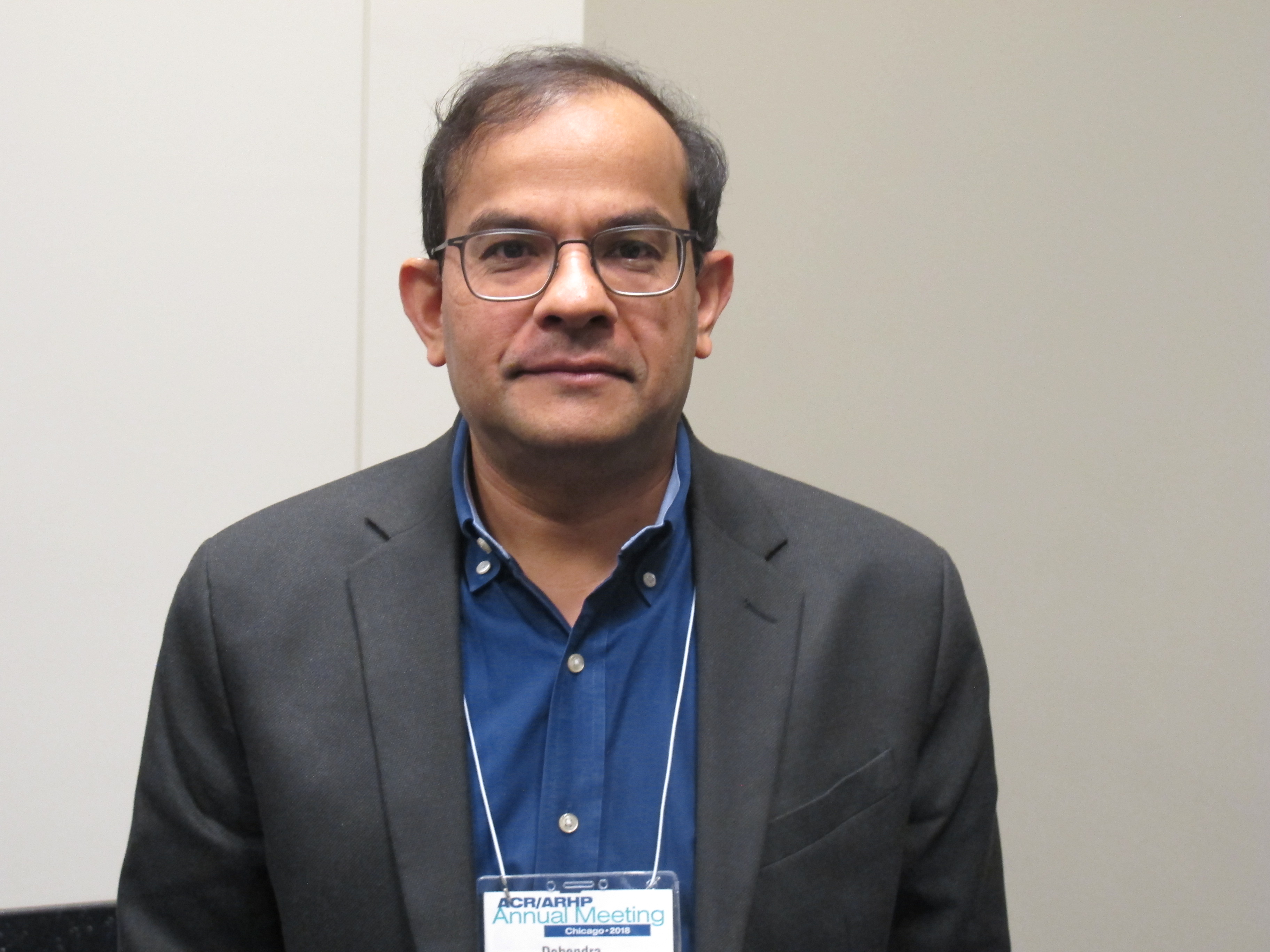
The findings are likely related to improvements in posttransplant immunosuppressive regimens, and may have implications for the timing of transplant going forward, Debendra N. Pattanaik, MD, said at the annual meeting of the American College of Rheumatology.
The biopsy-proven recurrence rate in 38 transplant recipients who received standard immunosuppression with prednisone, tacrolimus, and mycophenolate mofetil was 11%, and graft loss or death occurred in 26% at a median follow-up of 1,230 days, said Dr. Pattanaik, a rheumatologist at UTHSC, Memphis.
Patients with recurrence showed a trend for increased risk for graft loss or death, compared with recipients without recurrence (hazard ratio = 3.14), he noted during a press briefing at the meeting.
Lupus nephritis is a severe complication occurring in more than half of all patients with systemic lupus erythematosus (SLE), and despite a great deal of progress over the years, 10%-30% develop end-stage renal disease and require dialysis and/or transplant, he said, noting that studies have shown that transplant recipients do better over time than do those who remain on dialysis.
“So renal transplant is an important modality of treatment for end-stage renal disease from lupus nephritis,” he added.
However, recurrence of lupus nephritis in the graft is a concern, he said.
In previous eras – prior to improvements in immunosuppressive regimens for transplant recipients – studies showed variable rates of lupus nephritis recurrence, with some reporting rates up to 50% depending on the patient populations and protocols, he noted.
The rates in recent years at UTHSC seemed lower than that, so he and his colleagues looked more closely at the outcomes.
Case patients included all those with end-stage renal disease secondary to lupus nephritis who were transplanted between 2006 and 2017 at the center. Medical records of all 38 were reviewed along with information from the United Network for Organ Sharing Network. The mean age of the patients at baseline was 42 years, 89% were women, 89% were African American, and previous time on dialysis was a median of 4 years. Most (80%) received hemodialysis, and nearly one-third (31%) received living donor transplantation, Dr. Pattanaik said.
The main difference in the past decade compared with those previous eras is the use of posttransplant immunosuppressive regimens consisting of tacrolimus and mycophenolate mofetil rather than cyclosporine and azathioprine in addition to prednisone, he explained.
Previous reports showing higher recurrence rates were from studies in which patients received cyclosporine and azathioprine as part of the posttransplant regimen, he said.
“Our next question is whether patients can be transplanted early,” he said, explaining that transplant is often delayed for many months or years until SLE is in remission, but if the new regimens are reducing recurrence risk, early transplant may be feasible.
Dr. Pattanaik reported having no disclosures.
SOURCE: Pattanaik D et al. Arthritis Rheumatol. 2018;70(Suppl 10): Abstract 711.
CHICAGO – Lupus nephritis recurrence rates in kidney transplant recipients declined over the past decade, compared with rates seen in earlier studies, according to a review of cases at the University of Tennessee Health Science Center (UTHSC).
The findings are likely related to improvements in posttransplant immunosuppressive regimens, and may have implications for the timing of transplant going forward, Debendra N. Pattanaik, MD, said at the annual meeting of the American College of Rheumatology.
The biopsy-proven recurrence rate in 38 transplant recipients who received standard immunosuppression with prednisone, tacrolimus, and mycophenolate mofetil was 11%, and graft loss or death occurred in 26% at a median follow-up of 1,230 days, said Dr. Pattanaik, a rheumatologist at UTHSC, Memphis.
Patients with recurrence showed a trend for increased risk for graft loss or death, compared with recipients without recurrence (hazard ratio = 3.14), he noted during a press briefing at the meeting.
Lupus nephritis is a severe complication occurring in more than half of all patients with systemic lupus erythematosus (SLE), and despite a great deal of progress over the years, 10%-30% develop end-stage renal disease and require dialysis and/or transplant, he said, noting that studies have shown that transplant recipients do better over time than do those who remain on dialysis.
“So renal transplant is an important modality of treatment for end-stage renal disease from lupus nephritis,” he added.
However, recurrence of lupus nephritis in the graft is a concern, he said.
In previous eras – prior to improvements in immunosuppressive regimens for transplant recipients – studies showed variable rates of lupus nephritis recurrence, with some reporting rates up to 50% depending on the patient populations and protocols, he noted.
The rates in recent years at UTHSC seemed lower than that, so he and his colleagues looked more closely at the outcomes.
Case patients included all those with end-stage renal disease secondary to lupus nephritis who were transplanted between 2006 and 2017 at the center. Medical records of all 38 were reviewed along with information from the United Network for Organ Sharing Network. The mean age of the patients at baseline was 42 years, 89% were women, 89% were African American, and previous time on dialysis was a median of 4 years. Most (80%) received hemodialysis, and nearly one-third (31%) received living donor transplantation, Dr. Pattanaik said.
The main difference in the past decade compared with those previous eras is the use of posttransplant immunosuppressive regimens consisting of tacrolimus and mycophenolate mofetil rather than cyclosporine and azathioprine in addition to prednisone, he explained.
Previous reports showing higher recurrence rates were from studies in which patients received cyclosporine and azathioprine as part of the posttransplant regimen, he said.
“Our next question is whether patients can be transplanted early,” he said, explaining that transplant is often delayed for many months or years until SLE is in remission, but if the new regimens are reducing recurrence risk, early transplant may be feasible.
Dr. Pattanaik reported having no disclosures.
SOURCE: Pattanaik D et al. Arthritis Rheumatol. 2018;70(Suppl 10): Abstract 711.
REPORTING FROM THE ACR ANNUAL MEETING
Key clinical point:
Major finding: The biopsy-proven recurrence rate was 11%.
Study details: A review of 38 cases at one center.
Disclosures: Dr. Pattanaik reported having no disclosures.
Source: Pattanaik D et al. Arthritis Rheumatol. 2018;70(Suppl 10): Abstract 711.
Ablation plus transplant for severe scleroderma shows 11-year benefits
CHICAGO – Follow-up out to as long as 11 years from treatment confirmed the long-term efficacy and safety of myeloablative autologous stem cell transplantation for patients with severe scleroderma.
This extended follow-up comprised 43 survivors from the 75 patients originally randomized in a controlled, 6-year trial. Follow-up showed that, among the patients who underwent myeloablation and autologous transplant with hematopoietic stem cells, there were no long-term deaths or cancers, there was an 88% survival rate, and 92% remained off disease-modifying treatment, Keith M. Sullivan, MD, said at the annual meeting of the American College of Rheumatology.
Long-term survival among patients randomized to the study’s control arm, who received treatment with cyclophosphamide, was 53%.
Patients with severe scleroderma with significant internal organ damage who “are improved and off of disease-modifying antirheumatic drugs after 10 or more years from treatment is something new in autoimmune disease,” said Dr. Sullivan, a professor of medicine at Duke University, Durham, N.C.
Based on accumulated data from this randomized trial and other studies, the American Society for Blood and Marrow Transplantation issued a position statement in June 2018 that endorsed autologous hematopoietic stem cell transplantation as “standard of care” for systemic sclerosis (Biol Blood Marrow Transplant. 2018 June 25. doi: 10.1016/j.bbmt.2018.06.025), Dr. Sullivan noted in a video interview.
The SCOT (Scleroderma: Cyclophosphamide or Transplantation) trial randomized 75 patients with severe scleroderma and substantial internal organ involvement to receive treatment with either cyclophosphamide or myeloablative radiation followed by immune reconstitution with an autologous hematopoietic stem cell transplant. The trial’s primary endpoint, the global rank composite score at 54 months, showed the superiority of transplantation over standard treatment (N Engl J Med. 2018 Jan 4;378[1]:35-47).
Dr. Sullivan and his associates ran their long-term follow-up study on 43 of these 75 patients (25 from the transplanted group and 18 controls), excluding 21 patients who died during the original study, 4 additional patients from the control arm who died following the end of the original SCOT protocol, and 7 patients either lost to follow-up or who refused to participate in follow-up. Among the 25 transplanted patients, none died during the extended follow-up, 2 experienced cardiac failure, and 23 remained off of any disease-modifying antirheumatic drugs. Among the 18 survivors in the control arm, 3 had cardiac failure, 3 had respiratory failure, and 7 were on treatment with disease-modifying drugs, Dr. Sullivan reported.
In addition, 23 of the 25 (92%) transplanted patients had normal performance status by the Eastern Cooperative Oncology Group criteria, compared with 11 of the 18 controls (61%). A total of 14 (56%) transplant patients were employed, compared with 6 of the 18 controls (33%).
Patients who were transplanted “have their life back, are doing well, and are off treatment,” Dr. Sullivan noted.
Myeloablation and transplant is appropriate for scleroderma patients with significant internal organ involvement, about half of all patients with this disease. The best gauge of severe organ involvement is a pulmonary function test, with a forced vital capacity of 70% or less of predicted as a flag for patients who should consider transplantation, Dr. Sullivan said. He recommended monitoring lung function every 3 months in scleroderma patients because it can deteriorate very suddenly and quickly.
SCOT received no commercial funding. Dr. Sullivan had no disclosures to report.
SOURCE: Sullivan KM et al. ACR Annual Meeting, Abstract 1820.
REPORTING FROM THE ACR ANNUAL MEETING
Key clinical point:
Major finding: Survival after 11 years was 88% among transplanted patients and 53% among control patients treated with cyclophosphamide.
Study details: A long-term follow-up of 43 of the 75 patients enrolled in the SCOT trial.
Disclosures: SCOT received no commercial funding. Dr. Sullivan had no disclosures to report.
Source: Sullivan KM et al. ACR Annual Meeting, Abstract 1820.
Genetic risk factor found for RA-associated interstitial lung disease
CHICAGO – Rheumatoid arthritis–associated interstitial lung disease and idiopathic pulmonary fibrosis without RA share a common genetic underpinning whose hallmark is a gain-of-function MUC5B gene promoter variant that cranks up mucin production in the lungs, Pierre-Antoine Juge, MD, reported at the annual meeting of the American College of Rheumatology.

He presented a seven-country genetic case-control study of 620 patients with RA-associated interstitial lung disease (RA-ILD), 614 with RA but no ILD, and 5,448 unaffected controls. The key finding was that the MUC5B promoter variant rs35705950, already known to be the strongest genetic risk factor for idiopathic pulmonary fibrosis (IPF), also contributes substantially to the risk of RA-ILD.
Indeed, the presence of the MUC5B promoter variant in patients with RA proved to be associated with substantially higher risk of RA-ILD than the previously recognized risk factors for RA-ILD, including cigarette smoking and the human leukocyte antigen locus for RA, according to Dr. Juge, a rheumatologist at Bichat Hospital–Claude Bernard and Paris Diderot University.
MUC5B encodes for mucin production in the lungs. The increased risk of RA-ILD conferred by the presence of the MUC5B promoter variant was confined to the 41% of RA-ILD patients with a pattern of usual interstitial pneumonia (UIP) or possible UIP on high-resolution CT. The presence of the MUC5B promoter variant in RA patients was independently associated with an adjusted 6.1-fold increased risk of ILD with a UIP pattern on imaging – marked by honeycombing, reticular abnormalities, and subpleural involvement – compared with RA patients who didn’t possess the gain-of-function MUC5B variant. The risk of other types of RA-ILD wasn’t affected by the presence or absence of the MUC5B variant.
The MUC5B promoter variant was not a risk factor for development of RA.
These findings have potentially important implications for clinical practice, given that clinically significant ILD is present in about 10% of all RA patients and occult ILD is detectable using high-resolution CT in up to half of individuals with RA, Dr. Juge observed. Detection of the MUC5B promoter variant could be used to screen patients with RA for preclinical ILD. Also, there is now a sound rationale to study drugs known to be effective for IPF as potential treatments for RA-ILD, he said.
Dr. Juge reported having no financial conflicts regarding the study, which was sponsored by the National Institutes of Health, the U.S. Department of Defense, the French Rheumatology Society, the Japanese Society for the Promotion of Science, Fondation Arthritis, and the Nina Ireland Program for Lung Health.
In conjunction with his presentation in Chicago, the study was published online in the New England Journal of Medicine (doi: 10.1056/NEJMoa1801562).
SOURCE: Juge P-A et al. Arthritis Rheumatol. 2018;70(Suppl 10): Abstract 1819.
CHICAGO – Rheumatoid arthritis–associated interstitial lung disease and idiopathic pulmonary fibrosis without RA share a common genetic underpinning whose hallmark is a gain-of-function MUC5B gene promoter variant that cranks up mucin production in the lungs, Pierre-Antoine Juge, MD, reported at the annual meeting of the American College of Rheumatology.
He presented a seven-country genetic case-control study of 620 patients with RA-associated interstitial lung disease (RA-ILD), 614 with RA but no ILD, and 5,448 unaffected controls. The key finding was that the MUC5B promoter variant rs35705950, already known to be the strongest genetic risk factor for idiopathic pulmonary fibrosis (IPF), also contributes substantially to the risk of RA-ILD.
Indeed, the presence of the MUC5B promoter variant in patients with RA proved to be associated with substantially higher risk of RA-ILD than the previously recognized risk factors for RA-ILD, including cigarette smoking and the human leukocyte antigen locus for RA, according to Dr. Juge, a rheumatologist at Bichat Hospital–Claude Bernard and Paris Diderot University.
MUC5B encodes for mucin production in the lungs. The increased risk of RA-ILD conferred by the presence of the MUC5B promoter variant was confined to the 41% of RA-ILD patients with a pattern of usual interstitial pneumonia (UIP) or possible UIP on high-resolution CT. The presence of the MUC5B promoter variant in RA patients was independently associated with an adjusted 6.1-fold increased risk of ILD with a UIP pattern on imaging – marked by honeycombing, reticular abnormalities, and subpleural involvement – compared with RA patients who didn’t possess the gain-of-function MUC5B variant. The risk of other types of RA-ILD wasn’t affected by the presence or absence of the MUC5B variant.
The MUC5B promoter variant was not a risk factor for development of RA.
These findings have potentially important implications for clinical practice, given that clinically significant ILD is present in about 10% of all RA patients and occult ILD is detectable using high-resolution CT in up to half of individuals with RA, Dr. Juge observed. Detection of the MUC5B promoter variant could be used to screen patients with RA for preclinical ILD. Also, there is now a sound rationale to study drugs known to be effective for IPF as potential treatments for RA-ILD, he said.
Dr. Juge reported having no financial conflicts regarding the study, which was sponsored by the National Institutes of Health, the U.S. Department of Defense, the French Rheumatology Society, the Japanese Society for the Promotion of Science, Fondation Arthritis, and the Nina Ireland Program for Lung Health.
In conjunction with his presentation in Chicago, the study was published online in the New England Journal of Medicine (doi: 10.1056/NEJMoa1801562).
SOURCE: Juge P-A et al. Arthritis Rheumatol. 2018;70(Suppl 10): Abstract 1819.
REPORTING FROM THE ACR ANNUAL MEETING
High-dose flu vaccine in RA patients beats standard dose
CHICAGO – The administration of high-dose vs. standard-dose influenza vaccine provided substantially better immune responses in seropositive rheumatoid arthritis patients in a randomized, active-controlled trial.
High-dose trivalent influenza vaccine is known to improve immune responses in the elderly, but the current findings, which were presented at the annual meeting of the American College of Rheumatology, are the first to document a successful intervention to enhance vaccine responses in immunocompromised patients, according to Inés Colmegna, MD, of McGill University, Montreal.
Dr. Colmegna and her colleagues assessed antibody responses to either standard-dose (15 mcg of hemagglutinin per strain) quadrivalent inactivated influenza vaccine (SD-QIV) or high-dose (60 mcg of hemagglutinin per strain) trivalent inactivated influenza vaccine (HD-TIV) in 140 and 139 patients, respectively.
Seroprotection rates prior to vaccination were comparable in the two groups, but the high-dose recipients had consistently higher overall responses to vaccination.
Seroconversion rates were 22.3% vs. 8.6% (odds ratio, 2.93) for the H3N2 strain (A/HongKong/4801/2014), and 44.6% vs. 28.6% (OR, 1.93) for the B Victoria Lin strain (B/Brisbane/60/2008). For the H1N1 strain A/California/7/2009 in 2016-2017 and closely related A/Michigan/45/2015 in 2017-2018, the seroconversion rates were 51.1% vs. 30.0% (OR, 2.91) and 46.4% vs. 24.6% (OR, 2.79), respectively. Seroprotection rates for the H3N2 strain were 48.5% vs. 30.9%, and for the B Victoria Lin strain, 60.0% vs. 50.7%. The seroprotection rates for the H1N1 strains together were and 80.4% vs. 73.5%, Dr. Colmegna said.
Seroconversion was defined as at least a fourfold serum hemagglutination inhibition (HI) antibody increase from prevaccination level (day 0), and seroprotection was defined as percent with HI titers of 1:40 or greater at postvaccination day 28.
After the researchers controlled for age, vaccine type, treatment type in the 3 months prior to vaccination and during the study period, Charlson comorbidity index, and RA duration, the only significant predictors of vaccine seroresponse were vaccine dose and age.
The findings are notable because RA patients have a nearly threefold increase in the risk of contracting influenza infection or related illness, compared with age-matched healthy controls, because of “inherent immune dysfunction associated with RA, comorbidities, the age of our patients, and immunosuppressive therapy,” Dr. Colmegna said.
For this reason, RA patients are a priority group for annual vaccination. However, while vaccination remains the most effective method for preventing influenza and its associated complications, vaccine-induced antibody responses and protection in RA are suboptimal, she explained, noting that this puts them at increased risk for severe influenza.
“There is a high priority to develop new approaches to try to decrease this risk,” she said.
It was unknown whether HD-TIV – the only currently available high-dose influenza vaccine – would safely enhance antibody production in RA as it does in the elderly, so she and her colleagues recruited patients from a tertiary care center during the 2016-2017 and 2017-2018 Northern Hemisphere influenza seasons for this study.
The mean age of the patients was 61 years, and 80% were women. All were on stable treatment with either disease-modifying antirheumatic drugs (DMARDs) or biologics for at least 3 months prior to vaccination; treatment types included DMARDs in 138 patients (49.5%), anticytokine therapy in 92 patients (33%), and anti-B-cell therapy and small molecules in 49 patients (17.6%). An analysis by treatment type showed a possible reduction in the rate of seroconversion in patients who received anti-B-cell therapy and small molecules, but the number of patients in the group was too small to make definitive conclusions, Dr. Colmegna said.
Treatment in all groups was safe, with no differences in adverse events between those receiving high- or standard-dose vaccine, and none of the adverse events were related to treatment.
Further, the high-dose vaccine was not associated with an increase in disease activity.
“We believe that these results will likely change clinical practice,” she concluded.
Dr. Colmegna reported having no disclosures.
SOURCE: Colmegna I et al. Arthritis Rheumatol. 2018;70(Suppl 10): Abstract 837.
REPORTING FROM THE ACR ANNUAL MEETING
Key clinical point: High- vs. standard-dose flu vaccine improves immune responses in RA patients.
Major finding: High-dose trivalent inactivated influenza vaccine was associated with greater odds of H3N2, B Victoria Lin, and H1N1 seroconversion.
Study details: A randomized, active-controlled trial of 279 RA patients
Disclosures: Dr. Colmegna reported having no disclosures.
Source: Colmegna I et al. Arthritis Rheumatol. 2018;70(Suppl 10): Abstract 837.
Multiple interventions boost RA treat-to-target success
CHICAGO – A novel disease-management approach that paired routine monitoring of the clinical status of patients with RA and training for their rheumatologists on how to refine their treat-to-target practices led to a modest but meaningful boost in the achievement of treat-to-target goals in a single-center study with 2,549 patients.
After 1 year, patients assigned to the intervention program had a 12% improvement in their treat-to-target implementation score, compared with patients who received standard care, an increase that was “clinically meaningful,” Cianna L. Leatherwood, MD, said while presenting a poster at the annual meeting of the American College of Rheumatology.
This increased score represented improvements in its four components: measuring disease activity, using a disease activity score in treatment decision making, documenting evidence for creating a treatment goal, and making shared decisions, Dr. Leatherwood explained in a video interview. Some of Dr. Leatherwood’s collaborators in this study from Brigham and Women’s Hospital in Boston developed and first introduced the treat-to-target implementation score a few years ago (Arthritis Rheum. 2017 July;69[7]:1374-80).
The major interventions used in this program included having patients complete the Routine Assessment of Patient Index Data 3 index (Rheum Dis Clin North Am. 2009 Nov;35[4]:773-8) at every clinic encounter, focus-group discussions with panels of patients both prior to and during the year-long program, and monthly “learning collaborative sessions” for the nine rheumatologists in the intervention arm to discuss and develop treat-to-target practices, said Dr. Leatherwood, a rheumatologist at Kaiser Permanente in Oakland, Calif. The physicians in the intervention group also received frequent email reminders about adopting a treat-to-target approach. The control arm of the study included 11 rheumatologists who did not participate in these sessions or receive the reminders.
Dr. Leatherwood expressed confidence that other health systems could now adapt and use the treatment model she and her associates developed and tested after tailoring it to better match their local conditions.
The study received partial funding from Pfizer. Dr. Leatherwood had no disclosures to report.
SOURCE: Leatherwood CL et al. ACR Annual Meeting, Abstract 326.
REPORTING FROM THE ACR ANNUAL MEETING
Key clinical point:
Major finding: After 1 year, the treat-to-target implementation score in the intervention patients was 12% higher than among the control patients.
Study details: A single-center, controlled study with 2,549 RA patients and 22 rheumatologists.
Disclosures: The study received partial funding from Pfizer. Dr. Leatherwood had no disclosures to report.
Source: Leatherwood CL et al. ACR Annual Meeting, Abstract 326.
PET/CT has good accuracy for diagnosing GCA
CHICAGO – Combined PET/CT has good diagnostic accuracy, including a 98% negative predictive value, when compared with temporal artery biopsy for suspected giant cell arteritis, according to findings from a study of 64 patients.
Study participants included patients with newly suspected giant cell arteritis (GCA) and all underwent PET/CT from the vertex to the diaphragm within 72 hours of starting corticosteroid therapy and prior to undergoing temporal artery biopsy (TAB). Two nuclear medicine physicians blinded to clinical and biopsy data identified GCA in the scans of 12 of 58 patients (21%) who ultimately underwent both PET/CT and TAB, Anthony M. Sammel, MBBS, reported at the annual meeting of the American College of Rheumatology.
Compared with TAB, which is the standard of care for diagnosing GCA in most centers, global GCA assessment by PET/CT had a sensitivity of 92%, specificity of 85%, and positive predictive value of 61%, said Dr. Sammel, a rheumatologist at Royal North Shore Hospital in Sydney.
The findings, and particularly the 98% negative predictive value, suggest that PET/CT could be used first line to rule out suspected GCA, although the sample size in the study was modest, he noted.
“I believe [the findings] would support PET/CT, when we include the head, neck, and chest, as a first-line test for patients newly suspected of having giant cell arteritis. I think we do need to be mindful that it’s not perfect,” he said, explaining that a TAB is warranted in a patient with a negative scan despite a clinician’s sense that there is a high likelihood of GCA.
However, the findings suggest that a negative study in a low to moderate risk patient would be “very, very reassuring,” and as such patients probably do not need a biopsy, he said in a video interview in which he also discussed cost-benefit issues with respect to PET/CT in this setting.
Of note, PET/CT diagnosed alternative conditions, including cancer and infections, that can mimic GCA in 13 study participants. At least one of those patients “may well have come to serious harm” on immunosuppressive therapy had his cervical spine infection gone undiagnosed, he said.
This study was funded by Arthritis Australia. Dr. Sammel reported having no relevant disclosures.
SOURCE: Sammel AM et al. ACR Annual Meeting, Abstract L15.
REPORTING FROM THE ACR ANNUAL MEETING
Key clinical point:
Major finding: PET/CT had 92% sensitivity, 85% specificity, 61% positive predictive value, and 98% negative predictive value.
Study details: A study of diagnostic accuracy of PET/CT for giant cell arteritis in 64 patients.
Disclosures: This study was funded by Arthritis Australia. Dr. Sammel reported having no relevant disclosures.
Source: Sammel AM et al. ACR Annual Meeting, Abstract L15.
Novel blood test brings RA prevention closer to reality
CHICAGO – A positive B cell clonality test in a peripheral blood sample predicts imminent onset of rheumatoid arthritis with a high degree of accuracy in at-risk individuals, Niek de Vries, MD, PhD, reported at the annual meeting of the American College of Rheumatology.

This finding, now confirmed twice over in separate validation studies, opens the door to trials of pharmacologic treatment aimed at preventing rheumatoid arthritis (RA).
“In my view, a positive test might be an indication for preventive treatment and retesting at 1 year to evaluate the treatment effect,” said Dr. de Vries, professor of rheumatology at the University of Amsterdam.
Many patients with RA experience a pre-RA phase marked by joint pain, the presence of RA-specific autoantibodies, IgM rheumatoid factor, and/or anticitrullinated protein antibodies, but no synovial inflammation. The challenge in attempting to develop RA preventive strategies targeting this population is that only about 28% of them go on to develop RA within 3 years. Exposing the entire preclinical-phase population to powerful antirheumatic drugs to try to prevent RA in the minority who are actually headed for overt disease is not an attractive strategy.
That’s why Dr. de Vries and his coinvestigators developed a method of B cell receptor (BCR) analysis using PCR and next-generation sequencing techniques. They determined that when a clone comprised more than 0.5% of the total B cell receptor population, it can be considered an expanded or dominant clone. They then demonstrated that when a patient in the pre-RA phase has five or more dominant clones in a peripheral blood sample, that can be considered a positive BCR test. In two published studies, they showed that a positive BCR test in the pre-RA stage accurately predicts onset of overt RA within the next several years (Ann Rheum Dis. 2017 Nov;76[11]:1924-30 and Ann Rheum Dis. 2018;77:151). They have also shown that at the time of RA onset, the BCR clones disappear from peripheral blood and reappear in the synovium.
At the ACR annual meeting, Dr. de Vries presented the results of a new BCR test validation study, this one involving 129 pre–RA-phase Dutch patients. The purpose of this study was to learn whether the BCR test is more predictive than clinical predictors such as the Risk Rule Model, and also to determine whether a higher number of dominant clones predicts RA onset even more accurately than the five-or-more clone threshold the investigators had been using. The answer to both questions proved to be yes.
Thirty-five percent of the 129 pre-RA subjects had a positive BCR test as defined by the presence of five or more expanded clones. A total of 75% of them went on to develop RA within the next 3 years. None of the BCR test-negative patients did. That result translated to a test sensitivity of 100%, a specificity of 87%, a positive predictive value of 71%, and a negative predictive value of 100%. A positive BCR blood test was associated with a 120-fold increased risk of an RA diagnosis within 3 years.
The investigators also compared outcomes in the 17% of study participants with a high degree of BCR test positivity, defined as the presence of nine or more expanded clones, versus the 18% of subjects whose positive BCR test had five to eight clones. Overall, 91% with a highly positive BCR test featuring nine or more clones developed RA within 3 years, compared with 55% of those with five to eight clones.
These findings permit categorization of pre-RA patients into three groups. Those with a negative BCR test can be reassured that their 3-year risk of developing RA is similar to the background risk in the general population. Those with a mid-range positive BCR test – that is, five to eight dominant clones – should probably be retested periodically, although the optimum interval is still under study. And patients with a highly positive BCR test might be candidates for preventive therapy.
Before RA-preventive therapy during the high-risk pre-RA phase can be introduced into routine clinical practice, however, several issues need to be resolved, Dr. de Vries continued. Although a single dose of rituximab (Rituxan) showed efficacy in a proof-of-concept study, that was off-label therapy. There is as yet no approved agent for prevention of RA in high-risk patients. Also, the risk/benefit ratio of preventive therapy will need to be determined. And rheumatologists will have to figure out how to identify these high-risk pre-RA individuals early, when preventive therapy is likely to most effective.
Several audience members observed that the Dutch investigators’ BCR test using PCR and next-generation sequencing is technically complex. They asked if the BCR results might correlate with any far more readily available serologic tests. The answer is no, according to Dr. de Vries.
“I think it’s very important to realize that what we test is the migration of B cells or plasmablast-like cells through the blood at the moment that we’re testing. This is completely different from a serological assessment of antibody production by plasma cells which are present in the bone marrow, which changes very little despite effective treatment. In contrast, if we test B cell migration while a patient gets corticosteroids we see an immediate disappearance of all these cells. So it’s a different parameter,” the rheumatologist explained.
The Dutch Arthritis Association funded the study. Dr. de Vries noted that he is a coinventor of the BCR test, the intellectual property rights for which belong to the University of Amsterdam. He receives research funding from Pfizer, Roche, Janssen, and GlaxoSmithKline.
SOURCE: de Vries N et al. Arthritis Rheumatol. 2018;70(Suppl 10). Abstract 835.
CHICAGO – A positive B cell clonality test in a peripheral blood sample predicts imminent onset of rheumatoid arthritis with a high degree of accuracy in at-risk individuals, Niek de Vries, MD, PhD, reported at the annual meeting of the American College of Rheumatology.
This finding, now confirmed twice over in separate validation studies, opens the door to trials of pharmacologic treatment aimed at preventing rheumatoid arthritis (RA).
“In my view, a positive test might be an indication for preventive treatment and retesting at 1 year to evaluate the treatment effect,” said Dr. de Vries, professor of rheumatology at the University of Amsterdam.
Many patients with RA experience a pre-RA phase marked by joint pain, the presence of RA-specific autoantibodies, IgM rheumatoid factor, and/or anticitrullinated protein antibodies, but no synovial inflammation. The challenge in attempting to develop RA preventive strategies targeting this population is that only about 28% of them go on to develop RA within 3 years. Exposing the entire preclinical-phase population to powerful antirheumatic drugs to try to prevent RA in the minority who are actually headed for overt disease is not an attractive strategy.
That’s why Dr. de Vries and his coinvestigators developed a method of B cell receptor (BCR) analysis using PCR and next-generation sequencing techniques. They determined that when a clone comprised more than 0.5% of the total B cell receptor population, it can be considered an expanded or dominant clone. They then demonstrated that when a patient in the pre-RA phase has five or more dominant clones in a peripheral blood sample, that can be considered a positive BCR test. In two published studies, they showed that a positive BCR test in the pre-RA stage accurately predicts onset of overt RA within the next several years (Ann Rheum Dis. 2017 Nov;76[11]:1924-30 and Ann Rheum Dis. 2018;77:151). They have also shown that at the time of RA onset, the BCR clones disappear from peripheral blood and reappear in the synovium.
At the ACR annual meeting, Dr. de Vries presented the results of a new BCR test validation study, this one involving 129 pre–RA-phase Dutch patients. The purpose of this study was to learn whether the BCR test is more predictive than clinical predictors such as the Risk Rule Model, and also to determine whether a higher number of dominant clones predicts RA onset even more accurately than the five-or-more clone threshold the investigators had been using. The answer to both questions proved to be yes.
Thirty-five percent of the 129 pre-RA subjects had a positive BCR test as defined by the presence of five or more expanded clones. A total of 75% of them went on to develop RA within the next 3 years. None of the BCR test-negative patients did. That result translated to a test sensitivity of 100%, a specificity of 87%, a positive predictive value of 71%, and a negative predictive value of 100%. A positive BCR blood test was associated with a 120-fold increased risk of an RA diagnosis within 3 years.
The investigators also compared outcomes in the 17% of study participants with a high degree of BCR test positivity, defined as the presence of nine or more expanded clones, versus the 18% of subjects whose positive BCR test had five to eight clones. Overall, 91% with a highly positive BCR test featuring nine or more clones developed RA within 3 years, compared with 55% of those with five to eight clones.
These findings permit categorization of pre-RA patients into three groups. Those with a negative BCR test can be reassured that their 3-year risk of developing RA is similar to the background risk in the general population. Those with a mid-range positive BCR test – that is, five to eight dominant clones – should probably be retested periodically, although the optimum interval is still under study. And patients with a highly positive BCR test might be candidates for preventive therapy.
Before RA-preventive therapy during the high-risk pre-RA phase can be introduced into routine clinical practice, however, several issues need to be resolved, Dr. de Vries continued. Although a single dose of rituximab (Rituxan) showed efficacy in a proof-of-concept study, that was off-label therapy. There is as yet no approved agent for prevention of RA in high-risk patients. Also, the risk/benefit ratio of preventive therapy will need to be determined. And rheumatologists will have to figure out how to identify these high-risk pre-RA individuals early, when preventive therapy is likely to most effective.
Several audience members observed that the Dutch investigators’ BCR test using PCR and next-generation sequencing is technically complex. They asked if the BCR results might correlate with any far more readily available serologic tests. The answer is no, according to Dr. de Vries.
“I think it’s very important to realize that what we test is the migration of B cells or plasmablast-like cells through the blood at the moment that we’re testing. This is completely different from a serological assessment of antibody production by plasma cells which are present in the bone marrow, which changes very little despite effective treatment. In contrast, if we test B cell migration while a patient gets corticosteroids we see an immediate disappearance of all these cells. So it’s a different parameter,” the rheumatologist explained.
The Dutch Arthritis Association funded the study. Dr. de Vries noted that he is a coinventor of the BCR test, the intellectual property rights for which belong to the University of Amsterdam. He receives research funding from Pfizer, Roche, Janssen, and GlaxoSmithKline.
SOURCE: de Vries N et al. Arthritis Rheumatol. 2018;70(Suppl 10). Abstract 835.
REPORTING FROM THE ACR ANNUAL MEETING
Key clinical point: A peripheral blood sample showing a high level of expanded B cell receptor clones accurately predicts looming onset of rheumatoid arthritis.
Major finding: Ninety-one percent of patients with a highly positive B cell receptor clonality blood test developed rheumatoid arthritis within the next 3 years.
Study details: This study included 129 patients deemed at high risk for developing rheumatoid arthritis who were followed for 3 years.
Disclosures: The Dutch Arthritis Association funded the study. The presenter noted that he is a coinventor of the B cell receptor blood test, the intellectual property rights for which belong to the University of Amsterdam.
Source: De Vries N et al. Arthritis Rheumatol. 2018;70(Suppl 10): Abstract 835.