User login
American Academy of Neurology (AAN): Annual Meeting
AAN: Cleveland mobile stroke unit reduces time to thrombolysis
WASHINGTON – A mobile stroke unit significantly reduced the time to evaluation, diagnosis, and treatment when compared with standard emergency medical units for patients experiencing symptoms of an acute stroke, Dr. Ken Uchino reported at the annual meeting of the American Academy of Neurology.
In a 1-year prospective study conducted in Cleveland, patients who were evaluated at the mobile stroke unit and diagnosed with an acute ischemic stroke received treatment with intravenous tissue plasminogen activator (TPA) a median of about 26 minutes earlier than did patients transported via EMS to the hospital’s stroke center, a statistically significant difference, said Dr. Uchino of the Cleveland Clinic’s cerebrovascular center and the Cleveland Pre-Hospital Acute Stroke Treatment (PHAST) study group.

In addition, 25% of the patients who were having an acute ischemic stroke received TPA within the first hour of the onset of symptoms, “the golden hour of thrombolysis.” No one in the control group met that goal, he noted.
American Heart Association/American Stroke Association guidelines recommend a door-to-needle time of 60 minutes or less and set a goal for participating hospitals to administer TPA to at least 50% of their patients with acute ischemic stroke within 60 minutes of arriving at the hospital.
Of the 100 patients evaluated for stroke, 33 had a probable acute ischemic stroke and 16 of 19 patients who qualified received intravenous TPA.* One patient did not receive TPA because of crew error, and there were problems with intravenous access in two patients. Another 30 were diagnosed with a possible acute ischemic stroke. Of the remainder, 4 were diagnosed with a transient ischemic attack, 5 were diagnosed with intracerebral hemorrhage, and 28 were classified as “other.”
One patient with an intracerebral hemorrhage, a 93-year-old woman on warfarin, was successfully treated with prothrombin complex concentrate for warfarin reversal before arriving at the hospital.
Further study is needed to obtain data on the outcomes and cost-effectiveness of the mobile stroke units, he said.
The mobile unit is a modified emergency response vehicle staffed by a nurse, paramedic, emergency medical technician, and a CT technologist. The unit has a mobile CT scanner and telemedicine equipment with point-of-care lab testing; a vascular neurologist and a neuroradiologist work remotely to evaluate patients and images. The protocol starts with the 911 call and activation of the emergency medical system. An algorithm is used to identify patients who might be having a stroke.
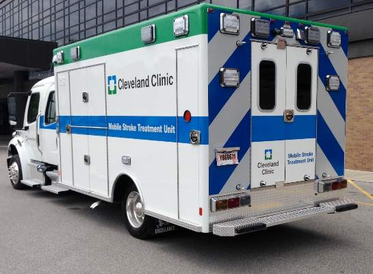
Once the mobile stroke unit reaches the patient, the team performs a CT scan and lab testing and the vascular neurologist interacts remotely with the patient and the unit’s personnel. If a diagnosis is confirmed, intravenous TPA is initiated and the patient is triaged to the appropriate hospital, Dr. Uchino explained.The study compared the times to treatment for patients experiencing acute stroke symptoms who were treated at the mobile stroke unit in the city (100 patients) with those presenting to Cleveland Clinic hospital emergency departments with a stroke alert called within 30 minutes of arrival to the hospital (53 patients), from Jan. 1, 2014, through December 2014. The mean age of patients was 63 years, about 56% were females. Intravenous thrombolysis was administered to 16 patients (16%) of those in the mobile stroke unit group and 12 (almost 23%) of those in the control group, which was not a significant difference.
The door-to-CT completion time was a median of 13 minutes (range, 9-21 minutes) in the mobile stroke unit group and 18 minutes (range, 12-26 minutes) among controls (P = .0026).
The door-to-CT read time was a median of 25 minutes in both groups. The door-to-international normalized ratio result time was a median of 13 minutes (range, 7-18) in the mobile stroke unit group and 44 minutes (range, 36-61) among controls, a significant difference (P < .001). The median time for door-to-video login was 11 minutes (range, 7-17) for the mobile stroke unit group and 20 minutes (range, 14-27) for controls.
The door-to-intravenous thrombolysis time was a median of 32 minutes (range, 24-47) in the mobile unit group and 58 minutes (range, 52-66) among controls, a 26-minute difference that was statistically significant, (P = .0012), Dr. Uchino said. The median time from the onset of symptoms to thrombolysis was 97 minutes (range, 61-144) in the mobile stroke unit group and 123 minutes (range, 110-176) in the controls (P = .0485).
Dr. Uchino and his associates at the Cleveland Clinic are part of the Cleveland PHAST study group; other participants include Case Western Reserve University. He had no disclosures. The mobile unit received funding from the Milton and Tamar Maltz Family Foundation and the Cleveland Clinic.
*CORRECTION, 4/27/15: The numbers of patients who actually received TPA out of those who qualified were accidentally transposed.
WASHINGTON – A mobile stroke unit significantly reduced the time to evaluation, diagnosis, and treatment when compared with standard emergency medical units for patients experiencing symptoms of an acute stroke, Dr. Ken Uchino reported at the annual meeting of the American Academy of Neurology.
In a 1-year prospective study conducted in Cleveland, patients who were evaluated at the mobile stroke unit and diagnosed with an acute ischemic stroke received treatment with intravenous tissue plasminogen activator (TPA) a median of about 26 minutes earlier than did patients transported via EMS to the hospital’s stroke center, a statistically significant difference, said Dr. Uchino of the Cleveland Clinic’s cerebrovascular center and the Cleveland Pre-Hospital Acute Stroke Treatment (PHAST) study group.
In addition, 25% of the patients who were having an acute ischemic stroke received TPA within the first hour of the onset of symptoms, “the golden hour of thrombolysis.” No one in the control group met that goal, he noted.
American Heart Association/American Stroke Association guidelines recommend a door-to-needle time of 60 minutes or less and set a goal for participating hospitals to administer TPA to at least 50% of their patients with acute ischemic stroke within 60 minutes of arriving at the hospital.
Of the 100 patients evaluated for stroke, 33 had a probable acute ischemic stroke and 16 of 19 patients who qualified received intravenous TPA.* One patient did not receive TPA because of crew error, and there were problems with intravenous access in two patients. Another 30 were diagnosed with a possible acute ischemic stroke. Of the remainder, 4 were diagnosed with a transient ischemic attack, 5 were diagnosed with intracerebral hemorrhage, and 28 were classified as “other.”
One patient with an intracerebral hemorrhage, a 93-year-old woman on warfarin, was successfully treated with prothrombin complex concentrate for warfarin reversal before arriving at the hospital.
Further study is needed to obtain data on the outcomes and cost-effectiveness of the mobile stroke units, he said.
The mobile unit is a modified emergency response vehicle staffed by a nurse, paramedic, emergency medical technician, and a CT technologist. The unit has a mobile CT scanner and telemedicine equipment with point-of-care lab testing; a vascular neurologist and a neuroradiologist work remotely to evaluate patients and images. The protocol starts with the 911 call and activation of the emergency medical system. An algorithm is used to identify patients who might be having a stroke.
Once the mobile stroke unit reaches the patient, the team performs a CT scan and lab testing and the vascular neurologist interacts remotely with the patient and the unit’s personnel. If a diagnosis is confirmed, intravenous TPA is initiated and the patient is triaged to the appropriate hospital, Dr. Uchino explained.The study compared the times to treatment for patients experiencing acute stroke symptoms who were treated at the mobile stroke unit in the city (100 patients) with those presenting to Cleveland Clinic hospital emergency departments with a stroke alert called within 30 minutes of arrival to the hospital (53 patients), from Jan. 1, 2014, through December 2014. The mean age of patients was 63 years, about 56% were females. Intravenous thrombolysis was administered to 16 patients (16%) of those in the mobile stroke unit group and 12 (almost 23%) of those in the control group, which was not a significant difference.
The door-to-CT completion time was a median of 13 minutes (range, 9-21 minutes) in the mobile stroke unit group and 18 minutes (range, 12-26 minutes) among controls (P = .0026).
The door-to-CT read time was a median of 25 minutes in both groups. The door-to-international normalized ratio result time was a median of 13 minutes (range, 7-18) in the mobile stroke unit group and 44 minutes (range, 36-61) among controls, a significant difference (P < .001). The median time for door-to-video login was 11 minutes (range, 7-17) for the mobile stroke unit group and 20 minutes (range, 14-27) for controls.
The door-to-intravenous thrombolysis time was a median of 32 minutes (range, 24-47) in the mobile unit group and 58 minutes (range, 52-66) among controls, a 26-minute difference that was statistically significant, (P = .0012), Dr. Uchino said. The median time from the onset of symptoms to thrombolysis was 97 minutes (range, 61-144) in the mobile stroke unit group and 123 minutes (range, 110-176) in the controls (P = .0485).
Dr. Uchino and his associates at the Cleveland Clinic are part of the Cleveland PHAST study group; other participants include Case Western Reserve University. He had no disclosures. The mobile unit received funding from the Milton and Tamar Maltz Family Foundation and the Cleveland Clinic.
*CORRECTION, 4/27/15: The numbers of patients who actually received TPA out of those who qualified were accidentally transposed.
AT THE AAN 2015 ANNUAL MEETING
Key clinical point: Mobile stroke units could allow more patients to meet a door-to-needle (DTN) time of under 60 minutes.
Major finding: Those treated at a mobile stroke unit received thrombolysis about 30 minutes faster than patients treated by EMS only.
Data source: A prospective 1-year study conducted in the city of Cleveland compared the time to thrombolysis and other outcomes in 100 patients with stroke symptoms treated at a mobile stroke unit and 53 treated by the EMS.
Disclosures: Dr. Uchino had no disclosures. The mobile unit received funding from the Milton and Tamar Maltz Family Foundation and the Cleveland Clinic.

Comorbidities in migraine should guide treatment course
PHILADELPHIA – Anxiety and mood disorders, certain cardiovascular conditions, epilepsy, and poor sleep are all comorbidities in migraineurs that must be considered before choosing a patient’s treatment plan, according to Dr. Deborah I. Friedman.
Treating these comorbidities while minimizing risk factors is essential to chronic migraine prevention, Dr. Friedman told an audience at the Emerging Concepts in Headache Therapy session during this year’s annual meeting of the American Academy of Neurology.

Modifiable risk factors include the frequency of attack, caffeine intake, medication overuse, obesity, and sleep apnea.
Depression, anxiety, and suicide
Screening for anxiety and depression in migraine patients is important, given the close association between these psychiatric diagnoses and migraine, she said.
"Mood and anxiety disorders are anywhere from 2 to 10 times more prevalent in migraineurs," said Dr. Friedman, professor of neurology at the University of Texas Southwestern Medical Center at Dallas. "And 25% of patients with migraine meet [the] criteria for mood and anxiety disorders."
But teasing out which condition begets which comorbidity can be difficult; each might be a risk factor for the other.
"We know that depression, anxiety, and phobias are bidirectionally linked to migraine. If you have one, you’re more likely to have the other, and vice versa," Dr. Friedman said.
One possible mechanism for this is shared etiologic factors such as genetics. However, whether there is a confounder is still unknown. In the case of adverse childhood experiences and stressful life events, for example, "we know there is a large association of an unfortunate occurrence and migraine," Dr. Friedman said. "Is there a link between these things and posttraumatic stress disorder?"
However, patients with episodic migraine have less risk of developing mood and anxiety disorders, compared with chronic daily headache sufferers, Dr. Friedman said. "The comorbidity may relate to disabling and recurrent pain more than to the migraine itself."
In addition, a population-based cohort study of migraine patients, people with nonmigraine severe headache, and controls with no history of severe headache Dr. Friedman cited indicated statistically significant odds of attempted suicide in migraine when there is depression at baseline (odds ratio, 3.18); anxiety (OR, 4.78); depression and anxiety (OR, 12.1); and a previous suicide attempt (OR, 54.94) (Headache 2012;52:723-31).
Although the link between migraine and depression is high, Dr. Friedman said the link with anxiety is "probably much more common," occurring at a rate of five times more than would be expected in the general population.
For patients deemed to have a psychiatric comorbidity, Dr. Friedman said in an interview that cognitive-behavioral therapy is her preferred first-line therapy, particularly for anxiety and panic.
"It’s easy to reach for a pill to treat anxiety, but in the long haul, I am not sure it really benefits the patient that much because they don’t really gain insight into what’s causing their situation," Dr. Friedman said in the interview. It also might not be possible to "get two birds with one stone" by prescribing one pill to treat headache and depression and/or anxiety, Dr. Friedman said.
PFO and Raynaud’s
The odds that patients with migraine also have patent foramen ovale (PFO) or a right-left shunt are nearly eight times greater than in the general public, according to Dr. Friedman. The prevalence of PFO in migraine with aura is more than twice the rate of the general population.
Some nonrandomized studies have shown benefit to migraine from PFO closure, while the 2006 sham-controlled MIST (Migraine Intervention With STARFlex Technology) trial in the United Kingdom failed to achieve either its primary or secondary endpoints.
"Perhaps those endpoints were not realistic, because they were complete resolution of headache days at 3 months," Dr. Friedman said.
Some posit that the relationship between migraine and PFO might explain the increased risk of ischemic stroke and white matter intensities; however, since results from the PREMIUM and PRIMA studies on PFO and migraine are still pending, Dr. Friedman said there are no clinical recommendations for treating PFO in the context of just migraine at this time.
Raynaud’s phenomenon is another "chicken-egg" dilemma, Dr. Friedman said. Raynaud’s is five times more likely in those with migraine than in the general population, and migraine has similar occurrence rates in Raynaud’s.
Clinicians should take note that in addition to avoiding triptans and ergot derivatives in migraine patients with Raynaud’s, "beta-blockers can induce Raynaud’s in patients who have actually never experienced it," Dr. Friedman said, "and they can also make Raynaud’s worse."
However, particularly in patients with migraine who also have prominent autonomic symptoms, calcium channel blockers can treat both conditions, Dr. Friedman said.
Epilepsy and ‘migralepsy’
The incidence of migraine is nearly 2.5 times greater in persons with epilepsy than in those without it, according to Dr. Friedman.
Perhaps because they share a paroxysmal nature, Dr. Friedman said it is interesting to note that the comorbidities for epilepsy mirror those in migraine, including most major psychiatric diagnoses except psychosis, sleep and movement disorders, fibromyalgia, and asthma.
Seizures known as "migralepsy" that are triggered by migraine with aura can occur in patients either during or within 1 hour of a migraine attack, Dr. Friedman said. The mechanism is thought to be the cortical spread of depression.
Comorbid migraine in epilepsy decreases the likelihood of early treatment response, shortens remission periods, and is associated with intractable epilepsy, requiring polytherapy, Dr. Friedman said.
For those reasons, she recommended that patients not decrease their seizure threshold, either by their behaviors or their medications, and that clinicians select medications that can prevent both migraine and seizures. "Most of the medications we use for migraine do not affect the seizure threshold," she said in a follow-up interview. "Bupropion, venlafaxine, tramadol, [and] some of the antipsychotics and various stimulants (such as attention-deficit/hyperactivity disorder drugs) can do it. "The tricyclic antidepressants have long been associated with lowering the seizure threshold, but there is actually no good evidence that this is true."
Sleep disorders
"Sleep is like caffeine [in migraine]; it’s a double-edged sword," Dr. Friedman said. "Sleeping well can trigger a migraine, but it can also get rid of a migraine. Sleep interferes with pain, but pain interferes with sleep."
There are some data linking somnambulism, nightmares, and bruxism to migraine. "I have been surprised in my own practice how many of my patients tell me they used to sleep walk as a child," he said.
Referring to a 2010 study, Dr. Friedman said severe sleep disturbance was associated with a five times greater frequency of headache than in controls, and that insomnia, sleep initiation, and excessive daytime sleepiness were the most common complaints (J. Headache Pain 2010;11:197-206).
Although snoring and sleep apnea are not considered comorbidities of migraine, they do heighten the risk of episodic migraines becoming chronic, which is why Dr. Friedman recommended screening patients for sleep apnea. Asking patients about their sleep hygiene and sleep histories, including in childhood if they are adults, is important, as is reviewing what they ingest, from caffeine in food and drink to medications.
"Incorporating sleep questionnaires into your practice is very helpful," Dr. Friedman said.
She said she had no relevant financial disclosures.
On Twitter @whitneymcknight
PHILADELPHIA – Anxiety and mood disorders, certain cardiovascular conditions, epilepsy, and poor sleep are all comorbidities in migraineurs that must be considered before choosing a patient’s treatment plan, according to Dr. Deborah I. Friedman.
Treating these comorbidities while minimizing risk factors is essential to chronic migraine prevention, Dr. Friedman told an audience at the Emerging Concepts in Headache Therapy session during this year’s annual meeting of the American Academy of Neurology.
Modifiable risk factors include the frequency of attack, caffeine intake, medication overuse, obesity, and sleep apnea.
Depression, anxiety, and suicide
Screening for anxiety and depression in migraine patients is important, given the close association between these psychiatric diagnoses and migraine, she said.
"Mood and anxiety disorders are anywhere from 2 to 10 times more prevalent in migraineurs," said Dr. Friedman, professor of neurology at the University of Texas Southwestern Medical Center at Dallas. "And 25% of patients with migraine meet [the] criteria for mood and anxiety disorders."
But teasing out which condition begets which comorbidity can be difficult; each might be a risk factor for the other.
"We know that depression, anxiety, and phobias are bidirectionally linked to migraine. If you have one, you’re more likely to have the other, and vice versa," Dr. Friedman said.
One possible mechanism for this is shared etiologic factors such as genetics. However, whether there is a confounder is still unknown. In the case of adverse childhood experiences and stressful life events, for example, "we know there is a large association of an unfortunate occurrence and migraine," Dr. Friedman said. "Is there a link between these things and posttraumatic stress disorder?"
However, patients with episodic migraine have less risk of developing mood and anxiety disorders, compared with chronic daily headache sufferers, Dr. Friedman said. "The comorbidity may relate to disabling and recurrent pain more than to the migraine itself."
In addition, a population-based cohort study of migraine patients, people with nonmigraine severe headache, and controls with no history of severe headache Dr. Friedman cited indicated statistically significant odds of attempted suicide in migraine when there is depression at baseline (odds ratio, 3.18); anxiety (OR, 4.78); depression and anxiety (OR, 12.1); and a previous suicide attempt (OR, 54.94) (Headache 2012;52:723-31).
Although the link between migraine and depression is high, Dr. Friedman said the link with anxiety is "probably much more common," occurring at a rate of five times more than would be expected in the general population.
For patients deemed to have a psychiatric comorbidity, Dr. Friedman said in an interview that cognitive-behavioral therapy is her preferred first-line therapy, particularly for anxiety and panic.
"It’s easy to reach for a pill to treat anxiety, but in the long haul, I am not sure it really benefits the patient that much because they don’t really gain insight into what’s causing their situation," Dr. Friedman said in the interview. It also might not be possible to "get two birds with one stone" by prescribing one pill to treat headache and depression and/or anxiety, Dr. Friedman said.
PFO and Raynaud’s
The odds that patients with migraine also have patent foramen ovale (PFO) or a right-left shunt are nearly eight times greater than in the general public, according to Dr. Friedman. The prevalence of PFO in migraine with aura is more than twice the rate of the general population.
Some nonrandomized studies have shown benefit to migraine from PFO closure, while the 2006 sham-controlled MIST (Migraine Intervention With STARFlex Technology) trial in the United Kingdom failed to achieve either its primary or secondary endpoints.
"Perhaps those endpoints were not realistic, because they were complete resolution of headache days at 3 months," Dr. Friedman said.
Some posit that the relationship between migraine and PFO might explain the increased risk of ischemic stroke and white matter intensities; however, since results from the PREMIUM and PRIMA studies on PFO and migraine are still pending, Dr. Friedman said there are no clinical recommendations for treating PFO in the context of just migraine at this time.
Raynaud’s phenomenon is another "chicken-egg" dilemma, Dr. Friedman said. Raynaud’s is five times more likely in those with migraine than in the general population, and migraine has similar occurrence rates in Raynaud’s.
Clinicians should take note that in addition to avoiding triptans and ergot derivatives in migraine patients with Raynaud’s, "beta-blockers can induce Raynaud’s in patients who have actually never experienced it," Dr. Friedman said, "and they can also make Raynaud’s worse."
However, particularly in patients with migraine who also have prominent autonomic symptoms, calcium channel blockers can treat both conditions, Dr. Friedman said.
Epilepsy and ‘migralepsy’
The incidence of migraine is nearly 2.5 times greater in persons with epilepsy than in those without it, according to Dr. Friedman.
Perhaps because they share a paroxysmal nature, Dr. Friedman said it is interesting to note that the comorbidities for epilepsy mirror those in migraine, including most major psychiatric diagnoses except psychosis, sleep and movement disorders, fibromyalgia, and asthma.
Seizures known as "migralepsy" that are triggered by migraine with aura can occur in patients either during or within 1 hour of a migraine attack, Dr. Friedman said. The mechanism is thought to be the cortical spread of depression.
Comorbid migraine in epilepsy decreases the likelihood of early treatment response, shortens remission periods, and is associated with intractable epilepsy, requiring polytherapy, Dr. Friedman said.
For those reasons, she recommended that patients not decrease their seizure threshold, either by their behaviors or their medications, and that clinicians select medications that can prevent both migraine and seizures. "Most of the medications we use for migraine do not affect the seizure threshold," she said in a follow-up interview. "Bupropion, venlafaxine, tramadol, [and] some of the antipsychotics and various stimulants (such as attention-deficit/hyperactivity disorder drugs) can do it. "The tricyclic antidepressants have long been associated with lowering the seizure threshold, but there is actually no good evidence that this is true."
Sleep disorders
"Sleep is like caffeine [in migraine]; it’s a double-edged sword," Dr. Friedman said. "Sleeping well can trigger a migraine, but it can also get rid of a migraine. Sleep interferes with pain, but pain interferes with sleep."
There are some data linking somnambulism, nightmares, and bruxism to migraine. "I have been surprised in my own practice how many of my patients tell me they used to sleep walk as a child," he said.
Referring to a 2010 study, Dr. Friedman said severe sleep disturbance was associated with a five times greater frequency of headache than in controls, and that insomnia, sleep initiation, and excessive daytime sleepiness were the most common complaints (J. Headache Pain 2010;11:197-206).
Although snoring and sleep apnea are not considered comorbidities of migraine, they do heighten the risk of episodic migraines becoming chronic, which is why Dr. Friedman recommended screening patients for sleep apnea. Asking patients about their sleep hygiene and sleep histories, including in childhood if they are adults, is important, as is reviewing what they ingest, from caffeine in food and drink to medications.
"Incorporating sleep questionnaires into your practice is very helpful," Dr. Friedman said.
She said she had no relevant financial disclosures.
On Twitter @whitneymcknight
EXPERT ANALYSIS AT THE AAN 2014 ANNUAL MEETING

EMS staff missed stroke 41% of the time, study shows
PHILADELPHIA – Emergency personnel miss stroke in 41% of patients in their prehospital care, a study shows. In light of this, physicians should have a high degree of suspicion for stroke when treating patients presenting with elevated systolic blood pressure, a dispatch call of stroke, prior stroke or diabetes history, and unconsciousness.
Prehospital identification and notification of stroke have been associated with both shorter door-to-needle times and increased thrombolysis rates, but "the sensitivity and specificity of prehospital care providers [are] widely variable," Dr. Ethan Brandler, of the emergency medicine department at SUNY Downstate Medical Center in Brooklyn, N.Y., told a Platform Blitz audience at the annual meeting of the American Academy of Neurology.

To assess the rates of proper stroke diagnosis by emergency medical services (EMS), Dr. Brandler and his associates reviewed 10,384 electronic prehospital care reports of all patients delivered by EMS to a single major metropolitan site between Jan. 1, 2010, and Dec. 31, 2011.
Using the American Heart Association’s Get With the Guidelines stroke database, the investigators determined that of the 75 confirmed cases of stroke, half of which were in men aged 19-49 years, EMS personnel accurately diagnosed 44 patients.
"Forty-one percent of all stroke patients were missed by EMS," Dr. Brandler said. "And 51 patients were misdiagnosed as stroke when they had something else: stroke-mimic, sepsis, infection, [and] hypoglycemia, among other neurologic and systemic problems."
The sensitivity level of 59% and specificity level of 99.5% (95% confidence interval, 47-69 and 99.1-99.6, respectively), combined with the positive likelihood ratio of 119 (95% CI, 85-165) and the negative likelihood ratio of 0.41 (95% CI, 0.32-0.54), meant that "if a paramedic called us to say a stroke was going on, the likelihood was extremely high it was a true stroke," Dr. Brandler said. "However, when they didn’t think it was a stroke, it didn’t mean much."
Stroke risk increased 83% with each increase in prehospital systolic blood pressure quartile (P = .04), Dr. Brandler and his colleagues found.
Independent predictors of stroke being the official diagnosis included an emergency services dispatcher who reported the call as a stroke, even if EMS personnel did not ultimately diagnose it that way (age-adjusted odds ratio, 9.8; 95% CI, 2.2-43.7; P = .003]. "Perhaps [the dispatcher’s] information is better than the crew’s because it is gathered in a more systematic fashion," Dr. Brandler said.
Other independent risk factors included a history of diabetes (AAOR, 2.2; 95% CI, 1.1-4.6; P = .026) and a history of stroke (AAOR, 4.4; 95% CI, 1.3-15; P = .017).
Patients who arrived unconscious with unknown cause also were considered at higher risk for having had a stroke (AAOR, 4.4; 95% CI, 1.3-15; P = .017). This was particularly the case when patients presented with basilar artery inclusions, Dr. Brandler said.
The study was funded by grants from the National Institutes of Health. Dr. Brandler did not report any other relevant disclosures.
On Twitter @whitneymcknight
PHILADELPHIA – Emergency personnel miss stroke in 41% of patients in their prehospital care, a study shows. In light of this, physicians should have a high degree of suspicion for stroke when treating patients presenting with elevated systolic blood pressure, a dispatch call of stroke, prior stroke or diabetes history, and unconsciousness.
Prehospital identification and notification of stroke have been associated with both shorter door-to-needle times and increased thrombolysis rates, but "the sensitivity and specificity of prehospital care providers [are] widely variable," Dr. Ethan Brandler, of the emergency medicine department at SUNY Downstate Medical Center in Brooklyn, N.Y., told a Platform Blitz audience at the annual meeting of the American Academy of Neurology.
To assess the rates of proper stroke diagnosis by emergency medical services (EMS), Dr. Brandler and his associates reviewed 10,384 electronic prehospital care reports of all patients delivered by EMS to a single major metropolitan site between Jan. 1, 2010, and Dec. 31, 2011.
Using the American Heart Association’s Get With the Guidelines stroke database, the investigators determined that of the 75 confirmed cases of stroke, half of which were in men aged 19-49 years, EMS personnel accurately diagnosed 44 patients.
"Forty-one percent of all stroke patients were missed by EMS," Dr. Brandler said. "And 51 patients were misdiagnosed as stroke when they had something else: stroke-mimic, sepsis, infection, [and] hypoglycemia, among other neurologic and systemic problems."
The sensitivity level of 59% and specificity level of 99.5% (95% confidence interval, 47-69 and 99.1-99.6, respectively), combined with the positive likelihood ratio of 119 (95% CI, 85-165) and the negative likelihood ratio of 0.41 (95% CI, 0.32-0.54), meant that "if a paramedic called us to say a stroke was going on, the likelihood was extremely high it was a true stroke," Dr. Brandler said. "However, when they didn’t think it was a stroke, it didn’t mean much."
Stroke risk increased 83% with each increase in prehospital systolic blood pressure quartile (P = .04), Dr. Brandler and his colleagues found.
Independent predictors of stroke being the official diagnosis included an emergency services dispatcher who reported the call as a stroke, even if EMS personnel did not ultimately diagnose it that way (age-adjusted odds ratio, 9.8; 95% CI, 2.2-43.7; P = .003]. "Perhaps [the dispatcher’s] information is better than the crew’s because it is gathered in a more systematic fashion," Dr. Brandler said.
Other independent risk factors included a history of diabetes (AAOR, 2.2; 95% CI, 1.1-4.6; P = .026) and a history of stroke (AAOR, 4.4; 95% CI, 1.3-15; P = .017).
Patients who arrived unconscious with unknown cause also were considered at higher risk for having had a stroke (AAOR, 4.4; 95% CI, 1.3-15; P = .017). This was particularly the case when patients presented with basilar artery inclusions, Dr. Brandler said.
The study was funded by grants from the National Institutes of Health. Dr. Brandler did not report any other relevant disclosures.
On Twitter @whitneymcknight
AT THE AAN 2014 ANNUAL MEETING
Key clinical point: A higher degree of suspicion is warranted for stroke in patients with elevated systolic blood pressure, a dispatch call of stroke, prior stroke or diabetes history, and unconsciousness.
Major finding: EMS staff had 59% sensitivity and 99% specificity (95% confidence intervals, 47-69 and 99.4-99.6, respectively) when diagnosing stroke; and positive and negative likelihood ratios were 119 and 0.41 (95% CIs, 85-165 and 0.32-0.54). Stroke risk increased 83% with each increase in prehospital systolic blood pressure quartile (P = .04).
Data source: Two-year, retrospective analysis of data for 10,384 electronic prehospital care reports of patients brought by EMTs to a single metropolitan site.
Disclosures: The study was funded by grants from the National Institutes of Health. Dr. Brandler did not report any other relevant disclosures.

Imaging with florbetaben reliably excludes amyloid pathology
PHILADELPHIA – The radioimaging agent 18F-florbetaben reliably excluded amyloid pathology in a study of 74 end-of-life patients with dementia.
When used in PET brain scans, the agent showed 98% sensitivity for beta amyloid plaques, with a 96% negative predictive value, Dr. Marwan N. Sabbagh said at the annual meeting of the American Academy of Neurology.
"A negative scan should encourage the physician to search for other causes of cognitive decline and to tailor available treatment options," said Dr. Sabbagh, director of the Banner Sun Health Research Institute in Sun City, Ariz.
The study comprised 205 elderly patients who were near the end of life and had agreed to post mortem brain histopathology. The PET scans were read by three radiologists who had been trained in person on the use of the agent. Histopathology confirmed the scan results.

Of the group, 74 patients had both scans and histopathology verification. Most (57) had a clinical diagnosis of Alzheimer’s disease; three had been diagnosed with Lewy body dementia; and six were diagnosed with "other" dementia. Eight subjects were not demented at the time of death.
On histopathology, 47 of the patients showed amyloid pathology: 44 of the Alzheimer’s patients; 1 of the Lewy body patients; 1 of the "other dementia" patients; and 1 of the nondemented patients.
Florbetaben scans correctly identified amyloid plaques in 44 of these 47 patients, for a sensitivity of 98%. Scans from 24 of the 27 patients without amyloid pathology were correctly read as negative, for specificity of 89%. All but one of the 25 scans read as negative were confirmed negative by histopathology, for a negative predictive value of 96%.
These values are slightly better than those seen with 18F florbetapir (Amyvid), the PET imaging agent approved in 2012. It has a sensitivity of 92% and specificity of 95% for readers who received individual training in reading the scans. Both agents have a half-life of about 2 hours.
Florbetaben (NeuroCeq) received Food and Drug Administration approval in March, making it the third amyloid imaging compound approved for use in the United States.
A fourth, NAV4694, is being developed by Navidea Biopharmaceuticals. In early studies, the company said that the agent has a sensitivity, specificity, and overall accuracy ranging from 94% to 98%.
Avid Radiopharmaceuticals, the maker of 18F-florbetapir, is owned by Eli Lilly.
Piramal Imaging, maker of 18F-florbetaben, funded the study. Dr. Sabbagh has received grant money from the company as served as an adviser. He also disclosed that he has received grant money from Avid Radiopharmaceuticals.
On Twitter @alz_gal
PHILADELPHIA – The radioimaging agent 18F-florbetaben reliably excluded amyloid pathology in a study of 74 end-of-life patients with dementia.
When used in PET brain scans, the agent showed 98% sensitivity for beta amyloid plaques, with a 96% negative predictive value, Dr. Marwan N. Sabbagh said at the annual meeting of the American Academy of Neurology.
"A negative scan should encourage the physician to search for other causes of cognitive decline and to tailor available treatment options," said Dr. Sabbagh, director of the Banner Sun Health Research Institute in Sun City, Ariz.
The study comprised 205 elderly patients who were near the end of life and had agreed to post mortem brain histopathology. The PET scans were read by three radiologists who had been trained in person on the use of the agent. Histopathology confirmed the scan results.
Of the group, 74 patients had both scans and histopathology verification. Most (57) had a clinical diagnosis of Alzheimer’s disease; three had been diagnosed with Lewy body dementia; and six were diagnosed with "other" dementia. Eight subjects were not demented at the time of death.
On histopathology, 47 of the patients showed amyloid pathology: 44 of the Alzheimer’s patients; 1 of the Lewy body patients; 1 of the "other dementia" patients; and 1 of the nondemented patients.
Florbetaben scans correctly identified amyloid plaques in 44 of these 47 patients, for a sensitivity of 98%. Scans from 24 of the 27 patients without amyloid pathology were correctly read as negative, for specificity of 89%. All but one of the 25 scans read as negative were confirmed negative by histopathology, for a negative predictive value of 96%.
These values are slightly better than those seen with 18F florbetapir (Amyvid), the PET imaging agent approved in 2012. It has a sensitivity of 92% and specificity of 95% for readers who received individual training in reading the scans. Both agents have a half-life of about 2 hours.
Florbetaben (NeuroCeq) received Food and Drug Administration approval in March, making it the third amyloid imaging compound approved for use in the United States.
A fourth, NAV4694, is being developed by Navidea Biopharmaceuticals. In early studies, the company said that the agent has a sensitivity, specificity, and overall accuracy ranging from 94% to 98%.
Avid Radiopharmaceuticals, the maker of 18F-florbetapir, is owned by Eli Lilly.
Piramal Imaging, maker of 18F-florbetaben, funded the study. Dr. Sabbagh has received grant money from the company as served as an adviser. He also disclosed that he has received grant money from Avid Radiopharmaceuticals.
On Twitter @alz_gal
AT THE AAN 2014 ANNUAL MEETING
Key clinical point: Search for other causes of cognitive decline and ‘tailor available treatment options’ after getting a negative scan.
Major finding: The radioligand florbetaben showed a 98% sensitivity and 96% negative predictive value for amyloid brain plaques.
Data source: A prospective study of 74 patients who had PET scanning and brain histopathology.
Disclosures: Dr. Sabbagh has received grant money from, and served as an adviser to, 18F florbetaben’s manufacturer, Piramal Imaging, which funded the study. Dr. Sabbagh also disclosed ties with Avid Radiopharmaceuticals.
Orexin antagonist improved sleep in phase III studies
PHILADELPHIA – An orexin receptor antagonist – the first drug to affect a neural system that promotes wakefulness – has proven safe and effective for up to 1 year in a pooled analysis of three phase III studies of patients with insomnia.
The drug (suvorexant) significantly improved both subjective and objective measures of sleep, including sleep onset, total sleep time, and wakefulness after sleep, Dr. W. Joseph Herring said at the annual meeting of the American Academy of Neurology.
Based on these data, the manufacturer, Merck, proceeded last year to preliminary talks with the Food and Drug Administration. The committee considering suvorexant recommended that Merck focus on the smaller of two proposed dose ranges – 20 mg for people up to 64 years of age, and 15 mg for those aged 65 years and older.
The orexin neural system was discovered in the late 1990s. Orexin neurons release two neuropeptides that interact with downstream receptors that promote wakefulness. Secretion follows a circadian rhythm. Suvorexant orexin antagonists block the activity of this wake-signaling system, allowing sleep to occur.
So far, studies have investigated the drug in almost 3,000 patients; 160 of these were treated for at least a year. The three phase III trials comprised more than 275,000 exposure nights. Most of the patients were women; 46% of the patients were aged 65 years or older.
Sleep was measured by subjective assessment and objective scales, including polysomnography.
In the two efficacy studies, the higher doses decreased time to sleep onset by 15 minutes on the first night; the lower doses did so by 10 minutes. By 3 months, the high-dose group’s decreased time to sleep onset was 5 minutes; the low dose group’s time was lowered to 4 minutes.
On the first night, the high-dose drug reduced wakefulness after sleep by 40 minutes; the low-dose drug did so by 35 minutes. After 3 months, the reductions were similar (about 25 minutes).
When the night was divided into thirds, both doses decreased wakefulness significantly and about equally, especially in the second and third fractions of the night.
In the subgroup of 160 patients who were treated for at least 12 months, the drugs showed persistent efficacy overall, although the high doses were somewhat more effective, Dr. Herring, Merck’s executive director of neuroscience clinical researchDr. Herring said.
A multivariate regression analysis found that, compared with placebo, patients who took the high dose were 2.4 times more likely to be considered responders at 1 month and 1.8 times more likely to be responders at 3 months. Those who took the low dose were 1.8 times more likely to respond at 1 and 3 months.
"The drugs really proved about equal in the chances of a good response," said Dr. Herring.
Although there were more adverse events in the active groups than in the placebo groups – and more in the high-dose groups, compared with the low-dose groups – suvorexant was considered safe, he said. The discontinuation rates for drug-related adverse events in the high-dose, low-dose, and placebo groups were 4%, 3%, and 2%, respectively.
The most common issues were next-day somnolence (3% for placebo, 7% with the low dose, and 11% for the high dose, respectively); headache (6%, 7%, and 7%, respectively); and fatigue (2%, 2%, and 4%, respectively).
Few serious adverse events were reported. These included one case of suicidal ideation each in the placebo and low-dose groups, and eight cases in the high-dose group. Dr. Herring said these were mild to moderate and of short duration.
Neither of the doses was associated with withdrawal symptoms or clinically significant insomnia rebound during the washout periods. "The drug appears to have a low potential for abuse," Dr. Herring noted.
Merck sponsored the studies. Dr. Herring is a full-time employee of the company.
On Twitter @alz_gal
Key clinical point: Orexin antagonist suvorexant improved sleep based on both subjective and objective measures, with minimal side effects.
Major finding: In patients with insomnia, suvorexant reduced the time to sleep onset by up to 10 minutes, and wakefulness after sleep by up to 40 minutes.
Data source: Three phase III studies involving almost 3,000 patients.
Disclosures: Dr. Herring is director of neuroscience clinical research at Merck, which sponsored the studies.
Stress linked to frequency of tension, migraine headache
PHILADELPHIA – A 10-point increase in headache patients’ perception of life stress was associated with up to a 10% increase in the frequency of tension-type headache, according to Dr. Zaza Katsarava.
A somewhat attenuated, but still significant relationship was observed with migraine, said Dr. Katsarava of the University of Essen (Germany).

"This is a confirmation of what we see every day in clinical practice. It’s an added piece of information suggesting that we need a comprehensive approach in how we treat our headache patients," he said at the annual meeting of the American Academy of Neurology.
He discussed findings of the German Headache Consortium study, which followed more than 5,000 people for 2 years. During that time, the subjects, aged 21-71 years, completed surveys every 3 months. Once a year, this was a long survey that let researchers identify headaches as tension type, migraine with coexisting tension type, migraine only, or unclassifiable. The long surveys also inquired about age, sex, body mass index, socioeconomic status, depression, stress, and medications.
Every 3 months throughout the study period, respondents completed a short questionnaire asking whether they had experienced headache during the prior 3 months and if so, how many headache days occurred during that time. Subjects also rated their perceived stress level over the same period, using a 0- to 100-point visual analog scale. The data were adjusted for sex, age, frequent intake of acute pain drugs, drinking, smoking, body mass index, and education.
About a third of respondents (31%) reported tension-type headache, with a mean of 2 headache days per month and a mean stress level of 52. Migraine with coexisting tension-type headache was present in 11%, with a mean of 4 headache days per month and a mean stress level of 59. Migraine occurred in 14%, with a mean of 4.5 headache days per month and a mean stress level of 62. Headache was unclassifiable in 17%.
Among those with tension-type headache, every 10-point increase on the stress measure was associated with an overall 6% increase in monthly headache days. The association also occurred among those with migraine, although it was somewhat attenuated (4% overall).
There were some significant differences in the association when the groups were broken down by age, however. For those with tension-type headache, younger people experienced the biggest effect. For those in their 20s and 30s, every 10-point increase in perceived stress was associated with a 10% increase in headache days. For those in their 40s, headaches increased about 7% for every 10-point stress increase. The increase dropped to 6% for those in their 50s, and to 3% for those in their 60s.
Among those with migraine, the 10-point stress increase was associated with an 8% increase in headache days for those in their 20s, and a 5% increase for those in their 30s. The associated increase was 3% for those in their 40s, 6% for those in their 60s, and less than 1% for those in their 70s.
The findings beg that never-ending question of whether tension begets headaches, or headaches beget tension, Dr. Katsarava said. This study, with its cross-sectional design and lack of a control group, can’t answer that question. It does, however, paint a clear picture of a very real relationship, he concluded.
The study was sponsored by the German Ministry for Education and Research. Dr. Katsarava disclosed that he has received honoraria from Allergan and Amgen.
On Twitter @alz_gal
PHILADELPHIA – A 10-point increase in headache patients’ perception of life stress was associated with up to a 10% increase in the frequency of tension-type headache, according to Dr. Zaza Katsarava.
A somewhat attenuated, but still significant relationship was observed with migraine, said Dr. Katsarava of the University of Essen (Germany).
"This is a confirmation of what we see every day in clinical practice. It’s an added piece of information suggesting that we need a comprehensive approach in how we treat our headache patients," he said at the annual meeting of the American Academy of Neurology.
He discussed findings of the German Headache Consortium study, which followed more than 5,000 people for 2 years. During that time, the subjects, aged 21-71 years, completed surveys every 3 months. Once a year, this was a long survey that let researchers identify headaches as tension type, migraine with coexisting tension type, migraine only, or unclassifiable. The long surveys also inquired about age, sex, body mass index, socioeconomic status, depression, stress, and medications.
Every 3 months throughout the study period, respondents completed a short questionnaire asking whether they had experienced headache during the prior 3 months and if so, how many headache days occurred during that time. Subjects also rated their perceived stress level over the same period, using a 0- to 100-point visual analog scale. The data were adjusted for sex, age, frequent intake of acute pain drugs, drinking, smoking, body mass index, and education.
About a third of respondents (31%) reported tension-type headache, with a mean of 2 headache days per month and a mean stress level of 52. Migraine with coexisting tension-type headache was present in 11%, with a mean of 4 headache days per month and a mean stress level of 59. Migraine occurred in 14%, with a mean of 4.5 headache days per month and a mean stress level of 62. Headache was unclassifiable in 17%.
Among those with tension-type headache, every 10-point increase on the stress measure was associated with an overall 6% increase in monthly headache days. The association also occurred among those with migraine, although it was somewhat attenuated (4% overall).
There were some significant differences in the association when the groups were broken down by age, however. For those with tension-type headache, younger people experienced the biggest effect. For those in their 20s and 30s, every 10-point increase in perceived stress was associated with a 10% increase in headache days. For those in their 40s, headaches increased about 7% for every 10-point stress increase. The increase dropped to 6% for those in their 50s, and to 3% for those in their 60s.
Among those with migraine, the 10-point stress increase was associated with an 8% increase in headache days for those in their 20s, and a 5% increase for those in their 30s. The associated increase was 3% for those in their 40s, 6% for those in their 60s, and less than 1% for those in their 70s.
The findings beg that never-ending question of whether tension begets headaches, or headaches beget tension, Dr. Katsarava said. This study, with its cross-sectional design and lack of a control group, can’t answer that question. It does, however, paint a clear picture of a very real relationship, he concluded.
The study was sponsored by the German Ministry for Education and Research. Dr. Katsarava disclosed that he has received honoraria from Allergan and Amgen.
On Twitter @alz_gal
AT THE AAN 2014 ANNUAL MEETING
Key clinical point: A comprehensive approach might be needed in the way neurologists treat headache patients.
Major finding: Every 10-point increase in perceived life stress was associated with up to a 10% increase in monthly tension-type headache days.
Data source: The German Headache Consortium study followed 5,000 people for 2 years.
Disclosures: The study was sponsored by the German Ministry for Education and Research. Dr. Katsarava disclosed that he has received honoraria from Allergan and Amgen.

Psychiatric comorbidities add to migraineurs’ medical interventions
PHILADELPHIA – Patients who have migraines and comorbid psychiatric disorders visited the emergency department more, and received more brain imaging and narcotics, than patients who had only migraine.
The additional emergency visits and procedures – combined with significantly higher rates of hospital admissions and outpatient visits – run contrary to published recommendations, Dr. Mia Minen reported at the annual meeting of the American Academy of Neurology.
The imaging findings of her cross-sectional analysis are particularly troubling in light of current guidelines aimed at helping to minimize radiation exposure by avoiding head and brain imaging in patients with primary headache disorders, said Dr. Minen, a fellow at the Graham Headache Center, Boston.
"One of the AAN’s recommendations for the Choosing Wisely campaign was not to perform brain imaging for patients presenting to the ED with recurrence of their baseline primary headache disorder," she said. She added that the American College of Emergency Physicians has not found level A evidence supporting imaging in patients who present to the emergency department with headache, unless the headache is sudden and severe, or unless it’s accompanied by an abnormal neurological exam.
Her analysis looked at emergency treatment trends in a database of almost 3,000 headache patients seen over a 10-year period in a single hospital’s emergency department. The patients were a mean of 40 years old; most (80%) were women. About 2,000 had at least one psychiatric comorbidity; the most common psychiatric comorbidities were anxiety and depression.
Over the 10-year study period, migraine patients overall made an average of 11 ED visits, with 10 admissions and 26 outpatient visits. Those patients without a comorbid psychiatric diagnosis made significantly fewer visits in every category: 6 ED visits, 3 inpatient visits, and 11 outpatient visits.
Migraine patients with comorbidities presented a very different picture, Dr. Minen said. These patients had an average of 18 ED visits, 19 inpatient visits, and 45 outpatient visits over the study period.
Compared with migraineurs without psychiatric disorders, those with them were significantly more likely to undergo a CT of the head (relative risk, 1.4) and an MRI of the brain (RR, 1.5). They received narcotic treatment in the ED significantly more often as well.
"We need more studies to understand why this is the case," Dr. Minen said.
She added that the pharmacotherapy findings also were in contrast to recommendations in the AAN Choosing Wisely campaign.
"In 2013, one of the final recommendations was not to use opiates or butalbital for the treatment of migraine, except in rare circumstances."
Dr. Minen had no financial disclosures.
AT THE AAN 2014 ANNUAL MEETING
Key clinical point: More studies are needed to determine why migraineurs with psychiatric disorders are more likely than those without comorbidities to undergo brain imaging and to receive narcotics.
Major finding: Migraine patients with psychiatric comorbidities were 40% more likely to have head imaging and 50% more likely to receive narcotics than those without such conditions.
Data source: The cross-sectional analysis comprised almost 3,000 patients.
Disclosures: Dr. Minen had no financial disclosures.
Alemtuzumab found to reduce brain lesions, volume loss
PHILADELPHIA – The rates of new or growing brain lesions slowed in both treatment-naive and relapsed patients with relapse-remitting multiple sclerosis who took the monoclonal antibody alemtuzumab in two phase III trials with follow-up data out to 3 years.
Loss of brain volume also slowed over the 3-year period based on changes in brain parenchymal fraction (BPF), reported Dr. Douglas Arnold, a neurologist at the Montreal Neurological Institute and Hospital of McGill University.
The data are consistent with clinical disease activity endpoints previously reported in the CARE-MS I and II (Comparison of Alemtuzumab and Rebif Efficacy in Multiple Sclerosis) studies indicating that alemtuzumab could be used to reduce relapse rates in patients with first-line treatment-refractory relapsing-remitting multiple sclerosis (RRMS), and in those who are treatment naive.
At 3 years follow-up in the CARE-MS I study, 336 of 370 patients with RRMS who were treatment-naive and had received alemtuzumab 12 mg at baseline and again at 12 months in the randomized, controlled phase III trial were found to have no gadolinium-enhancing (Gd-enhancing) lesions. In this same cohort, 325 patients had no new or enlarging T2 lesions at year 3. Overall, no MRI activity was observed in 354 patients at 2 years and in 326 at 3 years.
The CARE-MS II study included 421 RRMS patients who had relapsed after undergoing another first-line therapy. At year3, 364 patients were free from Gd-enhancing lesions, and 361 had no new or enlarging T2 lesions. No MRI activity could be detected in 402 patients at 2 years and in 361 at 3 years.
In the CARE-MS I trial, 18% were re-treated with alemtuzumab in year 3; 20% of the CARE-MS II patients were re-treated with the monoclonal antibody during the third year, while less than 3% of all participants were treated with another disease-modifying therapy in year 3.
"Given that the majority of patients received no treatment for 2 years, the results support the durable efficacy of alemtuzumab in RRMS," Dr. Arnold said at the annual meeting of the American Academy of Neurology.
The treatment was also associated with a reduction in loss of brain volume over the 3-year study period, according to BPF data. In both patient populations, the median percentage reduction in BPF in the third year was less than observed in years 1 and 2: 0.19% and 0.10%, respectively, for year 3, compared with 0.59% and 0.48%, respectively, for the first year, and 0.25% and 0.22% in the second.
The data "support the positive benefit-risk profile of alemtuzumab in RRMS," Dr. Arnold said. Previous studies have shown that alemtuzumab had a slightly higher risk of infusion-associated reactions when compared with interferon beta-1a treatment: 77% of the alemtuzumab group vs. 66% in the interferon beta-1a group, although the infections were reported as mild to moderate.
Dr. Arnold disclosed he has received compensation from Bayer, Eli Lilly, Genentech, Genzyme, GlaxoSmithKline, Novartis, and others. Genzyme and Bayer supported CARE-MS I and II.
On Twitter @whitneymcknight
AT THE AAN 2014
Key clinical point: The benefit of alemtuzumab on MRI disease activity appears to extend for 2 years beyond the last dose.
Major finding: More than three-fourths of treatment-naive RRMS and relapsed RRMS patients showed activity-free MRIs at 3 years post treatment with alemtuzumab.
Data source: 370 and 421 patients, respectively, in the 3-year follow-up of the phase III, randomized, controlled CARE-MS I and II studies.
Disclosures: Dr. Arnold disclosed he has received compensation from Bayer, Eli Lilly, Genentech, Genzyme, GlaxoSmithKline, Novartis, and others. Genzyme and Bayer supported CARE-MS I and II.
Chronic migraine affects education, employment, and income
PHILADELPHIA – People with chronic migraine have higher levels of headache-related disability and unemployment and under-employment, which correspond to lower personal and household income.
Demographic findings of the CaMEO (Chronic Migraine Epidemiology and Outcomes) study confirm that chronic migraine exerts serious, long-lasting effects on personal and family well-being, Dr. Richard Lipton said at the annual meeting of the American Academy of Neurology.

CaMEO was a 13-month, Web-based survey that garnered input from almost 81,000 people who reported headache within the past year, said Dr. Lipton, director of the Montefiore Headache Center, Bronx, New York.
They completed a validated questionnaire that used diagnostic questions from the International Classification of Headache Disorders system to identify episodic migraine (less than 15 headache days/month) and chronic migraine (more than 15 headache days/month). After the initial assessment, they completed additional assessments every 3 months for the duration of the study.
Of the entire study population, 16,789 (21%) were found to have migraine. Of those, 1,476 (8.8%) had chronic migraine (CM). The rest had episodic migraine (EM).
Mean ages were similar in both groups (41 years). Those with CM were significantly more likely to be female (81% vs. 74%), obese (42% vs. 35%), and white (88% vs. 83%). The mean score on the MIDAS (Migraine Disability Assessment) test, a measure of headache-related disability, was significantly higher (worse) for those with CM (61 vs. 13).
Chronic migraine was associated with a significantly lower educational level, with 35% having at least a bachelor’s degree, compared with 46% of those with EM (odds ratio, 0.63). Full- or part-time employment was less common among those with CM (56% vs. 66%; OR, 0.66).
There were also significant differences in annual income. At every income level, those with CM had significantly higher odds of making less than did those with EM (OR, 0.65). People with CM also were significantly more likely to have low household income (OR, 0.63).
"The CM group was more likely to have individual incomes of less than $50,000/year (79% vs. 69%) and household incomes of less than $50,000 year (50% vs. 39%)," Dr. Lipton said.
"We have previously shown that people from low-income households and low educational backgrounds have a higher incidence of migraine onset," Dr. Lipton said. "This suggests there are some factors associated with low income, stressful life events, or even family history – you might come from a low-income family because your parents had migraine that was disabling to them."
In addition to baseline characteristics, the study is looking at barriers to care; migraine phenotypes and comorbidities; and family burden on both spouses and children.
Allergan Inc. sponsored the study. Dr. Lipton has received honoraria from the company.
On Twitter @alz_gal
PHILADELPHIA – People with chronic migraine have higher levels of headache-related disability and unemployment and under-employment, which correspond to lower personal and household income.
Demographic findings of the CaMEO (Chronic Migraine Epidemiology and Outcomes) study confirm that chronic migraine exerts serious, long-lasting effects on personal and family well-being, Dr. Richard Lipton said at the annual meeting of the American Academy of Neurology.
CaMEO was a 13-month, Web-based survey that garnered input from almost 81,000 people who reported headache within the past year, said Dr. Lipton, director of the Montefiore Headache Center, Bronx, New York.
They completed a validated questionnaire that used diagnostic questions from the International Classification of Headache Disorders system to identify episodic migraine (less than 15 headache days/month) and chronic migraine (more than 15 headache days/month). After the initial assessment, they completed additional assessments every 3 months for the duration of the study.
Of the entire study population, 16,789 (21%) were found to have migraine. Of those, 1,476 (8.8%) had chronic migraine (CM). The rest had episodic migraine (EM).
Mean ages were similar in both groups (41 years). Those with CM were significantly more likely to be female (81% vs. 74%), obese (42% vs. 35%), and white (88% vs. 83%). The mean score on the MIDAS (Migraine Disability Assessment) test, a measure of headache-related disability, was significantly higher (worse) for those with CM (61 vs. 13).
Chronic migraine was associated with a significantly lower educational level, with 35% having at least a bachelor’s degree, compared with 46% of those with EM (odds ratio, 0.63). Full- or part-time employment was less common among those with CM (56% vs. 66%; OR, 0.66).
There were also significant differences in annual income. At every income level, those with CM had significantly higher odds of making less than did those with EM (OR, 0.65). People with CM also were significantly more likely to have low household income (OR, 0.63).
"The CM group was more likely to have individual incomes of less than $50,000/year (79% vs. 69%) and household incomes of less than $50,000 year (50% vs. 39%)," Dr. Lipton said.
"We have previously shown that people from low-income households and low educational backgrounds have a higher incidence of migraine onset," Dr. Lipton said. "This suggests there are some factors associated with low income, stressful life events, or even family history – you might come from a low-income family because your parents had migraine that was disabling to them."
In addition to baseline characteristics, the study is looking at barriers to care; migraine phenotypes and comorbidities; and family burden on both spouses and children.
Allergan Inc. sponsored the study. Dr. Lipton has received honoraria from the company.
On Twitter @alz_gal
AT THE 2014 AAN ANNUAL MEETING
Key clinical point: Survey findings show that chronic migraine has serious negative effects on income, disability, and educational attainment.
Major finding: Compared with those with episodic migraine, people with chronic migraine were up to 38% less likely to be employed, have advanced degrees, and make more than $50,000/year.
Data source: CaMEO is a Web-based study of almost 17,000 migraineurs.
Disclosures: Allergan Inc. sponsored the study. Dr. Lipton has received honoraria from the company.

Lesion length, number correspond to neuromyelitis optica Dx
PHILADELPHIA – Magnetic resonance imaging of the optic nerve may help differentiate the causes of a first optic neuritis event, a study at Johns Hopkins University showed.
Optic neuritis can be a presenting symptom of both neuromyelitis optica and multiple sclerosis, Maureen Mealy, M.S.C.N., said at the annual meeting of the American Academy of Neurology. Serum antibodies can differentiate the two disorders as root causes. But early in the neuromyelitis optica (NMO) disease process, less than 20% of patients are positive for NMO immunoglobulin.

"It would be helpful if we could know whether the first incidence of optic neuritis was the presenting symptom of MS or NMO," said Ms. Mealy, clinical manager of the Johns Hopkins Transverse Myelitis Center and Neuromyelitis Optica Clinic, Baltimore.
To investigate the usefulness of MRI as a possible diagnostic tool, Ms. Mealy and her colleagues reviewed the records of 52 patients who presented with optic neuritis. Of these, 26 were later found to have NMO and 26 to have MS. The incident was a first event in 35% of the NMO patients and 61% of the MS patients.
Those with MS were a median of 32 years old, while those with NMO were a median of 36 years. At the time of the MRI, a test for NMO IgG was positive in 54% of those with NMO. None of the MS patients were positive for the antibody. Ms. Mealy noted that positive antibodies were more common among this NMO group than what is normally found in published studies, probably because the patients were seen at a major regional referral center.
The MRI was performed within 30 days of symptom onset. Investigators looked at some very specific regions of the optic nerve, Ms. Mealy said: the retrobulbar, canalicular, and prechiasmal segments; the chiasm; and the optic tract extending into the brain. Neuroradiologists measured the length of each lesion, and also scored each lesion by the number of involved segments. "There were a total of nine segments examined in each image – two retrobulbar, two canalicular, two prechiasmal, the chiasm, and the two optic tracts," she said.
Lesions were significantly longer among patients with NMO than among those with MS (30.5 vs. 13.5 mm). In addition, NMO-related events were significantly more likely to have multisegment lesions. Most MS-related events (19) had only one segment involved, whereas just 6 NMO events had only one segment involved. Eleven NMO events had three segments involved, two had four segments, and four had five or more segments. "There were no MS events with three or more involved segments," said Ms. Mealy, a multiple sclerosis certified nurse.
NMO-associated lesions were also more likely to be bilateral (25% vs. 0% MS lesions), and to involve the optic chiasm (25% vs. 7%) and optic tracts (18% vs. 4%).
MS lesions were more likely to be localized anteriorly in the retrobulbar and/or canalicular optic nerve (70% vs. 36% in NMO). "In contrast, 64% of NMO lesions involved the posterior pathways," she noted. "It seems that a long or extensive lesion that occurs more posteriorly could be an early biomarker for NMO."
She cautioned that the neuroradiologists were not blinded when reading the MRIs. This adds to the necessity for prospective blinded studies before imaging can be used as a predictor or diagnostic tool, she said.
Ms. Mealy said she had no financial disclosures.
On Twitter @Alz_Gal
PHILADELPHIA – Magnetic resonance imaging of the optic nerve may help differentiate the causes of a first optic neuritis event, a study at Johns Hopkins University showed.
Optic neuritis can be a presenting symptom of both neuromyelitis optica and multiple sclerosis, Maureen Mealy, M.S.C.N., said at the annual meeting of the American Academy of Neurology. Serum antibodies can differentiate the two disorders as root causes. But early in the neuromyelitis optica (NMO) disease process, less than 20% of patients are positive for NMO immunoglobulin.
"It would be helpful if we could know whether the first incidence of optic neuritis was the presenting symptom of MS or NMO," said Ms. Mealy, clinical manager of the Johns Hopkins Transverse Myelitis Center and Neuromyelitis Optica Clinic, Baltimore.
To investigate the usefulness of MRI as a possible diagnostic tool, Ms. Mealy and her colleagues reviewed the records of 52 patients who presented with optic neuritis. Of these, 26 were later found to have NMO and 26 to have MS. The incident was a first event in 35% of the NMO patients and 61% of the MS patients.
Those with MS were a median of 32 years old, while those with NMO were a median of 36 years. At the time of the MRI, a test for NMO IgG was positive in 54% of those with NMO. None of the MS patients were positive for the antibody. Ms. Mealy noted that positive antibodies were more common among this NMO group than what is normally found in published studies, probably because the patients were seen at a major regional referral center.
The MRI was performed within 30 days of symptom onset. Investigators looked at some very specific regions of the optic nerve, Ms. Mealy said: the retrobulbar, canalicular, and prechiasmal segments; the chiasm; and the optic tract extending into the brain. Neuroradiologists measured the length of each lesion, and also scored each lesion by the number of involved segments. "There were a total of nine segments examined in each image – two retrobulbar, two canalicular, two prechiasmal, the chiasm, and the two optic tracts," she said.
Lesions were significantly longer among patients with NMO than among those with MS (30.5 vs. 13.5 mm). In addition, NMO-related events were significantly more likely to have multisegment lesions. Most MS-related events (19) had only one segment involved, whereas just 6 NMO events had only one segment involved. Eleven NMO events had three segments involved, two had four segments, and four had five or more segments. "There were no MS events with three or more involved segments," said Ms. Mealy, a multiple sclerosis certified nurse.
NMO-associated lesions were also more likely to be bilateral (25% vs. 0% MS lesions), and to involve the optic chiasm (25% vs. 7%) and optic tracts (18% vs. 4%).
MS lesions were more likely to be localized anteriorly in the retrobulbar and/or canalicular optic nerve (70% vs. 36% in NMO). "In contrast, 64% of NMO lesions involved the posterior pathways," she noted. "It seems that a long or extensive lesion that occurs more posteriorly could be an early biomarker for NMO."
She cautioned that the neuroradiologists were not blinded when reading the MRIs. This adds to the necessity for prospective blinded studies before imaging can be used as a predictor or diagnostic tool, she said.
Ms. Mealy said she had no financial disclosures.
On Twitter @Alz_Gal
AT THE AAN 2014 ANNUAL MEETING
Key clinical point: Patients who present with optic neuritis and have multisegment lesions on MRI, particularly involving posterior pathways of the optic nerve, may be more likely to have NMO than MS.
Major finding: Optic nerve lesions associated with neuromyelitis optica were a mean of 30.5 mm long, compared with 13.5 mm for lesions associated with multiple sclerosis.
Data source: A retrospective study of 52 patients.
Disclosures: Ms. Mealy said she had no financial disclosures.