User login
Should healthy people take calcium and vitamin D to prevent fractures? What the US Preventive Services Task Force and others say
The United States preventive services task force (USPSTF) recently threw cold water on the use of calcium and vitamin D supplements to prevent fractures in adults, either finding inadequate evidence to make a recommendation or recommending against supplementation, depending on the population and the doses used.1
Complicating this issue, several recent studies have raised concern about the long-term cardiovascular risk of calcium supplementation.
With so many people taking calcium supplements, how do we put this into context for our patients? I believe that we need to consider the whole person when discussing these supplements, as there are data that they also help reduce the risk of falls, cancer, and even overall mortality rates.
THE USPSTF’S METHODS
The USPSTF bases its recommendations on explicit criteria2 developed by its Evidence-based Practice Center, which is under contract to the US Agency for Healthcare Research and Quality to conduct systematic reviews of the evidence on specific topics in clinical prevention. The USPSTF grades the strength of the evidence for the effectiveness of specific clinical preventive services as:
- A (strongly recommended)
- B (recommended)
- C (no recommendation)
- D (recommended against)
- I (insufficient evidence to make a recommendation for or against).
USPSTF recommendations consider the evidence of both benefit and harm of the intervention but do not include the cost of the intervention in the assessment.3
THE USPSTF’S GRADES ON CALCIUM AND VITAMIN D SUPPLEMENTATION
The USPSTF made the following recommendations in February 2013 about the use of calcium and vitamin D supplementation:
- For primary prevention of fractures in premenopausal women and men: grade I (current evidence is insufficient to assess the balance of the benefits and harms)
- For primary prevention of fractures in noninstitutionalized postmenopausal women, in daily doses greater than 400 IU of vitamin D and greater than 1,000 mg of calcium: also grade I
- For primary prevention of fractures in noninstitutionalized postmenopausal women, in daily doses of 400 IU or less of vitamin D and 1,000 mg or less of calcium: grade D (the USPSTF recommends against it, as these doses increase the incidence of renal stones and there is “adequate” evidence that these doses have no effect on the incidence of fractures).
WHAT THE USPSTF DID NOT DISCUSS
These recommendations do not apply to everybody. Rather, the document discusses “the effectiveness of specific clinical preventive services for patients without related signs or symptoms,”1 and states that the recommendations do not pertain to patients with osteoporosis or vitamin D deficiency or those who have had fractures.
Also, the document does not discuss the use of calcium supplementation by itself in fracture prevention. nor does it discuss possible benefits of calcium and vitamin D other than fracture prevention, such as reducing the risk of falls, cancer, or death. Further, the document states that “appropriate intake” of vitamin D and calcium is “essential to overall health”1 but does not state the amount that is considered appropriate.
The document does refer the reader to other USPSTF recommendations concerning screening for osteoporosis in women age 65 and older and in younger women who demonstrate the fracture risk of a 65-year-old woman,4 as well as to its recommendation for vitamin D supplementation to prevent falls in community-dwelling adults age 65 and older who are at higher risk of falls.5
Not included: A new meta-analysis
The USPSTF document also notes that after their review was completed, another metaanalysis concluded that fracture risk may be reduced by taking vitamin D in doses of 800 IU or higher.6
In that study, Bischoff-Ferrari et al6 performed a pooled analysis of vitamin D dose requirements for fracture prevention from 11 double-blind, randomized, controlled trials of oral vitamin D supplementation taken either daily or at weekly or 4-month intervals with or without calcium, compared with placebo or calcium alone in people age 65 and older. The primary end points were the incidence of hip fracture and any nonvertebral fracture according to Cox regression analysis, with adjustment for age, sex, community or institutional dwelling, and study. The aim was to evaluate actual vitamin D intake rather than the assigned dosage groups in the trials.
On the basis of actual vitamin D intake, the incidence of hip fracture was significantly (30%) lower in people with the highest actual intake (792–2,000 IU per day) than in controls. There was no reduction in the risk of hip fracture at any actual intake levels lower than 792 IU per day. Using this same analytic technique, the reduction in the incidence of nonvertebral fracture at the highest actual intake level was 16%.
Why were their findings different than those of the USPSTF? The authors hypothesized that some previous high-quality trials of vitamin D supplementation either showed no benefit because the participants were noncompliant and thus took less than the intended dose of vitamin D, or showed an unexpected benefit because the participants actually took more vitamin D than was specified in the study.
The USPSTF recommendations did not include studies of vitamin D without calcium, whereas Bischoff-Ferrari et al did, which could also explain some of the differences in the findings, as not all of the studies included in the two documents were the same. Several previous meta-analyses suggested that the dose of vitamin D was irrelevant when vitamin D was combined with calcium.
The data from Bischoff-Ferrari et al suggested that at the highest actual intake level of vitamin D, a smaller amount of calcium supplementation (< 1,000 mg daily) may be more beneficial in reducing fracture risk than a larger amount (≥ 1,000 mg daily). This is important, given the current level of concern initially raised by Bolland et al7 and others about the possible risks of higher doses of calcium supplements increasing cardiovascular risk. (More on this below.)
WHAT OTHER ORGANIZATIONS SAY
Both the National Osteoporosis Foundation and the American Society of Bone and Mineral Research suggest following the 2010 recommendations of the Institute of Medicine8 on calcium and vitamin D instead of those of the USPSTF, as the former address the overall health benefits of calcium and vitamin D in healthy individuals rather than only fracture prevention.
Neither the Institute of Medicine nor the USPSTF, however, addresses vitamin D requirements of people at high risk, such as those with vitamin D deficiency due to very little sun exposure, dark skin, problems absorbing dietary fat, or medications that interfere with vitamin D absorption, or those with osteoporosis.
The Institute of Medicine suggests that, for healthy adults under age 71, an adequate vitamin D intake is 600 IU daily, and for healthy adults age 71 and older it is 800 IU daily. They state that the safe upper limit for daily intake of vitamin D is 4,000 IU. As for adequate calcium intake, the daily recommendation is 1,200 mg for women ages 50 through 70, and 1,200 mg for all adults age 71 and older. As I have already discussed, the Institute of Medicine recommendations are based on the overall health benefits of calcium and vitamin D rather than solely on fracture prevention. Monitoring of vitamin D levels is not recommended unless the patient has osteoporosis or is at risk for vitamin D deficiency.
Risks of calcium supplementation
Much has been written recently about the risks of calcium supplementation.
This concern was first raised in 2008 by Bolland et al7 in a post hoc analysis of data collected to evaluate the effect of calcium supplements on bone density and fracture.7 More myocardial infarctions occurred in the calcium supplement group than in the placebo group, but the difference was not statistically significant, and the events occurred only in those who took more than 1,000 mg of calcium daily.
The same group reanalyzed data from the Women’s Health Initiative and found a 24% higher risk of myocardial infarction in women who took calcium with or without vitamin D, but only in those women assigned to take calcium supplementation who had not taken calcium supplements before the study began.9
More recently, Xiao et al10 evaluated the effect of both dietary and supplemental calcium on cardiovascular disease mortality rates.10 This was a prospective study of 388,229 men and women who participated in the National Institutes of Health-American Association of Retired Persons Diet and Heart Study. Supplemental calcium intake was associated with an elevated risk of cardiovascular disease in men, but not in women. Dietary calcium intake was unrelated to cardiovascular death.
The latest study to address this issue was from the Swedish Mammography Cohort, a population-based cohort that included 61,433 women born between 1914 and 1948, with a mean follow-up of 19 years.11 Diet was evaluated by food frequency questionnaires. A daily dietary intake of calcium below 600 mg was associated with higher risks of all-cause mortality, cardiovascular disease, ischemic heart disease, and stroke. However, compared with women whose daily calcium intake was between 600 and 999 mg, a dietary intake of more than 1,400 mg/day was associated with a higher death rate, with a hazard ratio for all-cause mortality of 1.40, cardiovascular disease 1.49, and ischemic heart disease 2.14.
Unfortunately, none of these studies were designed to assess the risk of cardiovascular disease related to calcium supplementation. Like the USPSTF, both the National Osteoporosis Foundation and the American Society of Bone and Mineral Research state that this type of study is needed to clarify both the benefit and risk of calcium supplementation.
Until these data are available, the American Society of Bone and Mineral Research has advised doctors and their patients “to discuss the best strategy for each individual patient, putting supplements as the last resort for healthier adults if they cannot reach recommended levels through the intake of calcium and vitamin rich foods.” For adults who cannot tolerate dairy products, calcium can be obtained from calcium-supplemented foods such as orange juice and Jello and from nondairy sources such as leafy green vegetables, almonds, garbanzo beans, tofu, and eggs.12
The National Osteoporosis Foundation suggests following the Institute of Medicine recommendations for adequate calcium and vitamin D rather than the USPSTF recommendations, most likely because the former are based on the overall health benefits of calcium and vitamin D rather than fracture prevention only. However, it reminds us that the Institute of Medicine recommendations do not apply to patients who are at the highest risk of fracture, ie, those with osteoporosis and vitamin D deficiency.
TAKE-HOME POINTS
- All medications, including those available over the counter, have benefits and risks.
- Even the USPSTF states that for a healthy lifestyle, the diet should contain adequate calcium and vitamin D intake.
- When following guidelines, practitioners should be certain that the guidelines pertain to the population they are treating—for example, not to apply the Institute of Medicine recommendations to a woman with a hip fracture, but that a healthy premenopausal woman who is taking calcium supplements should be advised to stop the supplements and focus on dietary sources of calcium.
- Only if individuals cannot obtain the recommended amount of calcium in their diet is it advisable for them to take a calcium supplement.
My recommendations
Based on the information summarized above, I recommend that my patients obtain as much calcium as possible from their diet—between 600 and 1,200 mg daily—and to take a calcium supplement only if they cannot obtain that amount of calcium in the diet. However, 24-hour calcium excretion is not recommended as a marker of calcium intake.
I also advise my patients to take a vitamin D supplement, per the Institute of Medicine report for overall good health. The USPSTF recommendations concerning vitamin D and calcium address only fracture prevention. As I am responsible for the overall health of my patients, not just fracture prevention, I choose to follow the National Osteoporosis Foundation and Institute of Medicine recommendations, not those of the USPSTF.
- Moyer VA, on behalf of the U.S. Preventive Services Task Force. Vitamin D and calcium supplementation to prevent fractures in adults: US Preventive Services Task Force Recommendation Statement. Ann Intern Med 2013; E-pub ahead of print. http://annals.org/article.aspx?articleid=1655858. Accessed April 23, 2013.
- Harris RP, Helfand M, Woolf SH, et al; Methods Work Group, Third US Preventive Services Task Force. Current methods of the US Preventive Services Task Force: a review of the process. Am J Prev Med 2001; 20(suppl 3):21–35.
- US Preventive Services Task Force. Procedure Manual. AHRQ Publication No. 08-05118-EF, July 2008. http://www.uspreventiveservicestaskforce.org/uspstf08/methods/procmanual.htm. Accessed April 22, 2013.
- Nelson HD, Haney EM, Dana T, Bougatsos C, Chou R. Screening for osteoporosis: an update for the US Preventive Services Task Force. Ann Intern Med 2010; 153:99–111.
- US Preventive Services Task Force. Prevention of Falls in Community-Dwelling Older Adults, Topic Page. http://www.uspreventiveservicestaskforce.org/uspstf/uspsfalls.htm. Accessed April 22, 2013.
- Bischoff-Ferrari HA, Willett WC, Orav EJ, et al. A pooled analysis of vitamin D dose requirements for fracture prevention. N Engl J Med 2012; 367:40–49.
- Bolland MJ, Barber PA, Doughty RN, et al. Vascular events in healthy older women receiving calcium supplementation: randomised controlled trial. BMJ 2008; 336:262–266.
- Standing Committee on the Scientific Evaluation of Dietary Reference Intakes. Food and Nutrition Board. Institute of Medicine. Dietary reference Intakes on Calcium and Vitamin D. Washington, DC: The National Academic Press; 2010.
- Bolland MJ, Avenell A, Baron JA, et al. Effect of calcium supplements on risk of myocardial infarction and cardiovascular events: meta-analysis. BMJ 2010; 341:c3691.
- Xiao Q, Murphy RA, Houston DK, Harris TB, Chow WH, Park Y. Dietary and Supplemental Calcium Intake and Cardiovascular Disease Mortality: The National Institutes of Health-AARP Diet and Health Study. JAMA Intern Med 2013:1–8.
- Michaëlsson K, Melhus H, Warensjö Lemming E, Wolk A, Byberg L. Long term calcium intake and rates of all cause and cardiovascular mortality: community based prospective longitudinal cohort study. BMJ 2013; 346:f228.
- National Osteoporosis Foundation (NOF). A Guide to Calcium-Rich Foods. http://nof.org/articles/886. Accessed April 22, 2013.
The United States preventive services task force (USPSTF) recently threw cold water on the use of calcium and vitamin D supplements to prevent fractures in adults, either finding inadequate evidence to make a recommendation or recommending against supplementation, depending on the population and the doses used.1
Complicating this issue, several recent studies have raised concern about the long-term cardiovascular risk of calcium supplementation.
With so many people taking calcium supplements, how do we put this into context for our patients? I believe that we need to consider the whole person when discussing these supplements, as there are data that they also help reduce the risk of falls, cancer, and even overall mortality rates.
THE USPSTF’S METHODS
The USPSTF bases its recommendations on explicit criteria2 developed by its Evidence-based Practice Center, which is under contract to the US Agency for Healthcare Research and Quality to conduct systematic reviews of the evidence on specific topics in clinical prevention. The USPSTF grades the strength of the evidence for the effectiveness of specific clinical preventive services as:
- A (strongly recommended)
- B (recommended)
- C (no recommendation)
- D (recommended against)
- I (insufficient evidence to make a recommendation for or against).
USPSTF recommendations consider the evidence of both benefit and harm of the intervention but do not include the cost of the intervention in the assessment.3
THE USPSTF’S GRADES ON CALCIUM AND VITAMIN D SUPPLEMENTATION
The USPSTF made the following recommendations in February 2013 about the use of calcium and vitamin D supplementation:
- For primary prevention of fractures in premenopausal women and men: grade I (current evidence is insufficient to assess the balance of the benefits and harms)
- For primary prevention of fractures in noninstitutionalized postmenopausal women, in daily doses greater than 400 IU of vitamin D and greater than 1,000 mg of calcium: also grade I
- For primary prevention of fractures in noninstitutionalized postmenopausal women, in daily doses of 400 IU or less of vitamin D and 1,000 mg or less of calcium: grade D (the USPSTF recommends against it, as these doses increase the incidence of renal stones and there is “adequate” evidence that these doses have no effect on the incidence of fractures).
WHAT THE USPSTF DID NOT DISCUSS
These recommendations do not apply to everybody. Rather, the document discusses “the effectiveness of specific clinical preventive services for patients without related signs or symptoms,”1 and states that the recommendations do not pertain to patients with osteoporosis or vitamin D deficiency or those who have had fractures.
Also, the document does not discuss the use of calcium supplementation by itself in fracture prevention. nor does it discuss possible benefits of calcium and vitamin D other than fracture prevention, such as reducing the risk of falls, cancer, or death. Further, the document states that “appropriate intake” of vitamin D and calcium is “essential to overall health”1 but does not state the amount that is considered appropriate.
The document does refer the reader to other USPSTF recommendations concerning screening for osteoporosis in women age 65 and older and in younger women who demonstrate the fracture risk of a 65-year-old woman,4 as well as to its recommendation for vitamin D supplementation to prevent falls in community-dwelling adults age 65 and older who are at higher risk of falls.5
Not included: A new meta-analysis
The USPSTF document also notes that after their review was completed, another metaanalysis concluded that fracture risk may be reduced by taking vitamin D in doses of 800 IU or higher.6
In that study, Bischoff-Ferrari et al6 performed a pooled analysis of vitamin D dose requirements for fracture prevention from 11 double-blind, randomized, controlled trials of oral vitamin D supplementation taken either daily or at weekly or 4-month intervals with or without calcium, compared with placebo or calcium alone in people age 65 and older. The primary end points were the incidence of hip fracture and any nonvertebral fracture according to Cox regression analysis, with adjustment for age, sex, community or institutional dwelling, and study. The aim was to evaluate actual vitamin D intake rather than the assigned dosage groups in the trials.
On the basis of actual vitamin D intake, the incidence of hip fracture was significantly (30%) lower in people with the highest actual intake (792–2,000 IU per day) than in controls. There was no reduction in the risk of hip fracture at any actual intake levels lower than 792 IU per day. Using this same analytic technique, the reduction in the incidence of nonvertebral fracture at the highest actual intake level was 16%.
Why were their findings different than those of the USPSTF? The authors hypothesized that some previous high-quality trials of vitamin D supplementation either showed no benefit because the participants were noncompliant and thus took less than the intended dose of vitamin D, or showed an unexpected benefit because the participants actually took more vitamin D than was specified in the study.
The USPSTF recommendations did not include studies of vitamin D without calcium, whereas Bischoff-Ferrari et al did, which could also explain some of the differences in the findings, as not all of the studies included in the two documents were the same. Several previous meta-analyses suggested that the dose of vitamin D was irrelevant when vitamin D was combined with calcium.
The data from Bischoff-Ferrari et al suggested that at the highest actual intake level of vitamin D, a smaller amount of calcium supplementation (< 1,000 mg daily) may be more beneficial in reducing fracture risk than a larger amount (≥ 1,000 mg daily). This is important, given the current level of concern initially raised by Bolland et al7 and others about the possible risks of higher doses of calcium supplements increasing cardiovascular risk. (More on this below.)
WHAT OTHER ORGANIZATIONS SAY
Both the National Osteoporosis Foundation and the American Society of Bone and Mineral Research suggest following the 2010 recommendations of the Institute of Medicine8 on calcium and vitamin D instead of those of the USPSTF, as the former address the overall health benefits of calcium and vitamin D in healthy individuals rather than only fracture prevention.
Neither the Institute of Medicine nor the USPSTF, however, addresses vitamin D requirements of people at high risk, such as those with vitamin D deficiency due to very little sun exposure, dark skin, problems absorbing dietary fat, or medications that interfere with vitamin D absorption, or those with osteoporosis.
The Institute of Medicine suggests that, for healthy adults under age 71, an adequate vitamin D intake is 600 IU daily, and for healthy adults age 71 and older it is 800 IU daily. They state that the safe upper limit for daily intake of vitamin D is 4,000 IU. As for adequate calcium intake, the daily recommendation is 1,200 mg for women ages 50 through 70, and 1,200 mg for all adults age 71 and older. As I have already discussed, the Institute of Medicine recommendations are based on the overall health benefits of calcium and vitamin D rather than solely on fracture prevention. Monitoring of vitamin D levels is not recommended unless the patient has osteoporosis or is at risk for vitamin D deficiency.
Risks of calcium supplementation
Much has been written recently about the risks of calcium supplementation.
This concern was first raised in 2008 by Bolland et al7 in a post hoc analysis of data collected to evaluate the effect of calcium supplements on bone density and fracture.7 More myocardial infarctions occurred in the calcium supplement group than in the placebo group, but the difference was not statistically significant, and the events occurred only in those who took more than 1,000 mg of calcium daily.
The same group reanalyzed data from the Women’s Health Initiative and found a 24% higher risk of myocardial infarction in women who took calcium with or without vitamin D, but only in those women assigned to take calcium supplementation who had not taken calcium supplements before the study began.9
More recently, Xiao et al10 evaluated the effect of both dietary and supplemental calcium on cardiovascular disease mortality rates.10 This was a prospective study of 388,229 men and women who participated in the National Institutes of Health-American Association of Retired Persons Diet and Heart Study. Supplemental calcium intake was associated with an elevated risk of cardiovascular disease in men, but not in women. Dietary calcium intake was unrelated to cardiovascular death.
The latest study to address this issue was from the Swedish Mammography Cohort, a population-based cohort that included 61,433 women born between 1914 and 1948, with a mean follow-up of 19 years.11 Diet was evaluated by food frequency questionnaires. A daily dietary intake of calcium below 600 mg was associated with higher risks of all-cause mortality, cardiovascular disease, ischemic heart disease, and stroke. However, compared with women whose daily calcium intake was between 600 and 999 mg, a dietary intake of more than 1,400 mg/day was associated with a higher death rate, with a hazard ratio for all-cause mortality of 1.40, cardiovascular disease 1.49, and ischemic heart disease 2.14.
Unfortunately, none of these studies were designed to assess the risk of cardiovascular disease related to calcium supplementation. Like the USPSTF, both the National Osteoporosis Foundation and the American Society of Bone and Mineral Research state that this type of study is needed to clarify both the benefit and risk of calcium supplementation.
Until these data are available, the American Society of Bone and Mineral Research has advised doctors and their patients “to discuss the best strategy for each individual patient, putting supplements as the last resort for healthier adults if they cannot reach recommended levels through the intake of calcium and vitamin rich foods.” For adults who cannot tolerate dairy products, calcium can be obtained from calcium-supplemented foods such as orange juice and Jello and from nondairy sources such as leafy green vegetables, almonds, garbanzo beans, tofu, and eggs.12
The National Osteoporosis Foundation suggests following the Institute of Medicine recommendations for adequate calcium and vitamin D rather than the USPSTF recommendations, most likely because the former are based on the overall health benefits of calcium and vitamin D rather than fracture prevention only. However, it reminds us that the Institute of Medicine recommendations do not apply to patients who are at the highest risk of fracture, ie, those with osteoporosis and vitamin D deficiency.
TAKE-HOME POINTS
- All medications, including those available over the counter, have benefits and risks.
- Even the USPSTF states that for a healthy lifestyle, the diet should contain adequate calcium and vitamin D intake.
- When following guidelines, practitioners should be certain that the guidelines pertain to the population they are treating—for example, not to apply the Institute of Medicine recommendations to a woman with a hip fracture, but that a healthy premenopausal woman who is taking calcium supplements should be advised to stop the supplements and focus on dietary sources of calcium.
- Only if individuals cannot obtain the recommended amount of calcium in their diet is it advisable for them to take a calcium supplement.
My recommendations
Based on the information summarized above, I recommend that my patients obtain as much calcium as possible from their diet—between 600 and 1,200 mg daily—and to take a calcium supplement only if they cannot obtain that amount of calcium in the diet. However, 24-hour calcium excretion is not recommended as a marker of calcium intake.
I also advise my patients to take a vitamin D supplement, per the Institute of Medicine report for overall good health. The USPSTF recommendations concerning vitamin D and calcium address only fracture prevention. As I am responsible for the overall health of my patients, not just fracture prevention, I choose to follow the National Osteoporosis Foundation and Institute of Medicine recommendations, not those of the USPSTF.
- Moyer VA, on behalf of the U.S. Preventive Services Task Force. Vitamin D and calcium supplementation to prevent fractures in adults: US Preventive Services Task Force Recommendation Statement. Ann Intern Med 2013; E-pub ahead of print. http://annals.org/article.aspx?articleid=1655858. Accessed April 23, 2013.
- Harris RP, Helfand M, Woolf SH, et al; Methods Work Group, Third US Preventive Services Task Force. Current methods of the US Preventive Services Task Force: a review of the process. Am J Prev Med 2001; 20(suppl 3):21–35.
- US Preventive Services Task Force. Procedure Manual. AHRQ Publication No. 08-05118-EF, July 2008. http://www.uspreventiveservicestaskforce.org/uspstf08/methods/procmanual.htm. Accessed April 22, 2013.
- Nelson HD, Haney EM, Dana T, Bougatsos C, Chou R. Screening for osteoporosis: an update for the US Preventive Services Task Force. Ann Intern Med 2010; 153:99–111.
- US Preventive Services Task Force. Prevention of Falls in Community-Dwelling Older Adults, Topic Page. http://www.uspreventiveservicestaskforce.org/uspstf/uspsfalls.htm. Accessed April 22, 2013.
- Bischoff-Ferrari HA, Willett WC, Orav EJ, et al. A pooled analysis of vitamin D dose requirements for fracture prevention. N Engl J Med 2012; 367:40–49.
- Bolland MJ, Barber PA, Doughty RN, et al. Vascular events in healthy older women receiving calcium supplementation: randomised controlled trial. BMJ 2008; 336:262–266.
- Standing Committee on the Scientific Evaluation of Dietary Reference Intakes. Food and Nutrition Board. Institute of Medicine. Dietary reference Intakes on Calcium and Vitamin D. Washington, DC: The National Academic Press; 2010.
- Bolland MJ, Avenell A, Baron JA, et al. Effect of calcium supplements on risk of myocardial infarction and cardiovascular events: meta-analysis. BMJ 2010; 341:c3691.
- Xiao Q, Murphy RA, Houston DK, Harris TB, Chow WH, Park Y. Dietary and Supplemental Calcium Intake and Cardiovascular Disease Mortality: The National Institutes of Health-AARP Diet and Health Study. JAMA Intern Med 2013:1–8.
- Michaëlsson K, Melhus H, Warensjö Lemming E, Wolk A, Byberg L. Long term calcium intake and rates of all cause and cardiovascular mortality: community based prospective longitudinal cohort study. BMJ 2013; 346:f228.
- National Osteoporosis Foundation (NOF). A Guide to Calcium-Rich Foods. http://nof.org/articles/886. Accessed April 22, 2013.
In reply: Glucocorticoid-induced osteoporosis
In Reply: I could find references for the use of testosterone in glucocorticoid-induced osteoporosis and could not find any references for the use of estrogen in this condition, except for the outdated American College of Rheumatology guidelines from the 1990s, which included Dr. Nancy Lane’s work. So perhaps it is the research that is gender-biased rather than my article. I agree that in osteoporosis that is not glucocorticoid-induced, estrogen has great fracture efficacy even in those without osteoporosis, as you stated, but I tried to keep my article evidence-based and on-topic regarding glucocorticoid-induced osteoporosis. As usual, topics that involve estrogen are highly volatile, and I did not mean to fuel the fire.
In Reply: I could find references for the use of testosterone in glucocorticoid-induced osteoporosis and could not find any references for the use of estrogen in this condition, except for the outdated American College of Rheumatology guidelines from the 1990s, which included Dr. Nancy Lane’s work. So perhaps it is the research that is gender-biased rather than my article. I agree that in osteoporosis that is not glucocorticoid-induced, estrogen has great fracture efficacy even in those without osteoporosis, as you stated, but I tried to keep my article evidence-based and on-topic regarding glucocorticoid-induced osteoporosis. As usual, topics that involve estrogen are highly volatile, and I did not mean to fuel the fire.
In Reply: I could find references for the use of testosterone in glucocorticoid-induced osteoporosis and could not find any references for the use of estrogen in this condition, except for the outdated American College of Rheumatology guidelines from the 1990s, which included Dr. Nancy Lane’s work. So perhaps it is the research that is gender-biased rather than my article. I agree that in osteoporosis that is not glucocorticoid-induced, estrogen has great fracture efficacy even in those without osteoporosis, as you stated, but I tried to keep my article evidence-based and on-topic regarding glucocorticoid-induced osteoporosis. As usual, topics that involve estrogen are highly volatile, and I did not mean to fuel the fire.
In reply: How to prevent glucocorticoid-induced osteoporosis
In Reply: I thank Drs. Bachmeyer and Gauthier for their kind comments. My review was limited to therapies currently available by prescription in the United States; therefore, strontium ranelate was not included. I agree with their comment that prospective studies are required to consider strontium ranelate as an effective therapy for glucocortocoid-induced osteoporosis.
In Reply: I thank Drs. Bachmeyer and Gauthier for their kind comments. My review was limited to therapies currently available by prescription in the United States; therefore, strontium ranelate was not included. I agree with their comment that prospective studies are required to consider strontium ranelate as an effective therapy for glucocortocoid-induced osteoporosis.
In Reply: I thank Drs. Bachmeyer and Gauthier for their kind comments. My review was limited to therapies currently available by prescription in the United States; therefore, strontium ranelate was not included. I agree with their comment that prospective studies are required to consider strontium ranelate as an effective therapy for glucocortocoid-induced osteoporosis.
How to prevent glucocorticoid-induced osteoporosis
Although glucocorticoid drugs such as prednisone, methylprednisolone, and dexamethasone have many benefits, they are the number-one cause of secondary osteoporosis. 1 When prescribing them for long-term therapy, physicians should take steps to prevent bone loss and fractures.
Being inexpensive and potent anti-inflammatory drugs, glucocorticoids are widely used to treat many diseases affecting millions of Americans, such as dermatologic conditions, inflammatory bowel disease, pulmonary diseases (eg, asthma, chronic obstructive pulmonary disease, interstitial lung disease), renal diseases (eg, glomerulonephritis), rheumatologic disorders (eg, rheumatoid arthritis, lupus, vasculitis, polymyalgia rheumatica), and transplant rejection.
This article discusses the mechanisms of glucocorticoid-induced bone loss and guidelines for preventing and treating it.
GLUCOCORTICOIDS PROMOTE BONE LOSS DIRECTLY AND INDIRECTLY
The pathophysiology of glucocorticoid-induced osteoporosis is much more complicated than was previously thought.
The older view was that these drugs mostly affect bone indirectly by inhibiting calcium absorption, causing secondary hyperparathyroidism. Indeed, they do inhibit calcium absorption from the gastrointestinal tract and induce renal calcium loss. However, most patients do not have elevated levels of parathyroid hormone.
Now, reduced bone formation rather than increased bone resorption is thought to be the predominant effect of glucocorticoids on bone turnover, as these drugs suppress the number and the activity of osteoblasts.
Direct effects on bone
Glucocorticoids directly affect bone cells in a number of ways—eg, by stimulating osteoclastogenesis, decreasing osteoblast function and life span, increasing osteoblast apoptosis, and impairing preosteoblast formation.2
Glucocorticoids also increase osteocyte apoptosis.3 Osteocytes, the most numerous bone cells, are thought to be an integral part of the “nervous system” of bone, directing bone-remodeling units to locations where repair of bone microfractures or removal of bone is needed. Osteocyte apoptosis caused by glucocorticoids may disrupt the signaling process, resulting in increased osteoclast activity in an area of apoptotic osteocytes and the inability to directly repair bone, thus impairing the bone’s ability to preserve its strength and architecture. Such disruption may affect bone quality and increase the risk of fracture independent of any decrease in bone mineral density. 4
Direct molecular effects
Glucocorticoids have been found to:
- Block the stimulatory effect of insulin-like growth factor 1 on bone formation5
- Oppose Wnt/beta-catenin signaling, resulting in decreased bone formation6
- Affect stromal cell differentiation, shunting cell formation towards more adipocyte formation so that fewer osteoblasts and chondrocytes are formed, resulting in less bone formation
- Increase levels of receptor activator of nuclear factor kappa (RANK) ligand and macrophage colony-stimulating factor and decrease levels of osteoprotegerin, resulting in increased osteoclastogenesis and increased bone resorption7
- Decrease estrogen, testosterone, and adrenal androgen levels, which also have adverse effects on bone cells.8
Inflammatory diseases also affect bone
Furthermore, many patients taking glucocorticoids are already at risk of osteoporosis because many of the diseases that require these drugs for treatment are associated with bone loss due to their inflammatory nature. In rheumatoid arthritis, RANK ligand, one of the cytokines involved in inflammation, causes bony erosions and also causes localized osteopenia. The malabsorption of calcium and vitamin D in inflammatory bowel disease is a cause of secondary osteoporosis.
Trabecular bone is affected first
The degree of bone loss in patients receiving glucocorticoids can vary markedly, depending on the skeletal site. Initially, these drugs affect trabecular bone because of its higher metabolic activity, but with prolonged use cortical bone is also affected.2 Greater trabecular thinning is seen in glucocorticoid-induced osteoporosis than in postmenopausal osteoporosis, in which more trabecular perforations are seen.9
Bone loss occurs rapidly during the first few months of glucocorticoid therapy, followed by a slower but continued loss with ongoing use.
FRACTURE RISK INCREASES RAPIDLY
With this decrease in bone mass comes a rapid increase in fracture risk, which correlates with the dose of glucocorticoids and the duration of use.10 Vertebral fractures resulting from prolonged cortisone use were first described in 1954.11
A dosage of 5 mg or more of prednisolone or its equivalent per day decreases bone mineral density and rapidly increases the risk of fracture over 3 to 6 months. The relative risks12:
- Any fracture—1.33 to 1.91
- Hip fracture—1.61 to 2.01
- Vertebral fracture—2.60 to 2.86
- Forearm fracture—1.09 to 1.13.
These risks are independent of age, sex, and underlying disease.12
Patients receiving glucocorticoids may suffer vertebral and hip fractures at higher bone mineral density values than patients with postmenopausal osteoporosis. In 2003, van Staa et al13 reported that, at any given bone mineral density, the incidence of new vertebral fracture in postmenopausal women receiving glucocorticoids was higher than in nonusers. This suggests that glucocorticoids have both a qualitative and a quantitative effect on bone.
Glucocorticoids also cause a form of myopathy, which increases the propensity to fall, further increasing the risk of fractures.
Fracture risk declines after oral glucocorticoids are stopped, reaching a relative risk of 1 approximately 2 years later.12 However, keep in mind that the underlying conditions being treated by the glucocorticoids also increase the patient’s fracture risk. Therefore, the patient’s risk of fracture needs to be evaluated even after stopping the glucocorticoid.
INHALED STEROIDS IN HIGH DOSES MAY ALSO INCREASE RISK
Although inhaled glucocorticoids are generally believed not to affect bone, some evidence suggests that in high doses (> 2,000 μg/day) they may result in significant osteoporosis over several years.14,15
In a retrospective cohort study, van Staa et al15 compared the risk of fracture in 171,000 patients taking the inhaled glucocorticoids fluticasone (Flovent), budesonide (Pulmicort), or beclomethasone (Beconase); 109,000 patients taking inhaled nonglucocorticoid bronchodilators; and 171,000 controls not using inhalers. They found no differences between the inhaled glucocorticoid and nonglucocorticoid bronchodilator groups in the risk of nonvertebral fracture. Users of inhaled glucocorticoids had a higher risk of fracture, particularly of the hip and spine, than did controls, but this may have been related more to the severity of the underlying respiratory disease than to the inhaled glucocorticoids.
Weldon et al16 suggested preventive measures to prevent glucocorticoid-induced effects on bone metabolism when prescribing inhaled glucocorticoids to children. They stated that prophylaxis against osteoporosis requires suspicion, assessment of bone density, supplemental calcium and vitamin D, and, if indicated, bisphosphonates to prevent bone fractures that could compromise the patient’s quality of life.
PREVENTING AND TREATING BONE LOSS DUE TO GLUCOCORTICOIDS
Effective options are available to prevent the deleterious effects of glucocorticoids on bone.
A plethora of guidelines offer direction on how to reduce fracture risk—ie, how to maintain bone mineral density while preventing additional bone loss, alleviating pain associated with existing fractures, maintaining and increasing muscle strength, and initiating lifestyle changes as needed.17,18 Guidelines from the American College of Rheumatology (ACR),17 published in 2001, are being updated. United Kingdom (UK) guidelines,18 published in December 2002, differ slightly from those of the ACR.
Limit exposure to glucocorticoids
Oral glucocorticoids should be given in the lowest effective dose for the shortest possible time. However, there is no safe oral glucocorticoid dose with respect to bone. Alternate-day dosing suppresses the adrenal axis less but has the same effect as daily dosing with regard to bone.
Recommend lifestyle measures from day 1
All guidelines recommend that as soon as a patient is prescribed a glucocorticoid, the clinician should prescribe certain preventive measures, including:
- Smoking cessation
- Weight-bearing and strength-building exercises
- Calcium intake of 1,000 to 1,500 mg per day
- Vitamin D 800 to 1,000 IU per day.
Calcium and vitamin D for all
The Cochrane Database of Systematic Reviews19 evaluated the data supporting the recommendation to use calcium and vitamin D as preventive therapy in patients receiving glucocorticoids. Five trials with 274 patients were included in the meta-analysis. At 2 years after starting calcium and vitamin D, there was a significant weighted mean difference of 2.6% (95% confidence interval [CI] 0.7–4.5) between the treatment and control groups in lumbar spine bone mineral density.
The authors concluded that because calcium and vitamin D have low toxicity and are inexpensive, all patients starting glucocorticoids should also take a calcium and a vitamin D supplement prophylactically.
Bisphosphonates are effective and recommended
The ACR17 and UK18 guidelines said that bisphosphonates are effective for preventing and treating bone loss in patients receiving glucocorticoids.
More recently, Stoch et al20 evaluated the efficacy and safety of alendronate (Fosamax) 70 mg weekly for preventing and treating bone loss in patients on glucocorticoid therapy. At 12 months, bone mineral density in the lumbar spine, trochanter, and total hip had increased from baseline in the alendronate group and was significantly higher than in the placebo group. At the same time, levels of biochemical markers of bone remodeling were significantly lower than at baseline in the alendronate group.
For premenopausal women, postmenopausal women on estrogen replacement therapy, and men, the ACR17 recommends risedronate (Actonel) 5 mg per day or alendronate 5 mg per day; for postmenopausal women not on estrogen, risedronate 5 mg per day or alendronate 10 mg per day is recommended.
Who should receive a bisphosphonate?
In men and postmenopausal women, the ACR17 recommends a bisphosphonate for patients starting long-term glucocorticoid treatment (ie, expected to last 3 months or more) in doses of 5 mg or more per day of prednisone or its equivalent, irrespective of bone mineral density values.
In patients already taking glucocorticoids, a bisphosphonate should be started if the bone mineral density is below a certain threshold. The rationale for using bone mineral thresholds instead of giving bisphosphonates to all is that these drugs have potentially significant side effects and so should not be prescribed if not needed. The appropriate threshold at which intervention should be considered in glucocorticoid-treated patients is a matter of controversy. Based on evidence that fractures occur at a higher bone mineral density in glucocorticoid-treated patients than in postmenopausal women, the UK guidelines18 recommend starting a bisphosphonate if the T score is less than −1.5 at the spine or hip, but the ACR17 guidelines propose a T-score cutoff of −1.0. Whichever cutoff is chosen, its significance in terms of absolute fracture risk will differ according to the age of the patient. Therefore, use of T scores as an intervention threshold is not advisable.
The ACR and the UK guidelines both recommend measuring the bone mineral density by dual-energy x-ray absorptiometry at baseline (even though preventive therapy is not based on this value) and repeating it 6 months later and then yearly.
In premenopausal women, bisphosphonates should be used with caution, as they cross the placenta and are teratogenic in animals. Nevertheless, the ACR guidelines17 state they can be given after appropriate counseling and instruction about contraception.
The UK guidelines18 note that in the large clinical trials of alendronate and risedronate, the incidence of vertebral fractures was low in premenopausal women, indicating a very low fracture risk. Therefore, the UK guidelines state that bone-active drugs should be reserved for premenopausal women who have very low bone mineral density or who suffer fragility fractures or who have other strong risk factors for fracture.
In children and adolescents, the data are insufficient to produce evidence-based guidelines for the prevention and treatment of glucocorticoid-induced osteoporosis. General measures include using the lowest effective dose of glucocorticoids for the shortest period of time, and considering alternate therapies, calcium and vitamin D supplementation, weight-bearing exercise, and proper nutrition.
Bisphosphonates are recommended when bone mineral density is falling despite these general measures and when “high-dose” glucocorticoids are likely to be used for a “prolonged” time, or in patients who have already had a fracture.21
Weekly doses may improve compliance
Risedronate is approved by the US Food and Drug Administration (FDA) for the prevention of glucocorticoid-induced osteoporosis, and both risedronate and alendronate are approved for its treatment.
The ACR guidelines recommend the FDA-approved (ie, daily) doses of alendronate and risedronate for glucocorticoid-induced osteoporosis. Most patients, however, are pre-scribed weekly doses of these two agents, as compliance is much greater with this schedule of administration.
Estrogen is being used more selectively
The 2001 ACR guidelines said that, although there were no randomized controlled trials of hormone replacement (or testosterone) therapy to prevent glucocorticoid-induced bone loss, patients receiving long-term glucocorticoid therapy who are hypogonadal should be offered hormone replacement therapy.17
In 2002, the principal results of the Women’s Health Initiative22 showed that hormone replacement therapy with estrogen and progesterone was associated with a higher risk of breast cancer. Since then, the consensus has been that hormone replacement therapy should be restricted to women with menopausal symptoms or to older women who cannot tolerate other therapies or who express a strong preference for hormone replacement therapy despite being informed about potential adverse events.23
A role for testosterone?
Since a daily dose of more than 5 to 7.5 mg of prednisone increases the risk of gonadotropin and testosterone suppression,24 testosterone replacement therapy has been used to treat glucocorticoid-induced osteoporosis in men.
In two placebo-controlled trials in men receiving glucocorticoid therapy for bronchial asthma or chronic obstructive pulmonary disease, testosterone therapy was associated with a significant 4% increase (95% CI 2–7) in bone mineral density in the lumbar spine.25,26
While these studies cannot be considered conclusive in view of their small size and the lack of fracture data, the Endocrine Society currently recommends that men with chronic obstructive pulmonary disease who are receiving glucocorticoids, are hypogonadal, and have no contraindications to androgen replacement therapy (eg, prostate cancer) be offered testosterone therapy to preserve lean body mass and bone mineral density.27
Calcitonin is not a first-line therapy
Neither the ACR nor the UK guidelines recommended calcitonin as first-line therapy.
A Cochrane systematic review28 evaluated the data on the use of calcitonin to prevent and treat glucocorticoid-induced osteoporosis. Nine trials met the inclusion criteria, and included 221 patients randomized to receive calcitonin and 220 patients who received placebo. Calcitonin was more effective than placebo in preserving bone density in the lumbar spine, with a weighted mean difference of 2.8% (95% CI 1.4–4.3) at 6 months and 3.2% (95% CI 0.3–6.1) at 12 months. However, at 24 months, the lumbar spine bone mineral density was not statistically different between groups, nor was the relative risk of fractures. Calcitonin was given subcutaneously in one trial, in which it showed a substantially greater degree of prevention of bone loss than in the other trials, in which it was given nasally.
NEWLY APPROVED AND INVESTIGATIONAL AGENTS
Zoledronic acid once a year
Zoledronic acid (Reclast), a bisphosphonate given intravenously once a year, was approved for glucocorticoid-induced osteoporosis after the ACR and UK guidelines were published.
Zoledronic acid underwent a randomized multicenter, double-blind, active control trial29 in 833 men and women, age range 18 to 85 years, who had glucocorticoid-induced osteoporosis (they had been treated with 7.5 mg per day or more of prednisone or its equivalent). Of these patients, 416 received a single infusion of 5 mg of zoledronic acid and daily oral placebo, and 417 received a single placebo infusion and daily oral risedronate 5 mg as an active control. All patients also received 1,000 mg of calcium and 400 to 1,000 IU of vitamin D per day. The study duration was 1 year.
Of those who had received a glucocorticoid for more than 3 months, those who received zoledronic acid had a significantly greater mean increase in lumbar spine bone mineral density compared with those in the oral risedronate group: 4.1% vs 2.7%, an absolute difference of 1.4% (P < .0001).
In those who had received a glucocorticoid for 3 months or less, those who received zoledronic acid also had a significantly greater mean increase in lumbar spine bone mineral density compared with those in the risedronate group at 1 year: 2.6% vs 0.6%, a treatment difference of 2% (P < .0001).
Bone biopsy specimens were obtained from 23 patients, 12 in the zoledronic acid group and 11 in the risedronate group.30 Qualitative assessment showed normal bone architecture and quality without mineralization defects. Apparent reductions in activation frequency and remodeling rates were seen when compared with the histomorphometric results in the zoledronic acid postmenopausal osteoporosis population.31 The long-term consequences of this degree of suppression of bone remodeling in the glucocorticoid-treated patients are unknown.
The overall safety and tolerability of zoledronic acid in the glucocorticoid-induced osteoporosis population was similar to that in the postmenopausal osteoporosis clinical trial.29,31 Adverse reactions reported in at least 2% of patients that were either not reported in the postmenopausal osteoporosis trial or were reported more frequently in the glucocorticoid-induced trial included the following: abdominal pain, musculoskeletal pain, nausea, and dyspepsia. The incidence of serious adverse events was similar in the zoledronic acid and the active control groups. In the zoledronic acid group, 2.2% of the patients withdrew from the study due to adverse events vs 1.4% in the active control group.
Teriparatide, a parathyroid hormone drug
Teriparatide (Forteo) consists of a fragment of the human parathyroid hormone molecule. It is given once daily by subcutaneous injection. It was also approved for treating glucocorticoid-induced osteoporosis after the current guidelines were written.
Teriparatide was compared with alendronate in a randomized, double-blind trial in patients with glucocorticoid-induced osteoporosis. 32 Entry criteria were treatment with at least 5 mg of prednisone per day for at least 3 months before screening and a T score of −2.0 or less in the lumbar spine, total hip, or femoral neck, or −1.0 or less plus one or more fragility fractures.
Eighty-three men and 345 women ages 21 or older were enrolled and randomized to receive injectable teriparatide 20 μg per day plus oral placebo or oral alendronate 10 mg per day plus injectable placebo. All of them also received calcium 1,000 mg per day and vitamin D 800 IU per day.
At 18 months, the bone mineral density had increased significantly more in the teriparatide group than in the alendronate group in the lumbar spine (P < .001) and in the total hip (P < .01). As expected, markers of bone turnover were suppressed in the alendronate group but were increased in the teriparatide group.
New vertebral fractures were found on radiography in 10 of 165 patients in the alendronate group vs 1 of 171 patients in the teriparatide group (P = .004). Clinical vertebral fractures occurred in 3 of 165 patients treated with alendronate but in none of the teriparatide-treated patients (P = .07). Nonvertebral fractures occurred in 8 of 214 patients treated with alendronate and 12 of 214 patients treated with teriparatide (P = .362). Three of 214 patients treated with alendronate suffered nonvertebral fragility fractures, compared with 5 of 214 patients treated with teriparatide (P = .455).
Denosumab, an antibody to RANK ligand
Denosumab (Prolia) is a fully human monoclonal antibody to RANK ligand. (Recall that glucocorticoids are associated with increases in RANK ligand and decreases in osteoprotegerin.) Denosumab is given subcutaneously in a dosage of 60 mg every 6 months. It was recently approved for the treatment of postmenopausal osteoporosis.
In a phase 2 study of denosumab33 in men and women with rheumatoid arthritis (an independent risk factor for bone loss), the bone mineral density of the lumbar spine increased irrespective of whether the patients were treated with bisphosphonates and glucocorticoids.
ADHERENCE TO GUIDELINES IS POOR
Unfortunately, prevention and treatment in actual clinical practice still lag behind what is recommended in the current guidelines, even though multiple therapies are available.
In 2005, Blalock et al34 expressed concerns about patients’ knowledge, beliefs, and behavior and the prevention and treatment of glucocorticoid-induced osteoporosis. They found that most patients taking oral glucocorticoids are not adequately educated about the prevention of osteoporosis, stating that “patients either are not being counseled or they are being counseled in a manner that is not sufficient to promote subsequent recall and behavior change.”34 They concluded that research is needed to develop effective ways to educate patients about how to prevent glucocorticoid-induced osteoporosis.
Also in 2005, Curtis et al35 reviewed the records of managed-care patients taking glucocorticoids, comparing the prescription of antiresorptive therapy and the use of over-the-counter calcium or vitamin D or both in the periods 2001 to 2003 vs 1995 to 1998. The frequency of bone mineral density measurement in 2001 to 2003 had increased threefold compared with 1995 to 1998, and the use of a prescription antiresorptive drug had increased approximately twofold. However, only 42% of the patients underwent bone mineral density testing or were prescribed bone-protective medicine. The rates were lowest for men, at 25%.
A CALL TO ACTION
Evidenced-based guidelines exist to guide the clinician in an attempt to prevent the deleterious effects of glucocorticoids on bone. Physicians, physician assistants, nurse practitioners, and pharmacists need to coordinate their effects to ensure that adherence to these guidelines improves. Only then will the bone health of patients treated with glucocorticoids improve.
- Bouvard B, Legrand E, Audran M, Chappard D. Glucocorticoid-induced osteoporosis: a review. Clin Rev Bone Miner Metab 2010; 8:15–26.
- Yao W, Cheng Z, Busse C, Pham A, Nakamura MC, Lane NE. Glucocorticoid excess in mice results in early activation of osteoclastogenesis and adipogenesis and prolonged suppression of osteogenesis: a longitudinal study of gene expression in bone tissue from glucocorticoid-treated mice. Arthritis Rheum 2008; 58:1674–1686.
- Manolagas SC. Corticosteroids and fractures: a close encounter of the third cell kind. J Bone Miner Res 2000; 15:1001–1005.
- Manolagas SC, Weinstein RS. New developments in the pathogenesis and treatment of steroid-induced osteoporosis. J Bone Miner Res 1999; 14:1061–1066.
- Canalis E, Bilezikian JP, Angeli A, Giustina A. Perspectives on glucocorticoid-induced osteoporosis. Bone 2004; 34:593–598.
- Ohnaka K, Tanabe M, Kawate H, Nawata H, Takayanagi R. Glucocorticoid suppresses the canonical Wnt signal in cultured human osteoblasts. Biochem Biophys Res Commun 2005; 329:177–181.
- Deal C. Potential new drug targets for osteoporosis. Nat Clin Pract Rheumatol 2009; 5:20–27.
- Lane NE, Lukert B. The science and therapy of glucocorticoid-induced bone loss. Endocrinol Metab Clin North Am 1998; 27:465–483.
- Dalle Carbonare L, Arlot ME, Chavassieux PM, Roux JP, Portero NR, Meunier PJ. Comparison of trabecular bone microarchitecture and remodeling in glucocorticoid-induced and postmenopausal osteoporosis. J Bone Miner Res 2001; 16:97–103.
- van Staa TP, Leufkens HG, Abenhaim L, Begaud B, Zhang B, Cooper C. Use of oral corticosteroids in the United Kingdom. QJM 2000; 93:105–111.
- Curtiss PH, Clark WS, Herndon CH. Vertebral fractures resulting from prolonged cortisone and corticotropin therapy. J Am Med Assoc 1954; 156:467–469.
- van Staa TP, Leufkens HG, Cooper C. The epidemiology of corticosteroid-induced osteoporosis: a meta-analysis. Osteoporos Int 2002; 13:777–787.
- van Staa TP, Laan RF, Barton IP, Cohen S, Reid DM, Cooper C. Bone density threshold and other predictors of vertebral fracture in patients receiving oral glucocorticoid therapy. Arthritis Rheum 2003; 48:3224–3229.
- Wong CA, Walsh LJ, Smith CJ, et al. Inhaled corticosteroid use and bone-mineral density in patients with asthma. Lancet 2000; 355:1399–1403.
- van Staa TP, Leufkens HG, Cooper C. Use of inhaled corticosteroids and risk of fractures. J Bone Miner Res 2001; 16:581–588.
- Weldon D. The effects of corticosteroids on bone growth and bone density. Ann Allergy Asthma Immunol 2009; 103:3–11.
- American College of Rheumatology Ad Hoc Committee on Glucocorticoid-Induced Osteoporosis. Recommendations for the prevention and treatment of glucocorticoid-induced osteoporosis: 2001 update. Arthritis Rheum 2001; 44:1496–1503.
- Compston J, Barlow D, Brown P, et al. Glucocorticoid-induced osteoporosis. Guidelines for prevention and treatment. London: Royal College of Physicians; 2002. http://www.rcplondon.ac.uk/pubs/books/glucocorticoid/Glucocorticoid.pdf. Accessed 5/20/2010.
- Homik J, Suarez-Almazor ME, Shea B, Cranney A, Wells G, Tugwell P. Calcium and vitamin D for corticosteroid-induced osteoporosis. Cochrane Database Syst Rev 2000; ( 2):CD000952.
- Stoch SA, Saag KG, Greenwald M, et al. Once-weekly oral alendronate 70 mg in patients with glucocorticoid-induced bone loss: a 12-month randomized, placebocontrolled clinical trial. J Rheumatol 2009; 36:1705–1714.
- Bianchi ML. Glucorticoids and bone: some general remarks and some special observations in pediatric patients. Calcif Tissue Int 2002; 70:384–390.
- Writing Group for the Women’s Health Initiative Investigators. Risks and benefits of estrogen plus progestin in healthy postmenopausal women. Principal results from the Women’s Health Initiative Randomized Controlled Trial. JAMA 2002; 288:321–333.
- Compston JE. The risks and benefits of HRT. J Musculoskelet Neuronal Interact 2004; 4:187–190.
- Reid IR, Ibbertson HK, France JT, Pybus J. Plasma testosterone concentrations in asthmatic men treated with glucocorticoids. Br Med J (Clin Res Ed) 1985; 291:574.
- Reid IR, Wattie DJ, Evans MC, Stapleton JP. Testosterone therapy in glucocorticoid-treated men. Arch Intern Med 1996; 156:1173–1177.
- Crawford BA, Liu PY, Kean MT, Bleasel JF, Handelsman DJ. Randomized placebo-controlled trial of androgen effects on muscle and bone in men requiring long-term systemic glucocorticoid treatment. J Clin Endocrinol Metab 2003; 88:3167–3176.
- Bhasin S, Cunningham GR, Hayes FJ, et al. Testosterone therapy in adult men with androgen deficiency syndromes: an Endocrine Society clinical practice guideline. J Clin Endocrinol Metab 2006; 91:1995–2010.
- Cranney A, Welch V, Adachi J, et al. Calcitonin for the treatment and prevention of corticosteroid-induced osteoporosis. Cochrane Database Syst Rev 2000; ( 2):CD0019830.
- Reid DM, Devogelaer JP, Saag K, et al; HORIZON investigators. Zoledronic acid and risedronate in the prevention and treatment of glucocorticoid-induced osteoporosis (HORIZON): a multicentre, double-blind, double-dummy, randomised controlled trial. Lancet 2009; 373:1253–1263.
- Recker RR, Delmas PD, Halse J, et al. Effects of intravenous zoledronic acid once yearly on bone remodeling and bone structure. J Bone Miner Res 2008; 23:6–16.
- Black DM, Delmas PD, Eastell R, et al. Once-yearly zoledronic acid for treatment of postmenopausal osteoporosis. N Engl J Med 2007; 356:1809–1822.
- Saag KG, Shane E, Boonen S, et al. Teriparatide or alendronate in glucocorticoid-induced osteoporosis. N Engl J Med 2007; 357:2028–2039.
- Dore RK, Cohen SB, Lane NE, et al; Denosumab RA Study Group. Effects of denosumab on bone mineral density and bone turnover in patients with rheumatoid arthritis receiving concurrent glucocorticoids or bisphosphonates. Ann Rheum Dis 2010; 69:872–875.
- Blalock SJ, Norton LL, Patel RA, Dooley MA. Patient knowledge, beliefs, and behavior concerning the prevention and treatment of glucocorticoid-induced osteoporosis. Arthritis Rheum 2005; 53:732–739.
- Curtis JR, Westfall AO, Allison JJ, et al. Longitudinal patterns in the prevention of osteoporosis in glucocorticoid-treated patients. Arthritis Rheum 2005; 52:2485–2494.
Although glucocorticoid drugs such as prednisone, methylprednisolone, and dexamethasone have many benefits, they are the number-one cause of secondary osteoporosis. 1 When prescribing them for long-term therapy, physicians should take steps to prevent bone loss and fractures.
Being inexpensive and potent anti-inflammatory drugs, glucocorticoids are widely used to treat many diseases affecting millions of Americans, such as dermatologic conditions, inflammatory bowel disease, pulmonary diseases (eg, asthma, chronic obstructive pulmonary disease, interstitial lung disease), renal diseases (eg, glomerulonephritis), rheumatologic disorders (eg, rheumatoid arthritis, lupus, vasculitis, polymyalgia rheumatica), and transplant rejection.
This article discusses the mechanisms of glucocorticoid-induced bone loss and guidelines for preventing and treating it.
GLUCOCORTICOIDS PROMOTE BONE LOSS DIRECTLY AND INDIRECTLY
The pathophysiology of glucocorticoid-induced osteoporosis is much more complicated than was previously thought.
The older view was that these drugs mostly affect bone indirectly by inhibiting calcium absorption, causing secondary hyperparathyroidism. Indeed, they do inhibit calcium absorption from the gastrointestinal tract and induce renal calcium loss. However, most patients do not have elevated levels of parathyroid hormone.
Now, reduced bone formation rather than increased bone resorption is thought to be the predominant effect of glucocorticoids on bone turnover, as these drugs suppress the number and the activity of osteoblasts.
Direct effects on bone
Glucocorticoids directly affect bone cells in a number of ways—eg, by stimulating osteoclastogenesis, decreasing osteoblast function and life span, increasing osteoblast apoptosis, and impairing preosteoblast formation.2
Glucocorticoids also increase osteocyte apoptosis.3 Osteocytes, the most numerous bone cells, are thought to be an integral part of the “nervous system” of bone, directing bone-remodeling units to locations where repair of bone microfractures or removal of bone is needed. Osteocyte apoptosis caused by glucocorticoids may disrupt the signaling process, resulting in increased osteoclast activity in an area of apoptotic osteocytes and the inability to directly repair bone, thus impairing the bone’s ability to preserve its strength and architecture. Such disruption may affect bone quality and increase the risk of fracture independent of any decrease in bone mineral density. 4
Direct molecular effects
Glucocorticoids have been found to:
- Block the stimulatory effect of insulin-like growth factor 1 on bone formation5
- Oppose Wnt/beta-catenin signaling, resulting in decreased bone formation6
- Affect stromal cell differentiation, shunting cell formation towards more adipocyte formation so that fewer osteoblasts and chondrocytes are formed, resulting in less bone formation
- Increase levels of receptor activator of nuclear factor kappa (RANK) ligand and macrophage colony-stimulating factor and decrease levels of osteoprotegerin, resulting in increased osteoclastogenesis and increased bone resorption7
- Decrease estrogen, testosterone, and adrenal androgen levels, which also have adverse effects on bone cells.8
Inflammatory diseases also affect bone
Furthermore, many patients taking glucocorticoids are already at risk of osteoporosis because many of the diseases that require these drugs for treatment are associated with bone loss due to their inflammatory nature. In rheumatoid arthritis, RANK ligand, one of the cytokines involved in inflammation, causes bony erosions and also causes localized osteopenia. The malabsorption of calcium and vitamin D in inflammatory bowel disease is a cause of secondary osteoporosis.
Trabecular bone is affected first
The degree of bone loss in patients receiving glucocorticoids can vary markedly, depending on the skeletal site. Initially, these drugs affect trabecular bone because of its higher metabolic activity, but with prolonged use cortical bone is also affected.2 Greater trabecular thinning is seen in glucocorticoid-induced osteoporosis than in postmenopausal osteoporosis, in which more trabecular perforations are seen.9
Bone loss occurs rapidly during the first few months of glucocorticoid therapy, followed by a slower but continued loss with ongoing use.
FRACTURE RISK INCREASES RAPIDLY
With this decrease in bone mass comes a rapid increase in fracture risk, which correlates with the dose of glucocorticoids and the duration of use.10 Vertebral fractures resulting from prolonged cortisone use were first described in 1954.11
A dosage of 5 mg or more of prednisolone or its equivalent per day decreases bone mineral density and rapidly increases the risk of fracture over 3 to 6 months. The relative risks12:
- Any fracture—1.33 to 1.91
- Hip fracture—1.61 to 2.01
- Vertebral fracture—2.60 to 2.86
- Forearm fracture—1.09 to 1.13.
These risks are independent of age, sex, and underlying disease.12
Patients receiving glucocorticoids may suffer vertebral and hip fractures at higher bone mineral density values than patients with postmenopausal osteoporosis. In 2003, van Staa et al13 reported that, at any given bone mineral density, the incidence of new vertebral fracture in postmenopausal women receiving glucocorticoids was higher than in nonusers. This suggests that glucocorticoids have both a qualitative and a quantitative effect on bone.
Glucocorticoids also cause a form of myopathy, which increases the propensity to fall, further increasing the risk of fractures.
Fracture risk declines after oral glucocorticoids are stopped, reaching a relative risk of 1 approximately 2 years later.12 However, keep in mind that the underlying conditions being treated by the glucocorticoids also increase the patient’s fracture risk. Therefore, the patient’s risk of fracture needs to be evaluated even after stopping the glucocorticoid.
INHALED STEROIDS IN HIGH DOSES MAY ALSO INCREASE RISK
Although inhaled glucocorticoids are generally believed not to affect bone, some evidence suggests that in high doses (> 2,000 μg/day) they may result in significant osteoporosis over several years.14,15
In a retrospective cohort study, van Staa et al15 compared the risk of fracture in 171,000 patients taking the inhaled glucocorticoids fluticasone (Flovent), budesonide (Pulmicort), or beclomethasone (Beconase); 109,000 patients taking inhaled nonglucocorticoid bronchodilators; and 171,000 controls not using inhalers. They found no differences between the inhaled glucocorticoid and nonglucocorticoid bronchodilator groups in the risk of nonvertebral fracture. Users of inhaled glucocorticoids had a higher risk of fracture, particularly of the hip and spine, than did controls, but this may have been related more to the severity of the underlying respiratory disease than to the inhaled glucocorticoids.
Weldon et al16 suggested preventive measures to prevent glucocorticoid-induced effects on bone metabolism when prescribing inhaled glucocorticoids to children. They stated that prophylaxis against osteoporosis requires suspicion, assessment of bone density, supplemental calcium and vitamin D, and, if indicated, bisphosphonates to prevent bone fractures that could compromise the patient’s quality of life.
PREVENTING AND TREATING BONE LOSS DUE TO GLUCOCORTICOIDS
Effective options are available to prevent the deleterious effects of glucocorticoids on bone.
A plethora of guidelines offer direction on how to reduce fracture risk—ie, how to maintain bone mineral density while preventing additional bone loss, alleviating pain associated with existing fractures, maintaining and increasing muscle strength, and initiating lifestyle changes as needed.17,18 Guidelines from the American College of Rheumatology (ACR),17 published in 2001, are being updated. United Kingdom (UK) guidelines,18 published in December 2002, differ slightly from those of the ACR.
Limit exposure to glucocorticoids
Oral glucocorticoids should be given in the lowest effective dose for the shortest possible time. However, there is no safe oral glucocorticoid dose with respect to bone. Alternate-day dosing suppresses the adrenal axis less but has the same effect as daily dosing with regard to bone.
Recommend lifestyle measures from day 1
All guidelines recommend that as soon as a patient is prescribed a glucocorticoid, the clinician should prescribe certain preventive measures, including:
- Smoking cessation
- Weight-bearing and strength-building exercises
- Calcium intake of 1,000 to 1,500 mg per day
- Vitamin D 800 to 1,000 IU per day.
Calcium and vitamin D for all
The Cochrane Database of Systematic Reviews19 evaluated the data supporting the recommendation to use calcium and vitamin D as preventive therapy in patients receiving glucocorticoids. Five trials with 274 patients were included in the meta-analysis. At 2 years after starting calcium and vitamin D, there was a significant weighted mean difference of 2.6% (95% confidence interval [CI] 0.7–4.5) between the treatment and control groups in lumbar spine bone mineral density.
The authors concluded that because calcium and vitamin D have low toxicity and are inexpensive, all patients starting glucocorticoids should also take a calcium and a vitamin D supplement prophylactically.
Bisphosphonates are effective and recommended
The ACR17 and UK18 guidelines said that bisphosphonates are effective for preventing and treating bone loss in patients receiving glucocorticoids.
More recently, Stoch et al20 evaluated the efficacy and safety of alendronate (Fosamax) 70 mg weekly for preventing and treating bone loss in patients on glucocorticoid therapy. At 12 months, bone mineral density in the lumbar spine, trochanter, and total hip had increased from baseline in the alendronate group and was significantly higher than in the placebo group. At the same time, levels of biochemical markers of bone remodeling were significantly lower than at baseline in the alendronate group.
For premenopausal women, postmenopausal women on estrogen replacement therapy, and men, the ACR17 recommends risedronate (Actonel) 5 mg per day or alendronate 5 mg per day; for postmenopausal women not on estrogen, risedronate 5 mg per day or alendronate 10 mg per day is recommended.
Who should receive a bisphosphonate?
In men and postmenopausal women, the ACR17 recommends a bisphosphonate for patients starting long-term glucocorticoid treatment (ie, expected to last 3 months or more) in doses of 5 mg or more per day of prednisone or its equivalent, irrespective of bone mineral density values.
In patients already taking glucocorticoids, a bisphosphonate should be started if the bone mineral density is below a certain threshold. The rationale for using bone mineral thresholds instead of giving bisphosphonates to all is that these drugs have potentially significant side effects and so should not be prescribed if not needed. The appropriate threshold at which intervention should be considered in glucocorticoid-treated patients is a matter of controversy. Based on evidence that fractures occur at a higher bone mineral density in glucocorticoid-treated patients than in postmenopausal women, the UK guidelines18 recommend starting a bisphosphonate if the T score is less than −1.5 at the spine or hip, but the ACR17 guidelines propose a T-score cutoff of −1.0. Whichever cutoff is chosen, its significance in terms of absolute fracture risk will differ according to the age of the patient. Therefore, use of T scores as an intervention threshold is not advisable.
The ACR and the UK guidelines both recommend measuring the bone mineral density by dual-energy x-ray absorptiometry at baseline (even though preventive therapy is not based on this value) and repeating it 6 months later and then yearly.
In premenopausal women, bisphosphonates should be used with caution, as they cross the placenta and are teratogenic in animals. Nevertheless, the ACR guidelines17 state they can be given after appropriate counseling and instruction about contraception.
The UK guidelines18 note that in the large clinical trials of alendronate and risedronate, the incidence of vertebral fractures was low in premenopausal women, indicating a very low fracture risk. Therefore, the UK guidelines state that bone-active drugs should be reserved for premenopausal women who have very low bone mineral density or who suffer fragility fractures or who have other strong risk factors for fracture.
In children and adolescents, the data are insufficient to produce evidence-based guidelines for the prevention and treatment of glucocorticoid-induced osteoporosis. General measures include using the lowest effective dose of glucocorticoids for the shortest period of time, and considering alternate therapies, calcium and vitamin D supplementation, weight-bearing exercise, and proper nutrition.
Bisphosphonates are recommended when bone mineral density is falling despite these general measures and when “high-dose” glucocorticoids are likely to be used for a “prolonged” time, or in patients who have already had a fracture.21
Weekly doses may improve compliance
Risedronate is approved by the US Food and Drug Administration (FDA) for the prevention of glucocorticoid-induced osteoporosis, and both risedronate and alendronate are approved for its treatment.
The ACR guidelines recommend the FDA-approved (ie, daily) doses of alendronate and risedronate for glucocorticoid-induced osteoporosis. Most patients, however, are pre-scribed weekly doses of these two agents, as compliance is much greater with this schedule of administration.
Estrogen is being used more selectively
The 2001 ACR guidelines said that, although there were no randomized controlled trials of hormone replacement (or testosterone) therapy to prevent glucocorticoid-induced bone loss, patients receiving long-term glucocorticoid therapy who are hypogonadal should be offered hormone replacement therapy.17
In 2002, the principal results of the Women’s Health Initiative22 showed that hormone replacement therapy with estrogen and progesterone was associated with a higher risk of breast cancer. Since then, the consensus has been that hormone replacement therapy should be restricted to women with menopausal symptoms or to older women who cannot tolerate other therapies or who express a strong preference for hormone replacement therapy despite being informed about potential adverse events.23
A role for testosterone?
Since a daily dose of more than 5 to 7.5 mg of prednisone increases the risk of gonadotropin and testosterone suppression,24 testosterone replacement therapy has been used to treat glucocorticoid-induced osteoporosis in men.
In two placebo-controlled trials in men receiving glucocorticoid therapy for bronchial asthma or chronic obstructive pulmonary disease, testosterone therapy was associated with a significant 4% increase (95% CI 2–7) in bone mineral density in the lumbar spine.25,26
While these studies cannot be considered conclusive in view of their small size and the lack of fracture data, the Endocrine Society currently recommends that men with chronic obstructive pulmonary disease who are receiving glucocorticoids, are hypogonadal, and have no contraindications to androgen replacement therapy (eg, prostate cancer) be offered testosterone therapy to preserve lean body mass and bone mineral density.27
Calcitonin is not a first-line therapy
Neither the ACR nor the UK guidelines recommended calcitonin as first-line therapy.
A Cochrane systematic review28 evaluated the data on the use of calcitonin to prevent and treat glucocorticoid-induced osteoporosis. Nine trials met the inclusion criteria, and included 221 patients randomized to receive calcitonin and 220 patients who received placebo. Calcitonin was more effective than placebo in preserving bone density in the lumbar spine, with a weighted mean difference of 2.8% (95% CI 1.4–4.3) at 6 months and 3.2% (95% CI 0.3–6.1) at 12 months. However, at 24 months, the lumbar spine bone mineral density was not statistically different between groups, nor was the relative risk of fractures. Calcitonin was given subcutaneously in one trial, in which it showed a substantially greater degree of prevention of bone loss than in the other trials, in which it was given nasally.
NEWLY APPROVED AND INVESTIGATIONAL AGENTS
Zoledronic acid once a year
Zoledronic acid (Reclast), a bisphosphonate given intravenously once a year, was approved for glucocorticoid-induced osteoporosis after the ACR and UK guidelines were published.
Zoledronic acid underwent a randomized multicenter, double-blind, active control trial29 in 833 men and women, age range 18 to 85 years, who had glucocorticoid-induced osteoporosis (they had been treated with 7.5 mg per day or more of prednisone or its equivalent). Of these patients, 416 received a single infusion of 5 mg of zoledronic acid and daily oral placebo, and 417 received a single placebo infusion and daily oral risedronate 5 mg as an active control. All patients also received 1,000 mg of calcium and 400 to 1,000 IU of vitamin D per day. The study duration was 1 year.
Of those who had received a glucocorticoid for more than 3 months, those who received zoledronic acid had a significantly greater mean increase in lumbar spine bone mineral density compared with those in the oral risedronate group: 4.1% vs 2.7%, an absolute difference of 1.4% (P < .0001).
In those who had received a glucocorticoid for 3 months or less, those who received zoledronic acid also had a significantly greater mean increase in lumbar spine bone mineral density compared with those in the risedronate group at 1 year: 2.6% vs 0.6%, a treatment difference of 2% (P < .0001).
Bone biopsy specimens were obtained from 23 patients, 12 in the zoledronic acid group and 11 in the risedronate group.30 Qualitative assessment showed normal bone architecture and quality without mineralization defects. Apparent reductions in activation frequency and remodeling rates were seen when compared with the histomorphometric results in the zoledronic acid postmenopausal osteoporosis population.31 The long-term consequences of this degree of suppression of bone remodeling in the glucocorticoid-treated patients are unknown.
The overall safety and tolerability of zoledronic acid in the glucocorticoid-induced osteoporosis population was similar to that in the postmenopausal osteoporosis clinical trial.29,31 Adverse reactions reported in at least 2% of patients that were either not reported in the postmenopausal osteoporosis trial or were reported more frequently in the glucocorticoid-induced trial included the following: abdominal pain, musculoskeletal pain, nausea, and dyspepsia. The incidence of serious adverse events was similar in the zoledronic acid and the active control groups. In the zoledronic acid group, 2.2% of the patients withdrew from the study due to adverse events vs 1.4% in the active control group.
Teriparatide, a parathyroid hormone drug
Teriparatide (Forteo) consists of a fragment of the human parathyroid hormone molecule. It is given once daily by subcutaneous injection. It was also approved for treating glucocorticoid-induced osteoporosis after the current guidelines were written.
Teriparatide was compared with alendronate in a randomized, double-blind trial in patients with glucocorticoid-induced osteoporosis. 32 Entry criteria were treatment with at least 5 mg of prednisone per day for at least 3 months before screening and a T score of −2.0 or less in the lumbar spine, total hip, or femoral neck, or −1.0 or less plus one or more fragility fractures.
Eighty-three men and 345 women ages 21 or older were enrolled and randomized to receive injectable teriparatide 20 μg per day plus oral placebo or oral alendronate 10 mg per day plus injectable placebo. All of them also received calcium 1,000 mg per day and vitamin D 800 IU per day.
At 18 months, the bone mineral density had increased significantly more in the teriparatide group than in the alendronate group in the lumbar spine (P < .001) and in the total hip (P < .01). As expected, markers of bone turnover were suppressed in the alendronate group but were increased in the teriparatide group.
New vertebral fractures were found on radiography in 10 of 165 patients in the alendronate group vs 1 of 171 patients in the teriparatide group (P = .004). Clinical vertebral fractures occurred in 3 of 165 patients treated with alendronate but in none of the teriparatide-treated patients (P = .07). Nonvertebral fractures occurred in 8 of 214 patients treated with alendronate and 12 of 214 patients treated with teriparatide (P = .362). Three of 214 patients treated with alendronate suffered nonvertebral fragility fractures, compared with 5 of 214 patients treated with teriparatide (P = .455).
Denosumab, an antibody to RANK ligand
Denosumab (Prolia) is a fully human monoclonal antibody to RANK ligand. (Recall that glucocorticoids are associated with increases in RANK ligand and decreases in osteoprotegerin.) Denosumab is given subcutaneously in a dosage of 60 mg every 6 months. It was recently approved for the treatment of postmenopausal osteoporosis.
In a phase 2 study of denosumab33 in men and women with rheumatoid arthritis (an independent risk factor for bone loss), the bone mineral density of the lumbar spine increased irrespective of whether the patients were treated with bisphosphonates and glucocorticoids.
ADHERENCE TO GUIDELINES IS POOR
Unfortunately, prevention and treatment in actual clinical practice still lag behind what is recommended in the current guidelines, even though multiple therapies are available.
In 2005, Blalock et al34 expressed concerns about patients’ knowledge, beliefs, and behavior and the prevention and treatment of glucocorticoid-induced osteoporosis. They found that most patients taking oral glucocorticoids are not adequately educated about the prevention of osteoporosis, stating that “patients either are not being counseled or they are being counseled in a manner that is not sufficient to promote subsequent recall and behavior change.”34 They concluded that research is needed to develop effective ways to educate patients about how to prevent glucocorticoid-induced osteoporosis.
Also in 2005, Curtis et al35 reviewed the records of managed-care patients taking glucocorticoids, comparing the prescription of antiresorptive therapy and the use of over-the-counter calcium or vitamin D or both in the periods 2001 to 2003 vs 1995 to 1998. The frequency of bone mineral density measurement in 2001 to 2003 had increased threefold compared with 1995 to 1998, and the use of a prescription antiresorptive drug had increased approximately twofold. However, only 42% of the patients underwent bone mineral density testing or were prescribed bone-protective medicine. The rates were lowest for men, at 25%.
A CALL TO ACTION
Evidenced-based guidelines exist to guide the clinician in an attempt to prevent the deleterious effects of glucocorticoids on bone. Physicians, physician assistants, nurse practitioners, and pharmacists need to coordinate their effects to ensure that adherence to these guidelines improves. Only then will the bone health of patients treated with glucocorticoids improve.
- Bouvard B, Legrand E, Audran M, Chappard D. Glucocorticoid-induced osteoporosis: a review. Clin Rev Bone Miner Metab 2010; 8:15–26.
- Yao W, Cheng Z, Busse C, Pham A, Nakamura MC, Lane NE. Glucocorticoid excess in mice results in early activation of osteoclastogenesis and adipogenesis and prolonged suppression of osteogenesis: a longitudinal study of gene expression in bone tissue from glucocorticoid-treated mice. Arthritis Rheum 2008; 58:1674–1686.
- Manolagas SC. Corticosteroids and fractures: a close encounter of the third cell kind. J Bone Miner Res 2000; 15:1001–1005.
- Manolagas SC, Weinstein RS. New developments in the pathogenesis and treatment of steroid-induced osteoporosis. J Bone Miner Res 1999; 14:1061–1066.
- Canalis E, Bilezikian JP, Angeli A, Giustina A. Perspectives on glucocorticoid-induced osteoporosis. Bone 2004; 34:593–598.
- Ohnaka K, Tanabe M, Kawate H, Nawata H, Takayanagi R. Glucocorticoid suppresses the canonical Wnt signal in cultured human osteoblasts. Biochem Biophys Res Commun 2005; 329:177–181.
- Deal C. Potential new drug targets for osteoporosis. Nat Clin Pract Rheumatol 2009; 5:20–27.
- Lane NE, Lukert B. The science and therapy of glucocorticoid-induced bone loss. Endocrinol Metab Clin North Am 1998; 27:465–483.
- Dalle Carbonare L, Arlot ME, Chavassieux PM, Roux JP, Portero NR, Meunier PJ. Comparison of trabecular bone microarchitecture and remodeling in glucocorticoid-induced and postmenopausal osteoporosis. J Bone Miner Res 2001; 16:97–103.
- van Staa TP, Leufkens HG, Abenhaim L, Begaud B, Zhang B, Cooper C. Use of oral corticosteroids in the United Kingdom. QJM 2000; 93:105–111.
- Curtiss PH, Clark WS, Herndon CH. Vertebral fractures resulting from prolonged cortisone and corticotropin therapy. J Am Med Assoc 1954; 156:467–469.
- van Staa TP, Leufkens HG, Cooper C. The epidemiology of corticosteroid-induced osteoporosis: a meta-analysis. Osteoporos Int 2002; 13:777–787.
- van Staa TP, Laan RF, Barton IP, Cohen S, Reid DM, Cooper C. Bone density threshold and other predictors of vertebral fracture in patients receiving oral glucocorticoid therapy. Arthritis Rheum 2003; 48:3224–3229.
- Wong CA, Walsh LJ, Smith CJ, et al. Inhaled corticosteroid use and bone-mineral density in patients with asthma. Lancet 2000; 355:1399–1403.
- van Staa TP, Leufkens HG, Cooper C. Use of inhaled corticosteroids and risk of fractures. J Bone Miner Res 2001; 16:581–588.
- Weldon D. The effects of corticosteroids on bone growth and bone density. Ann Allergy Asthma Immunol 2009; 103:3–11.
- American College of Rheumatology Ad Hoc Committee on Glucocorticoid-Induced Osteoporosis. Recommendations for the prevention and treatment of glucocorticoid-induced osteoporosis: 2001 update. Arthritis Rheum 2001; 44:1496–1503.
- Compston J, Barlow D, Brown P, et al. Glucocorticoid-induced osteoporosis. Guidelines for prevention and treatment. London: Royal College of Physicians; 2002. http://www.rcplondon.ac.uk/pubs/books/glucocorticoid/Glucocorticoid.pdf. Accessed 5/20/2010.
- Homik J, Suarez-Almazor ME, Shea B, Cranney A, Wells G, Tugwell P. Calcium and vitamin D for corticosteroid-induced osteoporosis. Cochrane Database Syst Rev 2000; ( 2):CD000952.
- Stoch SA, Saag KG, Greenwald M, et al. Once-weekly oral alendronate 70 mg in patients with glucocorticoid-induced bone loss: a 12-month randomized, placebocontrolled clinical trial. J Rheumatol 2009; 36:1705–1714.
- Bianchi ML. Glucorticoids and bone: some general remarks and some special observations in pediatric patients. Calcif Tissue Int 2002; 70:384–390.
- Writing Group for the Women’s Health Initiative Investigators. Risks and benefits of estrogen plus progestin in healthy postmenopausal women. Principal results from the Women’s Health Initiative Randomized Controlled Trial. JAMA 2002; 288:321–333.
- Compston JE. The risks and benefits of HRT. J Musculoskelet Neuronal Interact 2004; 4:187–190.
- Reid IR, Ibbertson HK, France JT, Pybus J. Plasma testosterone concentrations in asthmatic men treated with glucocorticoids. Br Med J (Clin Res Ed) 1985; 291:574.
- Reid IR, Wattie DJ, Evans MC, Stapleton JP. Testosterone therapy in glucocorticoid-treated men. Arch Intern Med 1996; 156:1173–1177.
- Crawford BA, Liu PY, Kean MT, Bleasel JF, Handelsman DJ. Randomized placebo-controlled trial of androgen effects on muscle and bone in men requiring long-term systemic glucocorticoid treatment. J Clin Endocrinol Metab 2003; 88:3167–3176.
- Bhasin S, Cunningham GR, Hayes FJ, et al. Testosterone therapy in adult men with androgen deficiency syndromes: an Endocrine Society clinical practice guideline. J Clin Endocrinol Metab 2006; 91:1995–2010.
- Cranney A, Welch V, Adachi J, et al. Calcitonin for the treatment and prevention of corticosteroid-induced osteoporosis. Cochrane Database Syst Rev 2000; ( 2):CD0019830.
- Reid DM, Devogelaer JP, Saag K, et al; HORIZON investigators. Zoledronic acid and risedronate in the prevention and treatment of glucocorticoid-induced osteoporosis (HORIZON): a multicentre, double-blind, double-dummy, randomised controlled trial. Lancet 2009; 373:1253–1263.
- Recker RR, Delmas PD, Halse J, et al. Effects of intravenous zoledronic acid once yearly on bone remodeling and bone structure. J Bone Miner Res 2008; 23:6–16.
- Black DM, Delmas PD, Eastell R, et al. Once-yearly zoledronic acid for treatment of postmenopausal osteoporosis. N Engl J Med 2007; 356:1809–1822.
- Saag KG, Shane E, Boonen S, et al. Teriparatide or alendronate in glucocorticoid-induced osteoporosis. N Engl J Med 2007; 357:2028–2039.
- Dore RK, Cohen SB, Lane NE, et al; Denosumab RA Study Group. Effects of denosumab on bone mineral density and bone turnover in patients with rheumatoid arthritis receiving concurrent glucocorticoids or bisphosphonates. Ann Rheum Dis 2010; 69:872–875.
- Blalock SJ, Norton LL, Patel RA, Dooley MA. Patient knowledge, beliefs, and behavior concerning the prevention and treatment of glucocorticoid-induced osteoporosis. Arthritis Rheum 2005; 53:732–739.
- Curtis JR, Westfall AO, Allison JJ, et al. Longitudinal patterns in the prevention of osteoporosis in glucocorticoid-treated patients. Arthritis Rheum 2005; 52:2485–2494.
KEY POINTS
- Glucocorticoids have both direct and indirect effects on bone cells, and they both suppress bone formation and promote resorption.
- Patients who need glucocorticoids should receive the lowest effective dose for the shortest possible time. They should also be advised to undertake general health measures, including stopping smoking, reducing alcohol intake, exercising daily, and taking in adequate amounts of calcium and vitamin D.
- Bisphosphonates and teriparatide (Forteo) are approved for treating glucocorticoid-induced osteoporosis, but adherence to guidelines for managing this condition is far from optimal.
The gout diagnosis
The presence of urate crystals in synovial fluid is the gold standard for diagnosing gout,1 yet clinicians—both primary care physicians and rheumatologists—may not routinely perform synovial fluid analysis even when evaluating a patient who presents with an acute inflammatory arthritis.2 This paper discusses the various reasons why this is so and reviews several important resulting clinical issues: how a presumptive diagnosis of gout is made, when to measure the serum urate level, and special considerations in the differential diagnosis.
SYNOVIAL FLUID ANALYSIS: WHY IS THE GOLD STANDARD NOT MORE ROUTINE?
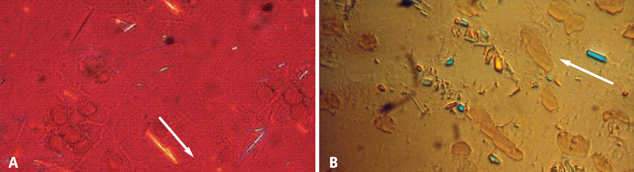
Occasionally, the aspirated joint does not appear to contain any joint fluid and the clinician may be concerned about the possibility of a “dry tap.” Other possible reasons include lack of experience with synovial fluid aspiration and evaluation, or limited access to the polarizing microscopes used to examine synovial fluid. Time is another factor; in a busy primary care practice, where patients are usually seen approximately every 7 to 11 minutes, there may not be time to aspirate a joint. The urgency of fluid examination is another issue, as synovial fluid must be examined immediately, since the crystals can become smaller, less numerous, and less birefringent with time.4
THE CLINICAL, OR PRESUMPTIVE, DIAGNOSIS
In the appropriate clinical scenario, a presumptive diagnosis of gout can be made on the basis of typical clinical features and the presence of hyperuricemia.1,2
Expert societies offer guidance, but no validation studies to date
Evidence-based recommendations for the diagnosis of gout from the European League Against Rheumatism (EULAR) state that in acute attacks, the rapid development of severe pain, swelling, and tenderness that peaks within 6 to 12 hours, especially with overlying erythema, is highly suggestive of crystal inflammation although not specific for gout.5 These recommendations further state that for typical presentations of gout (such as recurrent podagra [gouty pain in the great toe] with hyperuricemia), a clinical diagnosis alone is reasonably accurate.5
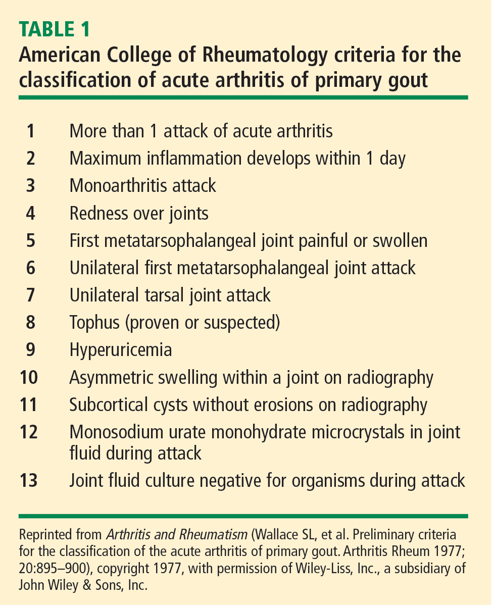
- The presence of urate crystals in joint fluid
- A tophus containing urate crystals
- Fulfillment of 6 or more of the criteria in Table 1.
No subsequent studies have been published on the validity or usefulness of any of these diagnostic criteria.
What must inform the presumptive diagnosis
Both the EULAR recommendations and the ACR criteria state that although the gold standard for diagnosing gout is the presence of urate crystals on synovial fluid analysis, a clinical diagnosis of gout can be made on the basis of certain patient criteria. This clinical, or presumptive, diagnosis of gout should be made based on the following:
- A careful patient and family history, including questions regarding comorbid conditions frequently associated with gout (such as hypertriglyceridemia, diabetes, coronary heart disease, hypertension, and the metabolic syndrome) and whether the patient has had previous similar episodes of acute joint pain and swelling in the absence of trauma
- Thorough identification of all current medications, some of which may be associated with hyperuricemia
- A thorough physical examination.
THE PHYSICAL EXAMINATION FOR GOUT
Examination of patients with a history suggestive of gout should include not only the joints but also the extensor surface of the forearms and feet. When patients are seen for a visit and gout is suspected, they should be instructed to remove their shoes and socks and roll up their sleeves to allow examination for evidence of tophi, which would suggest a past history of gouty arthritis. The ear, knee, and olecranon bursa are other common sites for tophi,3 so patients should also be asked to roll up their pants and sleeves and remove any head coverings.7 In the late stages of gouty arthritis, multiple joints may be involved, which can cause the condition to be confused with other diagnoses such as psoriatic arthritis or erosive osteoarthritis.7
ACUTE PRESENTATIONS OF GOUT
The typical gout presentation is remarkable for very intense pain that often occurs at night when the extremities are colder. Precipitation of urate in the distal extremities can occur when the extremities are horizontal and tend to become cold.8
Approximately 90% of initial gout attacks are monoarticular, leaving only 10% of cases that are oligoarticular or polyarticular.7 If more than one joint is involved, especially if the patient has a family history suggestive of gout or takes a medication that causes hyperuricemia, gout should be considered in the differential diagnosis even if the patient denies having a prior gout attack.
Frequently, patients will call their primary care physician during a gout attack but are not be able to schedule an appointment until after the attack has resolved. When possible, patients should be seen during the attack to confirm whether the attack is due to gout. A diagnosis of gout should not be made over the phone when a patient describes pain in the great toe, as only 50% of initial gout attacks occur in the great toe7 and it is not known what proportion of acute pain episodes in the great toe are attributable to gout. The most common cause of pain in the great toe is osteoarthritis.

SPECIAL CONSIDERATIONS FOR THE PRESUMPTIVE DIAGNOSIS OF GOUT
How long have acute attacks been occurring?
In a clinical scenario in which synovial fluid aspiration cannot be performed, the appropriateness of a presumptive diagnosis can be assessed by a discussion with the patient about how long he or she has been experiencing acute attacks of joint pain. If the attacks have occurred for more than 10 years, tophi will likely be present.3 After even longer periods, gout may become polyarticular.7 In postmenopausal women, the distal interphalangeal joints may be involved,3 which may lead to a misdiagnosis of osteoarthritis, as these joints are typically affected by osteoarthritis.
Is the patient taking a urate-raising medication?
Certain medications have been associated with hyper-uricemia, including cyclosporine and thiazide diuretics.9 If a patient has been taking one of these medications, gout should be considered in the differential diagnosis if the patient presents with acute joint pain.
It has been argued that a reduction in joint pain and swelling after the use of colchicine confirms a diagnosis of gout. However, other conditions—such as tendonitis, calcium pyrophosphate dihydrate (CPPD) crystal deposition disease (pseudogout),3 and rheumatoid arthritis (RA)—can also improve after treatment with colchicine.1
Be vigilant for fever
Another consideration in making a clinical diagnosis of gout is the association with a low-grade fever; these patients may feel as if they have the flu.8 Acute gout may also cause a high fever and an elevated white blood cell (WBC) count;3 in this situation, synovial fluid aspiration must be performed to exclude septic arthritis, either alone or in the presence of gouty arthritis. In situations where septic arthritis is suspected, an emergency visit to a rheumatologist is indicated for synovial fluid aspiration to be performed, as gout and sepsis can coexist.5 In such instances, Gram staining and culture of the synovial fluid should still be performed even if monosodium urate crystals are identified.5
MEASUREMENT OF SERUM URATE LEVELS
Measuring serum urate levels during an acute attack, treating the acute attack with anti-inflammatory medications, and reevaluating the patient in the office 2 weeks after the acute attack are all recommended in the management of a patient with gout. If the serum urate level was not elevated during the acute attack, it is likely to be elevated 2 weeks later if the patient has gout.10 Elevated levels of serum urate during the intercritical periods are predictive of future gout attacks.11 Measuring serum urate during the initial attack and then 2 weeks later yields two serum urate levels that can be compared to assist in considering a presumed diagnosis of gout. A study by Rigby and Wood concluded that in patients with low serum urate levels (< 4 mg/dL) 2 weeks following an inflammatory arthritis attack, a diagnosis of gout is unlikely.12
DIFFERENTIAL DIAGNOSIS OF GOUT
Rheumatoid arthritis
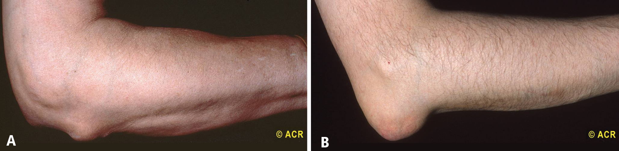
CPPD crystal deposition disease (pseudogout)
CPPD crystal deposition disease, or pseudogout, must also be included in the differential diagnosis of gout. This disease usually occurs in joints previously affected by osteoarthritis or joints that have been injured in the past.15 Attacks of CPPD crystal deposition disease commonly occur in the knee, in the wrist at the base of the thumb, or in the shoulder.15 Radiographic examination may reveal a line of calcification along the cartilage outlining the joint.15 Like gout, pseudogout attacks can occur spontaneously or after trauma, surgery, or a severe illness such as myocardial infarction or stroke.16
The presentation of pseudogout can be very similar to an acute attack of gout. The difference is seen when evaluating the crystals through a polarizing microscope. CPPD crystals are weakly positively birefringent (Figure 1B), in contrast to the negatively birefringent crystals seen with gout (Figure 1A).7 If a polarizing microscope is not available, the crystals usually can be distinguished by their differing shapes: urate crystals are fine and needlelike, whereas CPPD crystals are rhomboid (Figure 1).
Septic arthritis
When the differential diagnosis includes septic arthritis, the joint must be aspirated; a presumed diagnosis cannot be made. Among patients with an acute gouty attack, low-grade fever is reported during the attack in 29% of gout patients and 38% of patients with CPPD crystal deposition disease.14 Temperatures of 101°F or higher are not usually seen in patients with gout or CPPD crystal deposition disease and suggest an infection, although patients with septic arthritis may be afebrile, especially if they are taking immunosuppressive therapy or glucocorticoids, which can inhibit a febrile response. Synovial fluid analysis in patients with gout and septic arthritis can reveal WBC counts above 100,000 per mm3, whereas synovial fluid WBC counts above 50,000 per mm3 are more common in infection.
As noted earlier, gout and septic arthritis can coexist. In a patient presenting with a fever and a warm erythematous swollen joint, synovial fluid aspiration must be performed and evaluated for the presence of crystals and bacteria. The patient may require treatment for both causes of acute monoarticular arthritis.
In a patient undergoing renal dialysis, where gout or pseudogout can occur and where there is frequent intravascular manipulation, a septic joint can occur simultaneously.3,14 In this situation, not only must joint aspiration be performed, but the synovial fluid also needs to be evaluated for both crystals and bacteria. Again, the patient may require treatment for both causes of acute monoarticular arthritis.
CONCLUSIONS
The gold standard for diagnosing gout remains synovial fluid aspiration and analysis. In clinical situations when joint aspiration cannot be performed, the EULAR recommendations5 and the ACR criteria6 provide guidance for making a clinical or presumptive diagnosis of gout. A thorough patient history—both personal and family—and physical examination are critical in making a presumed diagnosis of gout. If the patient presents during an acute attack, serum urate measurement may be useful in making a clinical diagnosis if it reveals an elevated level. When the patient returns for follow-up 2 weeks later, a second serum urate measurement should be taken to allow comparison of the two levels. If the serum urate level is elevated at the follow-up visit, the EULAR recommendations state that a clinical diagnosis of gout can be made if the patient had an acute attack of arthritis in the great toe.
As noted in the EULAR recommendations, the future research agenda should include validating the clinical manifestations of gout against a diagnosis established by identification of urate crystals on synovial fluid analysis.5 Until this task can be completed, clinicians should become familiarized with the technique of joint aspiration so that in situations where a clinical or presumptive diagnosis of gout cannot be made—including cases where the differential diagnosis includes a septic joint—clinicians will be able to perform aspiration with confidence.
- Jelley MJ, Wortmann R. Practical steps in the diagnosis and management of gout. BioDrugs 2000; 14:99–107.
- Eggebeen AT. Gout: an update. Am Fam Physician 2007; 76:801–808, 811–812.
- Rott KT, Agudelo CA. Gout. JAMA 2003; 289:2857–2860.
- Poór G, Mituszova M. History, classification and epidemiology of crystal-related arthropathies. In: Hochberg MC, Silman AJ, Smolen JS, Weinblatt ME, Weisman MH, eds. Rheumatology. 3rd ed, vol 2. Edinburgh: Mosby; 2003:1893–1901.
- Zhang W, Doherty M, Pascual E, et al. EULAR evidence based recommendations for gout. Part I: Diagnosis. Report of a task force of the Standing Committee for International Clinical Studies Including Therapeutics (ESCISIT). Ann Rheum Dis 2006; 65:1301–1311.
- Wallace SL, Robinson H, Masi AT, Decker JL, McCarty DJ, Yü TF. Preliminary criteria for the classification of the acute arthritis of primary gout. Arthritis Rheum 1977; 20:895–900.
- Harris MD, Siegel LB, Alloway JA. Gout and hyperuricemia. Am Fam Physician 1999; 59:925–934.
- Saag KG, Mikuls TR. Recent advances in the epidemiology of gout. Curr Rheumatol Rep 2005; 7:235–241.
- Edwards NL. Gout. B. Clinical and laboratory features. In: Klippel JH, Crofford LJ, Stone JH, Weyand CM, eds. Primer on the Rheumatic Diseases. 12th ed. Atlanta, GA: Arthritis Foundation; 2001:313–319.
- Urano W, Yamanaka H, Tsutani H, et al. The inflammatory process in the mechanism of decreased serum uric acid concentrations during acute gouty arthritis. J Rheumatol 2002; 29:1950–1953.
- Shoji A, Yamanaka H, Kamatani N. A retrospective study of the relationship between serum urate level and recurrent attacks of gouty arthritis: evidence for reduction of recurrent gouty arthritis with anti-hyperuricemic therapy. Arthritis Rheum 2004; 51:321–325.
- Rigby AS, Wood PH. Serum uric acid levels and gout: what does this herald for the population? Clin Exp Rheumatol 1994; 12:395–400.
- George DL. Skin and rheumatic disease. In: Hochberg MC, Silman AJ, Smolen JS, Weinblatt ME, Weisman MH, eds. Rheumatology. 3rd ed, vol 1. Edinburgh: Mosby; 2003:279–292.
- Ho G Jr, DeNuccio M. Gout and pseudogout in hospitalized patients. Arch Intern Med 1993; 153:2787–2790.
- Agarwal AK. Gout and pseudogout. Prim Care 1993; 20:839–855.
- Halverson PB, Ryan LM. Arthritis associated with calcium-containing crystals. In: Klippel JH, Crofford LJ, Stone JH, Weyand CM, eds. Primer on the Rheumatic Diseases. 12th ed. Atlanta, GA: Arthritis Foundation; 2001:299–306.
The presence of urate crystals in synovial fluid is the gold standard for diagnosing gout,1 yet clinicians—both primary care physicians and rheumatologists—may not routinely perform synovial fluid analysis even when evaluating a patient who presents with an acute inflammatory arthritis.2 This paper discusses the various reasons why this is so and reviews several important resulting clinical issues: how a presumptive diagnosis of gout is made, when to measure the serum urate level, and special considerations in the differential diagnosis.
SYNOVIAL FLUID ANALYSIS: WHY IS THE GOLD STANDARD NOT MORE ROUTINE?
Occasionally, the aspirated joint does not appear to contain any joint fluid and the clinician may be concerned about the possibility of a “dry tap.” Other possible reasons include lack of experience with synovial fluid aspiration and evaluation, or limited access to the polarizing microscopes used to examine synovial fluid. Time is another factor; in a busy primary care practice, where patients are usually seen approximately every 7 to 11 minutes, there may not be time to aspirate a joint. The urgency of fluid examination is another issue, as synovial fluid must be examined immediately, since the crystals can become smaller, less numerous, and less birefringent with time.4
THE CLINICAL, OR PRESUMPTIVE, DIAGNOSIS
In the appropriate clinical scenario, a presumptive diagnosis of gout can be made on the basis of typical clinical features and the presence of hyperuricemia.1,2
Expert societies offer guidance, but no validation studies to date
Evidence-based recommendations for the diagnosis of gout from the European League Against Rheumatism (EULAR) state that in acute attacks, the rapid development of severe pain, swelling, and tenderness that peaks within 6 to 12 hours, especially with overlying erythema, is highly suggestive of crystal inflammation although not specific for gout.5 These recommendations further state that for typical presentations of gout (such as recurrent podagra [gouty pain in the great toe] with hyperuricemia), a clinical diagnosis alone is reasonably accurate.5
- The presence of urate crystals in joint fluid
- A tophus containing urate crystals
- Fulfillment of 6 or more of the criteria in Table 1.
No subsequent studies have been published on the validity or usefulness of any of these diagnostic criteria.
What must inform the presumptive diagnosis
Both the EULAR recommendations and the ACR criteria state that although the gold standard for diagnosing gout is the presence of urate crystals on synovial fluid analysis, a clinical diagnosis of gout can be made on the basis of certain patient criteria. This clinical, or presumptive, diagnosis of gout should be made based on the following:
- A careful patient and family history, including questions regarding comorbid conditions frequently associated with gout (such as hypertriglyceridemia, diabetes, coronary heart disease, hypertension, and the metabolic syndrome) and whether the patient has had previous similar episodes of acute joint pain and swelling in the absence of trauma
- Thorough identification of all current medications, some of which may be associated with hyperuricemia
- A thorough physical examination.
THE PHYSICAL EXAMINATION FOR GOUT
Examination of patients with a history suggestive of gout should include not only the joints but also the extensor surface of the forearms and feet. When patients are seen for a visit and gout is suspected, they should be instructed to remove their shoes and socks and roll up their sleeves to allow examination for evidence of tophi, which would suggest a past history of gouty arthritis. The ear, knee, and olecranon bursa are other common sites for tophi,3 so patients should also be asked to roll up their pants and sleeves and remove any head coverings.7 In the late stages of gouty arthritis, multiple joints may be involved, which can cause the condition to be confused with other diagnoses such as psoriatic arthritis or erosive osteoarthritis.7
ACUTE PRESENTATIONS OF GOUT
The typical gout presentation is remarkable for very intense pain that often occurs at night when the extremities are colder. Precipitation of urate in the distal extremities can occur when the extremities are horizontal and tend to become cold.8
Approximately 90% of initial gout attacks are monoarticular, leaving only 10% of cases that are oligoarticular or polyarticular.7 If more than one joint is involved, especially if the patient has a family history suggestive of gout or takes a medication that causes hyperuricemia, gout should be considered in the differential diagnosis even if the patient denies having a prior gout attack.
Frequently, patients will call their primary care physician during a gout attack but are not be able to schedule an appointment until after the attack has resolved. When possible, patients should be seen during the attack to confirm whether the attack is due to gout. A diagnosis of gout should not be made over the phone when a patient describes pain in the great toe, as only 50% of initial gout attacks occur in the great toe7 and it is not known what proportion of acute pain episodes in the great toe are attributable to gout. The most common cause of pain in the great toe is osteoarthritis.
SPECIAL CONSIDERATIONS FOR THE PRESUMPTIVE DIAGNOSIS OF GOUT
How long have acute attacks been occurring?
In a clinical scenario in which synovial fluid aspiration cannot be performed, the appropriateness of a presumptive diagnosis can be assessed by a discussion with the patient about how long he or she has been experiencing acute attacks of joint pain. If the attacks have occurred for more than 10 years, tophi will likely be present.3 After even longer periods, gout may become polyarticular.7 In postmenopausal women, the distal interphalangeal joints may be involved,3 which may lead to a misdiagnosis of osteoarthritis, as these joints are typically affected by osteoarthritis.
Is the patient taking a urate-raising medication?
Certain medications have been associated with hyper-uricemia, including cyclosporine and thiazide diuretics.9 If a patient has been taking one of these medications, gout should be considered in the differential diagnosis if the patient presents with acute joint pain.
It has been argued that a reduction in joint pain and swelling after the use of colchicine confirms a diagnosis of gout. However, other conditions—such as tendonitis, calcium pyrophosphate dihydrate (CPPD) crystal deposition disease (pseudogout),3 and rheumatoid arthritis (RA)—can also improve after treatment with colchicine.1
Be vigilant for fever
Another consideration in making a clinical diagnosis of gout is the association with a low-grade fever; these patients may feel as if they have the flu.8 Acute gout may also cause a high fever and an elevated white blood cell (WBC) count;3 in this situation, synovial fluid aspiration must be performed to exclude septic arthritis, either alone or in the presence of gouty arthritis. In situations where septic arthritis is suspected, an emergency visit to a rheumatologist is indicated for synovial fluid aspiration to be performed, as gout and sepsis can coexist.5 In such instances, Gram staining and culture of the synovial fluid should still be performed even if monosodium urate crystals are identified.5
MEASUREMENT OF SERUM URATE LEVELS
Measuring serum urate levels during an acute attack, treating the acute attack with anti-inflammatory medications, and reevaluating the patient in the office 2 weeks after the acute attack are all recommended in the management of a patient with gout. If the serum urate level was not elevated during the acute attack, it is likely to be elevated 2 weeks later if the patient has gout.10 Elevated levels of serum urate during the intercritical periods are predictive of future gout attacks.11 Measuring serum urate during the initial attack and then 2 weeks later yields two serum urate levels that can be compared to assist in considering a presumed diagnosis of gout. A study by Rigby and Wood concluded that in patients with low serum urate levels (< 4 mg/dL) 2 weeks following an inflammatory arthritis attack, a diagnosis of gout is unlikely.12
DIFFERENTIAL DIAGNOSIS OF GOUT
Rheumatoid arthritis
CPPD crystal deposition disease (pseudogout)
CPPD crystal deposition disease, or pseudogout, must also be included in the differential diagnosis of gout. This disease usually occurs in joints previously affected by osteoarthritis or joints that have been injured in the past.15 Attacks of CPPD crystal deposition disease commonly occur in the knee, in the wrist at the base of the thumb, or in the shoulder.15 Radiographic examination may reveal a line of calcification along the cartilage outlining the joint.15 Like gout, pseudogout attacks can occur spontaneously or after trauma, surgery, or a severe illness such as myocardial infarction or stroke.16
The presentation of pseudogout can be very similar to an acute attack of gout. The difference is seen when evaluating the crystals through a polarizing microscope. CPPD crystals are weakly positively birefringent (Figure 1B), in contrast to the negatively birefringent crystals seen with gout (Figure 1A).7 If a polarizing microscope is not available, the crystals usually can be distinguished by their differing shapes: urate crystals are fine and needlelike, whereas CPPD crystals are rhomboid (Figure 1).
Septic arthritis
When the differential diagnosis includes septic arthritis, the joint must be aspirated; a presumed diagnosis cannot be made. Among patients with an acute gouty attack, low-grade fever is reported during the attack in 29% of gout patients and 38% of patients with CPPD crystal deposition disease.14 Temperatures of 101°F or higher are not usually seen in patients with gout or CPPD crystal deposition disease and suggest an infection, although patients with septic arthritis may be afebrile, especially if they are taking immunosuppressive therapy or glucocorticoids, which can inhibit a febrile response. Synovial fluid analysis in patients with gout and septic arthritis can reveal WBC counts above 100,000 per mm3, whereas synovial fluid WBC counts above 50,000 per mm3 are more common in infection.
As noted earlier, gout and septic arthritis can coexist. In a patient presenting with a fever and a warm erythematous swollen joint, synovial fluid aspiration must be performed and evaluated for the presence of crystals and bacteria. The patient may require treatment for both causes of acute monoarticular arthritis.
In a patient undergoing renal dialysis, where gout or pseudogout can occur and where there is frequent intravascular manipulation, a septic joint can occur simultaneously.3,14 In this situation, not only must joint aspiration be performed, but the synovial fluid also needs to be evaluated for both crystals and bacteria. Again, the patient may require treatment for both causes of acute monoarticular arthritis.
CONCLUSIONS
The gold standard for diagnosing gout remains synovial fluid aspiration and analysis. In clinical situations when joint aspiration cannot be performed, the EULAR recommendations5 and the ACR criteria6 provide guidance for making a clinical or presumptive diagnosis of gout. A thorough patient history—both personal and family—and physical examination are critical in making a presumed diagnosis of gout. If the patient presents during an acute attack, serum urate measurement may be useful in making a clinical diagnosis if it reveals an elevated level. When the patient returns for follow-up 2 weeks later, a second serum urate measurement should be taken to allow comparison of the two levels. If the serum urate level is elevated at the follow-up visit, the EULAR recommendations state that a clinical diagnosis of gout can be made if the patient had an acute attack of arthritis in the great toe.
As noted in the EULAR recommendations, the future research agenda should include validating the clinical manifestations of gout against a diagnosis established by identification of urate crystals on synovial fluid analysis.5 Until this task can be completed, clinicians should become familiarized with the technique of joint aspiration so that in situations where a clinical or presumptive diagnosis of gout cannot be made—including cases where the differential diagnosis includes a septic joint—clinicians will be able to perform aspiration with confidence.
- Jelley MJ, Wortmann R. Practical steps in the diagnosis and management of gout. BioDrugs 2000; 14:99–107.
- Eggebeen AT. Gout: an update. Am Fam Physician 2007; 76:801–808, 811–812.
- Rott KT, Agudelo CA. Gout. JAMA 2003; 289:2857–2860.
- Poór G, Mituszova M. History, classification and epidemiology of crystal-related arthropathies. In: Hochberg MC, Silman AJ, Smolen JS, Weinblatt ME, Weisman MH, eds. Rheumatology. 3rd ed, vol 2. Edinburgh: Mosby; 2003:1893–1901.
- Zhang W, Doherty M, Pascual E, et al. EULAR evidence based recommendations for gout. Part I: Diagnosis. Report of a task force of the Standing Committee for International Clinical Studies Including Therapeutics (ESCISIT). Ann Rheum Dis 2006; 65:1301–1311.
- Wallace SL, Robinson H, Masi AT, Decker JL, McCarty DJ, Yü TF. Preliminary criteria for the classification of the acute arthritis of primary gout. Arthritis Rheum 1977; 20:895–900.
- Harris MD, Siegel LB, Alloway JA. Gout and hyperuricemia. Am Fam Physician 1999; 59:925–934.
- Saag KG, Mikuls TR. Recent advances in the epidemiology of gout. Curr Rheumatol Rep 2005; 7:235–241.
- Edwards NL. Gout. B. Clinical and laboratory features. In: Klippel JH, Crofford LJ, Stone JH, Weyand CM, eds. Primer on the Rheumatic Diseases. 12th ed. Atlanta, GA: Arthritis Foundation; 2001:313–319.
- Urano W, Yamanaka H, Tsutani H, et al. The inflammatory process in the mechanism of decreased serum uric acid concentrations during acute gouty arthritis. J Rheumatol 2002; 29:1950–1953.
- Shoji A, Yamanaka H, Kamatani N. A retrospective study of the relationship between serum urate level and recurrent attacks of gouty arthritis: evidence for reduction of recurrent gouty arthritis with anti-hyperuricemic therapy. Arthritis Rheum 2004; 51:321–325.
- Rigby AS, Wood PH. Serum uric acid levels and gout: what does this herald for the population? Clin Exp Rheumatol 1994; 12:395–400.
- George DL. Skin and rheumatic disease. In: Hochberg MC, Silman AJ, Smolen JS, Weinblatt ME, Weisman MH, eds. Rheumatology. 3rd ed, vol 1. Edinburgh: Mosby; 2003:279–292.
- Ho G Jr, DeNuccio M. Gout and pseudogout in hospitalized patients. Arch Intern Med 1993; 153:2787–2790.
- Agarwal AK. Gout and pseudogout. Prim Care 1993; 20:839–855.
- Halverson PB, Ryan LM. Arthritis associated with calcium-containing crystals. In: Klippel JH, Crofford LJ, Stone JH, Weyand CM, eds. Primer on the Rheumatic Diseases. 12th ed. Atlanta, GA: Arthritis Foundation; 2001:299–306.
KEY POINTS
- If the serum urate level was not elevated when measured during an acute attack of arthritis, it will likely be elevated at 2-week follow-up if the patient does indeed have gout.
- Gouty tophi are typically found in the olecranon bursa, whereas rheumatoid nodules are usually located on the extensor surface of the forearm.
- Urate crystals of gout are negatively bifringent and fine and needlelike in shape, whereas the crystals of pseudogout are weakly positively birefringent and rhomboid.
- Gout and septic arthritis can coexist; when the differential diagnosis includes septic arthritis, joint aspiration is required.
- Until criteria for the presumptive diagnosis of gout are validated, clinicians should become familiar with the technique of joint aspiration.