The United States has seen a decline in fatal myocardial infarctions, largely thanks to early detection of coronary artery disease. Current guidelines on assessment of cardiovascular risk still rely on the traditional 10-year risk model in clinical practice. However, the predictive value of this approach is only moderate, and many coronary events occur in people considered to be at low or intermediate risk.
Coronary artery calcium scoring has emerged as a means of risk stratification by direct measurement of disease. Primary care providers are either using it or are seeing it used by consulting physicians, and its relatively low cost and ease of performance have contributed to its widespread use. However, downstream costs, radiation exposure, and lack of randomized controlled trials have raised concerns.
This article reviews the usefulness and pitfalls of coronary artery calcium scoring, providing a better understanding of the test, its limitations, and the interpretation of results.
ATHEROSCLEROSIS AND CALCIUM
Figure 1. Pathogenic mechanism of atherosclerotic lesions and its relationship to the coronary artery calcium (CAC) score. A type 1 lesion (not depicted) contains lipoproteins that initiate an inflammatory response. A type 2 lesion contains an accumulation of foam cells. The type 3 lesion contains collections of extracellular lipid droplets. Eventually, these extracellular lipid pools form a lipid core, and a type 4 lesion is created. With time, this core develops a fibrous connective-tissue thickening that can calcify and give rise to a type 5 lesion detectable by imaging. Type 6 is a complicated lesion that can include thrombus from plaque rupture.Atherosclerosis begins in the first few decades of life with a fatty streak in which lipoproteins are deposited in the intimal and medial layers of blood vessels (Figure 1). Inflammatory cells such as macrophages and foam cells are then recruited to the areas of deposition where they cause apoptosis, creating a necrotic core with calcium deposits.1–3
As the calcium deposits grow, they can be detected by imaging tests such as computed tomography (CT), and quantified to assess the extent of disease.4
CALCIFICATION AND CORONARY ARTERY DISEASE
Coronary calcification occurs almost exclusively in atherosclerosis. Several autopsy studies5,6 and histopathologic studies7 have shown a direct relationship between the extent of calcification and atherosclerotic disease.
Sangiorgi et al7 performed a histologic analysis of 723 coronary artery segments. The amount of calcium correlated well with the area of plaque:
r = 0.89, P < .0001 in the left anterior descending artery
r = 0.7, P < .001 in the left circumflex artery
r = 0.89, P < .0001 in the right coronary artery.
Coronary artery calcium has also been associated with obstructive coronary artery disease in studies using intravascular ultrasonography and optical coherence tomography.8,9
TECHNICAL INFORMATION ABOUT THE TEST
First-generation CT scanners used for calcium scoring in the 1980s were electron-beam systems in which a stationary x-ray tube generated an oscillating electron beam, which was reflected around the patient table.10 A single, stationary detector ring captured the images.
These systems have been replaced by multidetector scanners, in which the x-ray tube and multiple rows of detectors are combined in a gantry that rotates at high speed around the patient.
Coronary calcium is measured by noncontrast CT of the heart. Thus, there is no risk of contrast-induced nephropathy or allergic reactions. Images are acquired while the patient holds his or her breath for 3 to 5 seconds. Electrocardiographic gating is used to reduce motion artifact.11,12 With modern scanners, the effective radiation dose associated with calcium testing is as low as 0.5 to 1.5 mSv,13,14 ie, about the same dose as that with mammography. The entire test takes 10 to 15 minutes.
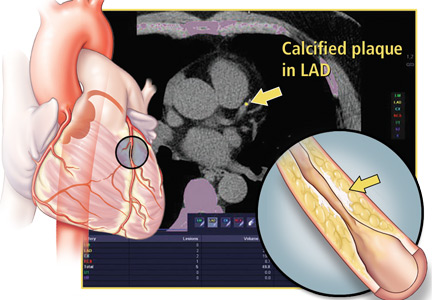
Figure 2. A screenshot from a standard calcium scoring program. The images show a small calcified plaque in the mid-left anterior descending artery (left upper panel, colored yellow), left circumflex artery (right upper panel, colored blue), and right coronary artery (right lower panel, colored red). The table in the left lower panel lists the results of the calcium score. (The Agatston score is listed as “Score”). The graph in the right lower panel shows the results of the individual patient relative to an age- and sex-matched patient population (eg, the MESA trial). This patient has a score of 62.2, indicating relatively mild calcification and falling on the 75% percentile for age, sex, and ethnicity in a control group.Coronary calcium on CT is most commonly quantified using the Agatston score. Calcification is defined as a hyperattenuating lesion above the threshold of 130 Hounsfield units with an area of 3 or more pixels (1 mm2). The score is calculated based on the area of calcification per coronary cross-section, multiplied by a factor that depends on the maximum amount of calcium in a cross-section (a weighted value system based on Hounsfield units of dense calcification in each major coronary artery).15 The sum of calcium in the right coronary, left anterior descending, and left circumflex arteries gives the total Agatston calcium score.
The results fall into 4 categories, which correlate with the severity of coronary artery disease, ranging from no significant disease to severe disease (Table 1). Other scores, which are not commonly used, include the calcium volume score16 and the calcium mass score.17Figure 2 shows a screenshot from a coronary artery calcium scoring program.
CALCIUM SCORING AS A DIAGNOSTIC TOOL
Early multicenter studies evaluated the utility of calcium scoring to predict coronary stenosis in patients who underwent both cardiac CT and coronary angiography. The sensitivity of calcium scoring for angiographically significant disease was high (95%), but its specificity was low (about 44%).18
Budoff et al,19 reviewing these and subsequent results, concluded that the value of calcium scoring is its high negative predictive value (about 98%); a negative score (no calcification) is strongly associated with the absence of obstructive coronary disease.
Blaha et al20 concluded that a score of 0 would indicate that the patient had a low risk of cardiovascular disease. A test with these characteristics is helpful in excluding cardiovascular disease or at least in determining that it is less likely to be present in a patient deemed to be at intermediate risk.
CALCIUM SCORING AS A PROGNOSTIC TOOL
Early on, investigators recognized the value of calcium scoring in predicting the risk of future cardiovascular events and death.21–25
Predicting cardiovascular events
Pletcher et al21 performed a meta-analysis of studies that measured calcification in asymptomatic patients with subsequent follow-up. The summary-adjusted relative risk of cardiac events such as myocardial infarction, coronary artery revascularization, and coronary heart disease-related death rose with the calcium score:
2.1 (95% confidence interval [CI] 1.6–2.9) with a score of 1 to 100
4.2 (95% CI 2.5–7.2) with scores of 101 to 400
7.2 (95% CI 3.9-13.0) with scores greater than 400.
The meta-analysis was limited in that it included only 4 studies, which were observational.
Kavousi et al,22 in a subsequent meta-analysis of 6,739 women at low risk of atherosclerotic cardiovascular disease based on the American College of Cardiology/American Heart Association (ACC/AHA) pooled cohort equation (10-year risk < 7.5%), found that 36.1% had calcium scores greater than 0. Compared with those whose score was 0, those with higher scores had a higher risk of atherosclerotic cardiovascular disease events. The incidence rates per 1,000 person-years were 1.41 vs 4.33 (relative risk 2.92, 95% CI 2.02–3.83; multivariable-adjusted hazard ratio 2.04, 95% CI 1.44–2.90). This study was limited because the population was mostly of European descent, making it less generalizable to non-European populations.
Calcium scoring has also been shown to be a strong predictor of incident cardiovascular events across different races beyond traditional risk factors such as hypertension, hyperlipidemia, and tobacco use.
Detrano et al,23 in a study of 6,722 patients with diverse ethnic backgrounds, found that the adjusted risk of a coronary event was increased by a factor of 7.73 for calcium scores between 101 and 300 and by a factor of 9.67 for scores above 300 (P < .001). A limitation of this study was that the patients and physicians were informed of the scores, which could have led to bias.
Carr et al24 found an association between calcium and coronary heart disease in a younger population (ages 32–46). In 12.5 years of follow-up, the hazard ratio for cardiovascular events increased exponentially with the calcium score:
2.6 (95% CI 1.0–5.7, P = .03) with calcium scores of 1 through 19
9.8 (95% CI 4.5–20.5, P < .001) with scores greater than 100.
Predicting mortality
Budoff et al,25 in an observational study of 25,253 patients, found coronary calcium to be an independent predictor of mortality in a multivariable model controlling for age, sex, ethnicity, and cardiac risk factors (model chi-square = 2,017, P < .0001). However, most of the patients were already known to have cardiac risk factors, making the study findings less generalizable to the general population.
Nasir et al26 found that mortality rates rose with the calcium score in a study with 44,052 participants. The annualized mortality rates per 1,000 person-years were:
0.87 (95% CI 0.72–1.06) with a score of 0
2.97 (95% CI 2.61–3.37) with scores of 1–100
6.90 (95% CI 6.02–7.90) with scores of 101–400
17.68 (95% CI 5.93–19.62) with scores higher than 400.
The mortality rate also rose with the number of traditional risk factors present, ie, current tobacco use, dyslipidemia, diabetes mellitus, hypertension, and family history of coronary artery disease. Interestingly, those with no risk factors but a calcium score greater than 400 had a higher mortality rate than those with no coronary calcium but more than 3 risk factors (16.89 per 1,000 person-years vs 2.72 per 1,000 person years). As in the previous study, the patient population that was analyzed was at high risk and therefore the findings are not generalizable.
Shaw et al27 found that patients without symptoms but with elevated coronary calcium scores had higher all-cause mortality rates at 15 years than those with a score of 0. The difference remained significant after Cox regression was performed, adjusting for traditional risk factors.
Coronary artery calcium scoring vs other risk-stratification methods
Current guidelines on assessing risk still rely on the traditional 10-year risk model in clinical practice.25 Patients are thus classified as being at low, intermediate, or high risk based on their probability of developing a cardiovascular event or cardiovascular disease-related death in the subsequent 10 years.
However, the predictive value of this approach is only moderate,28 and a significant number of cardiovascular events, including sudden cardiac death, occur in people who were believed to be at low or intermediate risk according to traditional risk factor-based predictions. Because risk scores are strongly influenced by age,29 they are least reliable in young adults.30
Akosah et al31 reviewed the records of 222 young adults (women age 55 or younger, men age 65 or younger) who presented with their first myocardial infarction, and found that only 25% would have qualified for primary prevention pharmacologic treatments according to the National Cholesterol Education Program III guidelines.32,33 Similar findings have been reported regarding previous versions of the risk scores.33
Thus, risk predictions based exclusively on traditional risk factors are not sensitive for detecting young individuals at increased risk, and lead to late treatment of young adults with atherosclerosis, which may be a less effective strategy.34
The reliance on age in risk algorithms also results in low specificity in elderly adults. Using risk scores, elderly adults are systematically stratified in higher risk categories, expanding the indication for statin therapy to almost all men age 65 or older regardless of their actual vascular health, according to current clinical practice guidelines.35,36
Risk scores are based on self-reported history and single-day measurements, since this kind of information is readily available to the physician in the clinic. Moreover, our knowledge about genetic and epigenetic factors associated with the development of atherosclerosis is still in its infancy, with current guidelines not supporting genetic testing as part of cardiovascular risk assessment.37 Thus, a reliable measure of an individual’s lifelong exposure to a number of environmental and genetic factors that may affect cardiovascular health appears unfeasible.
Atherosclerosis is a process in which interactions between genetic, epigenetic, environmental, and traditional risk factors result in subclinical inflammation that could develop into clinically significant disease. Therefore, subclinical coronary atherosclerosis has been shown to be a strong predictor of future incident cardiovascular disease events and death. Thus, alternative approaches that directly measure disease, such as calcium scoring, may help further refine risk stratification of cardiovascular disease.
The MESA trial (Multi-Ethnic Study of Atherosclerosis), for instance, in 6,814 participants, found coronary calcium to provide better discrimination and risk reclassification than the ankle-brachial index, high sensitivity C-reactive protein level, and family history.38 Coronary calcium also had the highest incremental improvement of the area under the receiver operating curve when added to the Framingham Risk Score (0.623 vs 0.784).
Reclassifying cardiovascular risk also has implications regarding whether to start therapies such as statins and aspirin.
For considering statin therapy
Nasir et al39 showed that, in patients eligible for statin therapy by the pooled cohort equation, the absence of coronary artery calcium reclassified approximately one-half of candidates as not eligible for statin therapy. The number needed to treat to prevent an atherosclerotic cardiovascular event in the population who were recommended a statin was 64 with a calcium score of 0, and 24 with a calcium score greater than 100. In the population for whom a statin was considered, the number needed to treat was 223 with a calcium score of 0 and 46 for those with a score greater than 100. Moreover, 57% of intermediate-risk patients and 41% of high-risk patients based on the Framingham Risk Score were found to have a calcium score of 0, implying that these patients may actually be at a lower risk.
The Society of Cardiovascular Computed Tomography guidelines40 say that statin therapy can be considered in patients who have a calcium score greater than 0.
For considering aspirin therapy
Miedema et al41 studied the role of coronary artery calcium in guiding aspirin therapy in 4,229 participants in the MESA trial who were not taking aspirin at baseline. Those with a calcium score higher than 100 had a number needed to treat of 173 in the group with a Framingham Risk Score less than 10% and 92 with a Framingham Risk Score of 10% or higher. The estimated number needed to harm for a major bleeding event was 442. For those who had a score of 0, the estimated number needed to treat was 2,036 for a Framingham Risk Score less than 10% and 808 for a Framingham Risk Score of 10% or higher, with an estimated number needed to harm of 442 for a major bleeding event.
The Society of Cardiovascular Computed Tomography guidelines40 recommend considering aspirin therapy for patients with a coronary calcium score of more than 100.
McClelland et al42 developed a MESA risk score to predict 10-year risk of coronary heart disease using the traditional risk factors along with coronary calcium. The score was validated externally with 2 separate longitudinal studies. Thus, this may serve as another tool to help providers further risk-stratify patients.
COST-EFFECTIVENESS OF THE TEST
As coronary calcium measurement began to be widely used, concerns were raised about the lack of data on its cost-effectiveness.
Cost-effectiveness depends not only on patient selection but also on the cost of therapy. For example, if the cost of a generic statin is $85 per year, then calcium scoring would not be beneficial. However, if the cost of a statin is more than $200, then calcium scoring would be much more cost-effective, offering a way to avoid treating some patients who do not need to be treated.43
Hong et al43 showed that coronary calcium testing was cost-effective when the patient and physician share decision-making about initiating statin therapy. This is especially important if the patient has financial limitations, is concerned about side effects, or wants to avoid taking unnecessary medications.
RISKS AND DOWNSIDES OF CALCIUM SCORING
According to some reports, $8.5 billion is spent annually for low-value care.44 Many of the 80 million CT scans performed annually in the United States are believed to be unnecessary and may lead to additional testing to investigate incidental findings.45
Growing use of coronary calcium measurement has raised similar concerns about radiation exposure, healthcare costs, and increased downstream testing triggered by the detection of incidental noncardiac findings. For instance, Onuma et al46 reported that, in 503 patients undergoing CT to evaluate coronary artery disease, noncardiac findings were seen in 58.1% of them, but only 22.7% of the 503 had clinically significant findings.
Some of these concerns have been addressed. Modern scanners can acquire images in only a few seconds, entailing lower radiation doses than in the past.13,14 The cost of the test is currently less than $100 in many US metropolitan areas.47 However, further studies are needed to adequately and cost-effectively guide follow-up imaging of incidental noncardiac findings.48
An important limitation of calcium scoring for risk assessment is that no randomized controlled trial has evaluated the impact of preventive interventions guided by calcium scores on hard event outcomes. It can be argued that there have been plenty of observational studies that have shown the benefit of coronary calcium scoring when judiciously done in the appropriate population.49 Similarly, no randomized controlled trial has tested the pooled cohort equation and the application of statins based on its use with the current guidelines. The feasibility and cost of a large randomized controlled trial to assess outcomes after coronary artery calcium measurement must also be considered.
Another limitation of coronary calcium scoring is that it cannot rule out the presence of noncalcified atherosclerotic plaque, which often is more unstable and prone to rupture.
In addition, calcification in the coronary vascular bed (even if severe) does not necessarily mean there is clinically relevant coronary stenosis. For instance, an asymptomatic patient could have a coronary artery calcium score higher than 100 and then get a coronary angiogram that reveals only a 30% lesion in the left anterior descending coronary artery. This is because accumulation of (calcified) plaque in the vessel wall is accommodated by expansion of vessel diameter, maintaining luminal dimensions (positive remodeling). By definition, this patient does have coronary artery disease but would be best served by medical management. This could have been determined without an invasive test in an otherwise asymptomatic patient. Thus, performing coronary angiography based on a coronary artery calcium score alone would not have changed this patient’s management and may have exposed the patient to risks of procedural complications, in addition to extra healthcare costs. Therefore, the presence or absence of symptoms should guide the clinician on whether to pursue stress testing for invasive coronary angiography based on the appropriate use criteria.50,51
WHO SHOULD BE TESTED?
In the ACC/AHA 2013 guidelines,37 coronary calcium scoring has a class IIB recommendation in scenarios where it may appear that the risk-based treatment decision is uncertain after formal risk estimation has been done. As discussed above, a score higher than 100 could be a rationale for starting aspirin therapy, and a score higher than 0 for statin therapy. The current guidelines also mention that the coronary calcium score is comparable to other predictors such as the C-reactive protein level and the ankle-brachial index.
Compared with the ACC/AHA guidelines, the 2016 Society of Cardiovascular Computed Tomography guidelines and expert consensus recently have added more specifics in terms of using this test for asymptomatic patients at intermediate risk (10-year risk of atherosclerotic cardiovascular disease 5%–20%) and in selected patients with a family history of premature coronary artery disease and 10-year risk less than 5%.40,52 The 2010 ACC/AHA guidelines were more specific, offering a class IIA recommendation for patients who were at intermediate risk (Framingham Risk Score 10%–20%).53
The ACC/AHA cited cost and radiation exposure as reasons they did not give coronary calcium measurement a stronger recommendation.37 However, as data continue to come in, the guidelines may change, especially since low-dose radiation tools are being used for cancer screening (lungs and breast) and since the cost has declined over the past decade.
OUR APPROACH
Given the negative predictive value of the coronary calcium score, our approach has been to use this test in asymptomatic patients who are found to be at intermediate risk of atherosclerotic cardiovascular disease based on the ACC/AHA risk calculation and are reluctant to start pharmacologic therapy, or who want a more personalized measure of coronary artery disease. This is preceded by a lengthy patient-physician discussion about the risks and benefits of the test.54
The patient’s risk can then be further clarified and possibly reclassified as either low or high if it doesn’t remain intermediate. A discussion can then take place on potentially starting pharmacologic therapy, intensive lifestyle modifications, or both.54,55 If an electronic medical record is available, CT results can be shown to the patient in the office to point out coronary calcifications. Seeing the lesions may serve an as additional motivating factor as patients embark on primary preventive efforts.56
Below, we describe cases of what we would consider appropriate and inappropriate use of coronary artery calcium scoring.
Example 1
A 55-year-old man presents for an annual physical and is found to have a 10-year risk of atherosclerotic cardiovascular disease of 7%, placing him in the intermediate-risk category. Despite an extensive conversation about lifestyle modifications and pharmacologic therapy, he is reluctant to initiate these measures. He is otherwise asymptomatic. Would calcium scoring be reasonable?
Yes, it would be reasonable to perform coronary artery calcium scoring in an otherwise asymptomatic man to help reclassify his risk for a coronary vascular event. The objective data provided by the test could motivate the patient to undertake primary prevention efforts or, if his score is 0, to show that he may not need drug therapy.
Example 2
A 55-year-old man who has a family history of coronary artery disease, is an active smoker, and has diabetes mellitus presents to the clinic with 2 months of exertional chest pain that resolves with rest. Would coronary artery calcium scoring be reasonable?
This patient is symptomatic and is at high risk of coronary artery disease. Statin therapy is already indicated in the AHA/ACC guidelines, since he has diabetes. Therefore, calcium scoring would not be helpful, as it would not change this patient’s management. Instead, he would be best served by stress testing or coronary angiography based on the stability of his symptoms and cardiac biomarkers.
Example 3
A 30-year-old woman with no medical history presents with on-and-off chest pain at both exertion and rest. Her electrocardiogram is unremarkable, and cardiac enzyme tests are negative. Would coronary calcium scoring be reasonable?
This young patient’s story is not typical for coronary artery disease. Therefore, she has a low pretest probability of obstructive coronary artery disease. Moreover, calcium scoring may not be helpful because at her young age there has not been enough time for calcification to develop (median age is the fifth decade of life). Thus, she would be exposed to radiation unnecessarily at a young age.
What to do with an elevated calcium score?
Coronary artery calcification is now being incidentally detected as patients undergo CT for other reasons such as screening for lung cancer based on the US Preventive Services Task Force guidelines. Patients may also get the test done on their own and then present to a provider with an elevated score.
It is important to consider the entire clinical scenario in such patients and not just the score. If a patient presents with an elevated calcium score but has no symptoms and falls in the intermediate-risk group, there is evidence to suggest that he or she should be started on statin or aspirin therapy or both.
As mentioned above, an abnormal test result does not mean that the patient should undergo more-invasive testing such as cardiac catheterization or even stress testing, especially if he or she has no symptoms. However, if the patient is symptomatic, then further cardiac evaluation would be recommended.
SUMMARY
Measuring coronary artery calcium has been found to be valuable in detecting coronary artery disease and in predicting cardiovascular events and death. The test is relatively easy to perform, with newer technology allowing for less radiation and cost. It serves as a more personalized measure of disease and can help facilitate patient-physician discussions about starting pharmacologic therapy, especially if a patient is reluctant.
Currently, coronary calcium scoring has a class IIB recommendation in scenarios in which the risk-based treatment decision is uncertain after formal risk estimation has been done according to the ACC/AHA guideline. The Society of Cardiovascular Computed Tomography guideline and expert consensus documents are more specific in recommending the test in asymptomatic patients in the intermediate-risk group.
Limitations of calcium scoring include the possibility of unnecessary cardiovascular testing such as cardiac catheterization or stress testing being driven by the calcium score alone, as well as the impact of incidental findings. With increased reporting of the coronary calcium score in patients undergoing CT for lung cancer screening, the score should be interpreted in view of the entire clinical scenario.
References
Hansson GK. Inflammation, atherosclerosis and coronary artery disease. N Engl J Med 2005; 352(16):1685–1695. doi:10.1056/NEJM199408183310709
Stary HC, Chandler AB, Dinsmore RE, et al. A definition of advanced types of atherosclerotic lesions and a histological classification of atherosclerosis: a report from the Committee on Vascular Lesions of the Council on Arteriosclerosis, American Heart Association. Arterioscler Thromb Vasc Biol 1995; 15(9):1512–1531. doi:10.1161/atvb.15.9.1512
Maurovich-Horvat P, Ferencik M, Voros S, Merkely B, Hoffmann U. Comprehensive plaque assessment by coronary CT angiography. Nat Rev Cardiol 2014; 11(7):390–402. doi:10.1038/nrcardio.2014.60
Eggen DA, Strong JP, McGill HC. Coronary calcification. Relationship to clinically significant coronary lesions and race, sex, and topographic distribution. Circulation 1965; 32(6):948–955. pmid:5845254
Oliver MF, Samuel E, Morley P, Young GB, Kapur PL. Detection of coronary-artery calcification during life. Lancet 1964; 283(7339):891–895. doi:10.1016/S0140-6736(64)91625-3
Sangiorgi G, Rumberger JA, Severson A, et al. Arterial calcification and not lumen stenosis is highly correlated with atherosclerotic plaque burden in humans: a histologic study of 723 coronary artery segments using nondecalcifying methodology. J Am Coll Cardiol 1998; 31(1):126–133. doi:10.1016/S0735-1097(97)00443-9
Baumgart D, Schmermund A, Goerge G, et al. Comparison of electron beam computed tomography with intracoronary ultrasound and coronary angiography for detection of coronary atherosclerosis. J Am Coll Cardiol 1997; 30(1):57–64. pmid:9207621
Krishnamoorthy P, Vengrenyuk Y, Ueda H, et al. Three-dimensional volumetric assessment of coronary artery calcification in patients with stable coronary artery disease by OCT. EuroIntervention 2017; 13(3):312–319. doi:10.4244/EIJ-D-16-00139
Budoff MJ, Achenbach S, Blumenthal RS, et al. Assessment of coronary artery disease by cardiac computed tomography: a scientific statement from the American Heart Association Committee on Cardiovascular Imaging and Intervention, Council on Cardiovascular Radiology and Intervention, and Committee on C. Circulation 2006; 114(16):1761–1791. doi:10.1161/CIRCULATIONAHA.106.178458
Schoepf UJ, Becker CR, Bruening RD, et al. Electrocardiographically gated thin-section CT of the lung. Radiology 1999; 212(3):649–654. doi:10.1148/radiology.212.3.r99se08649
Abbara S, Arbab-Zadeh A, Callister TQ, et al. SCCT guidelines for performance of coronary computed tomographic angiography: a report of the Society of Cardiovascular Computed Tomography Guidelines Committee. J Cardiovasc Comput Tomogr 2009; 3(3):190–204. doi:10.1016/j.jcct.2009.03.004
Nakazato R, Dey D, Gutstein A, et al. Coronary artery calcium scoring using a reduced tube voltage and radiation dose protocol with dual-source computed tomography. J Cardiovasc Comput Tomogr 2009; 3(6):394–400. doi:10.1016/j.jcct.2009.10.002
Hecht HS, De Siqueira MEM, Cham M, et al. Low- vs. standard-dose coronary artery calcium scanning. Eur Heart J Cardiovasc Imaging 2015; 16(4):358–363. doi:10.1093/ehjci/jeu218
Agatston AS, Janowitz WR, Hildner FJ, Zusmer NR, Viamonte M, Detrano R. Quantification of coronary artery calcium using ultrafast computed tomography. J Am Coll Cardiol 1990; 15(4):827–832. doi:10.1016/0735-1097(90)90282-T
Nezarat N, Kim M, Budoff M. Role of coronary calcium for risk stratification and prognostication. Curr Treat Options Cardiovasc Med 2017; 19(2):8. doi:10.1007/s11936-017-0509-7
Callister TQ, Cooil B, Raya SP, Lippolis NJ, Russo DJ, Raggi P. Coronary artery disease: improved reproducibility of calcium scoring with an electron-beam CT volumetric method. Radiology 1998; 208(3):807–814. doi:10.1148/radiology.208.3.9722864
Budoff MJ, Georgiou D, Brody A, et al. Ultrafast computed tomography as a diagnostic modality in the detection of coronary artery disease: a multicenter study. Circulation 1996; 93(5):898–904. pmid:8598080
Budoff MJ, Diamond GA, Raggi P, et al. Continuous probabilistic prediction of angiographically significant coronary artery disease using electron beam tomography. Circulation 2002; 105(15):1791–1796. doi:10.1161/01.CIR.0000014483.43921.8C
Blaha MJ, Cainzos-Achirica M, Greenland P, et al. Role of coronary artery calcium score of zero and other negative risk markers for cardiovascular disease: the Multi-Ethnic Study of Atherosclerosis (MESA). Circulation 2016; 133(9):849–858. doi:10.1161/CIRCULATIONAHA.115.018524
Pletcher MJ, Tice JA, Pignone M, Browner WS. Using the coronary artery calcium score to predict coronary heart disease events: a systematic review and meta-analysis. Arch Intern Med 2004; 164(12):1285–1292. doi:10.1001/archinte.164.12.1285
Kavousi M, Desai CS, Ayers C, et al. Prevalence and prognostic implications of coronary artery calcification in low-risk women: a meta-analysis. JAMA 2016; 316(20):2126–2134. doi:10.1001/jama.2016.17020
Detrano R, Guerci AD, Carr JJ, et al. Coronary calcium as a predictor of coronary events in four racial or ethnic groups. N Engl J Med 2008; 358(13):1336-1345. doi:10.1056/NEJMoa072100
Carr JJ, Jacobs DR, Terry JG, et al. Association of coronary artery calcium in adults aged 32 to 46 years with incident coronary heart disease and death. JAMA Cardiol 2017; 2(4):391–399. doi:10.1001/jamacardio.2016.5493
Budoff MJ, Shaw LJ, Liu ST, et al. Long-term prognosis associated with coronary calcification. Observations from a registry of 25,253 patients. J Am Coll Cardiol 2007; 49(18):1860–1870. doi:10.1016/j.jacc.2006.10.079
Nasir K, Rubin J, Blaha MJ, et al. Interplay of coronary artery calcification and traditional risk factors for the prediction of all-cause mortality in asymptomatic individuals. Circ Cardiovasc Imaging 2012; 5(4):467–473. doi:10.1161/CIRCIMAGING.111.964528
Shaw LJ, Giambrone AE, Blaha MJ, et al. Long-term prognosis after coronary artery calcification testing in asymptomatic patients: a cohort study. Ann Intern Med 2015; 163(1):14–21. doi:10.7326/M14-0612
Jackson G, Nehra A, Miner M, et al. The assessment of vascular risk in men with erectile dysfunction: the role of the cardiologist and general physician. Int J Clin Pract 2013; 67(11):1163–1172. doi:10.1111/ijcp.12200
Cook NR, Paynter NP, Eaton CB, et al. Comparison of the Framingham and Reynolds risk scores for global cardiovascular risk prediction in the multiethnic Women’s Health Initiative. Circulation 2012; 125(14):1748–1756. doi:10.1161/CIRCULATIONAHA.111.075929
Ford ES, Giles WH, Mokdad AH. The distribution of 10-year risk for coronary heart disease among U.S. adults: findings from the National Health and Nutrition Examination Survey III. J Am Coll Cardiol 2004; 43(10):1791–1796. doi:10.1016/j.jacc.2003.11.061
Akosah KO, Schaper A, Cogbill C, Schoenfeld P. Preventing myocardial infarction in the young adult in the first place: how do the National Cholesterol Education Panel III guidelines perform? J Am Coll Cardiol 2003;41(9):1475–1479. doi:10.1016/S0735-1097(03)00187-6
Lloyd-Jones DM, Leip EP, Larson MG, et al. Prediction of lifetime risk for cardiovascular disease by risk factor burden at 50 years of age. Circulation 2006;113(6):791–798. doi:10.1161/CIRCULATIONAHA.105.548206
Expert Panel on Detection and Treatment of High Blood Cholesterol in Adults. Executive summary of the third report of the National Cholesterol Education Program (NCEP) Expert Panel on Detection, Evaluation, and Treatment of High Blood Cholesterol in Adults (Adult Treatment Panel III). JAMA 2001; 285:24862497. pmid:11368702
Akosah KO, Gower E, Groon L, Rooney BL, Schaper A. Mild hypercholesterolemia and premature heart disease: do the national criteria underestimate disease risk? J Am Coll Cardiol 2000; 35(5):1178–1184. doi:10.1016/S0735-1097(00)00556-8
Steinberg D, Grundy SM. The case for treating hypercholesterolemia at an earlier age: moving toward consensus. J Am Coll Cardiol 2012; 60(25):2640–2641. doi:10.1016/j.jacc.2012.09.016
Martin SS, Blaha MJ, Blankstein R, et al. Dyslipidemia, coronary artery calcium, and incident atherosclerotic cardiovascular disease: Implications for statin therapy from the multi-ethnic study of atherosclerosis. Circulation 2014; 129(1):77–86. doi:10.1161/CIRCULATIONAHA.113.003625
Goff DC, Lloyd-Jones DM, Bennett G, et al. 2013 ACC/AHA guideline on the assessment of cardiovascular risk: a report of the American College of Cardiology/American Heart Association Task Force on Practice guidelines. Circulation 2014; 129(25 suppl 2):S49–S73. doi:10.1161/01.cir.0000437741.48606.98
Yeboah J, McClelland RL, Polonsky TS, et al. Comparison of novel risk markers for improvement in cardiovascular risk assessment in intermediate-risk individuals. JAMA 2012; 308(8):788–795. doi:10.1001/jama.2012.9624
Nasir K, Bittencourt MS, Blaha MJ, et al. Implications of coronary artery calcium testing among statin candidates according to American College of Cardiology/American Heart Association cholesterol management guidelines MESA (Multi-Ethnic Study of Atherosclerosis). J Am Coll Cardiol 2015; 66(15):1657–1668. doi:10.1016/j.jacc.2015.07.066
Hecht H, Blaha MJ, Berman DS, et al. Clinical indications for coronary artery calcium scoring in asymptomatic patients: expert consensus statement from the Society of Cardiovascular Computed Tomography. J Cardiovasc Comput Tomogr 2017; 11(2):157–168. doi:10.1016/j.jcct.2017.02.010
Miedema MD, Duprez DA, Misialek JR, et al. Use of coronary artery calcium testing to guide aspirin utilization for primary prevention: estimates from the multi-ethnic study of atherosclerosis. Circ Cardiovasc Qual Outcomes 2014; 7(3):453–460. doi:10.1161/CIRCOUTCOMES.113.000690
McClelland RL, Jorgensen NW, Budoff M, et al. 10-year coronary heart disease risk prediction using coronary artery calcium and traditional risk factors. J Am Coll Cardiol 2015; 66(15):1643–1653. doi:10.1016/j.jacc.2015.08.035
Hong JC, Blankstein R, Shaw LJ, et al. Implications of coronary artery calcium testing for treatment decisions among statin candidates according to the ACC/AHA cholesterol management guidelines: a cost-effectiveness analysis. JACC Cardiovasc Imaging 2017; 10(8):938–952. doi:10.1016/j.jcmg.2017.04.014
Schwartz AL, Landon BE, Elshaug AG, Chernew ME, McWilliams JM. Measuring low-value care in Medicare. JAMA Intern Med 2014; 174(7):1067–1076. doi:10.1001/jamainternmed.2014.1541
Lehnert BE, Bree RL. Analysis of appropriateness of outpatient CT and MRI referred from primary care clinics at an academic medical center: how critical is the need for improved decision support? J Am Coll Radiol 2010; 7(3):192–197. doi:10.1016/j.jacr.2009.11.010
Onuma Y, Tanabe K, Nakazawa G, et al. Noncardiac findings in cardiac imaging with multidetector computed tomography. J Am Coll Cardiol 2006; 48(2):402–406. doi:10.1016/j.jacc.2006.04.071
MacHaalany J, Yam Y, Ruddy TD, et al. Potential clinical and economic consequences of noncardiac incidental findings on cardiac computed tomography. J Am Coll Cardiol 2009; 54(16):1533–1541. doi:10.1016/j.jacc.2009.06.026
McEvoy JW, Martin SS, Blaha MJ, et al. The case for and against a coronary artery calcium trial: means, motive, and opportunity. JACC Cardiovasc Imaging 2016; 9(8):994–1002. doi:10.1016/j.jcmg.2016.03.012
Patel MR, Bailey SR, Bonow RO, et al. ACCF/SCAI/AATS/AHA/ASE/ASNC/HFSA/HRS/SCCM/SCCT/SCMR/STS 2012 appropriate use criteria for diagnostic catheterization. J Thorac Cardiovasc Surg 2012; 144(1):39–71. doi:10.1016/j.jtcvs.2012.04.013
Villines TC, Hulten EA, Shaw LJ, et al. Prevalence and severity of coronary artery disease and adverse events among symptomatic patients with coronary artery calcification scores of zero undergoing coronary computed tomography angiography. J Am Coll Cardiol 2011; 58(24):2533–2540. doi:10.1016/j.jacc.2011.10.851
Hecht HS, Cronin P, Blaha MJ, et al. 2016 SCCT/STR guidelines for coronary artery calcium scoring of noncontrast noncardiac chest CT scans: a report of the Society of Cardiovascular Computed Tomography and Society of Thoracic Radiology. J Thorac Imaging 2017; 32(5):W54–W66. doi:10.1097/RTI.0000000000000287
Greenland P, Alpert JS, Beller GA, et al. 2010 ACCF/AHA guideline for assessment of cardiovascular risk in asymptomatic adults: executive summary: a report of the American College of Cardiology foundation/American Heart Association task force on practice guidelines. Circulation 2010; 122(25):2748–2764. doi:10.1161/CIR.0b013e3182051bab
Martin SS, Sperling LS, Blaha MJ, et al. Clinician-patient risk discussion for atherosclerotic cardiovascular disease prevention: importance to implementation of the 2013 ACC/AHA guidelines. J Am Coll Cardiol 2015; 65(13):1361–1368. doi:10.1016/j.jacc.2015.01.043
Gupta A, Lau E, Varshney R, et al. The identification of calcified coronary plaque is associated with initiation and continuation of pharmacologic and lifestyle preventive therapies: a systematic review and meta-analysis. JACC Cardiovasc Imaging 2017; 10(8):833–842. doi:10.1016/j.jcmg.2017.01.030
Rozanski A, Gransar H, Shaw LJ, et al. Impact of coronary artery calcium scanning on coronary risk factors and downstream testing: The EISNER (Early Identification of Subclinical Atherosclerosis by Noninvasive Imaging Research) prospective randomized trial. J Am Coll Cardiol 2011; 57(15):1622–1632. doi:10.1016/j.jacc.2011.01.019
Parth Parikh, MD Department of Internal Medicine, Cleveland Clinic; Clinical Instructor of Medicine, Cleveland Clinic Lerner College of Medicine of Case Western Reserve University, Cleveland, OH
Nishant Shah, MD Department of Cardiovascular Medicine, Heart and Vascular Institute, Cleveland Clinic
Haitham Ahmed, MD, MPH Department of Cardiovascular Medicine, Heart and Vascular Institute, Cleveland Clinic; Assistant Professor, Cleveland Clinic Lerner College of Medicine of Case Western Reserve University, Cleveland, OH
Paul Schoenhagen, MD Department of Diagnostic Radiology, Imaging Institute, and Department of Cardiovascular Medicine, Heart and Vascular Institute, Cleveland Clinic; Professor, Cleveland Clinic Lerner College of Medicine of Case Western Reserve University, Cleveland, OH
Maan Fares, MD Department of Cardiovascular Medicine, Heart and Vascular Institute, Cleveland Clinic; Assistant Professor, Cleveland Clinic Lerner College of Medicine of Case Western Reserve University, Cleveland, OH
Address: Maan Fares, MD, Department of Cardiovascular Medicine, J2-4, Cleveland Clinic, 9500 Euclid Avenue, Cleveland, OH 44195; faresm@ccf.org
Author and Disclosure Information
Parth Parikh, MD Department of Internal Medicine, Cleveland Clinic; Clinical Instructor of Medicine, Cleveland Clinic Lerner College of Medicine of Case Western Reserve University, Cleveland, OH
Nishant Shah, MD Department of Cardiovascular Medicine, Heart and Vascular Institute, Cleveland Clinic
Haitham Ahmed, MD, MPH Department of Cardiovascular Medicine, Heart and Vascular Institute, Cleveland Clinic; Assistant Professor, Cleveland Clinic Lerner College of Medicine of Case Western Reserve University, Cleveland, OH
Paul Schoenhagen, MD Department of Diagnostic Radiology, Imaging Institute, and Department of Cardiovascular Medicine, Heart and Vascular Institute, Cleveland Clinic; Professor, Cleveland Clinic Lerner College of Medicine of Case Western Reserve University, Cleveland, OH
Maan Fares, MD Department of Cardiovascular Medicine, Heart and Vascular Institute, Cleveland Clinic; Assistant Professor, Cleveland Clinic Lerner College of Medicine of Case Western Reserve University, Cleveland, OH
Address: Maan Fares, MD, Department of Cardiovascular Medicine, J2-4, Cleveland Clinic, 9500 Euclid Avenue, Cleveland, OH 44195; faresm@ccf.org
The United States has seen a decline in fatal myocardial infarctions, largely thanks to early detection of coronary artery disease. Current guidelines on assessment of cardiovascular risk still rely on the traditional 10-year risk model in clinical practice. However, the predictive value of this approach is only moderate, and many coronary events occur in people considered to be at low or intermediate risk.
Coronary artery calcium scoring has emerged as a means of risk stratification by direct measurement of disease. Primary care providers are either using it or are seeing it used by consulting physicians, and its relatively low cost and ease of performance have contributed to its widespread use. However, downstream costs, radiation exposure, and lack of randomized controlled trials have raised concerns.
This article reviews the usefulness and pitfalls of coronary artery calcium scoring, providing a better understanding of the test, its limitations, and the interpretation of results.
ATHEROSCLEROSIS AND CALCIUM
Figure 1. Pathogenic mechanism of atherosclerotic lesions and its relationship to the coronary artery calcium (CAC) score. A type 1 lesion (not depicted) contains lipoproteins that initiate an inflammatory response. A type 2 lesion contains an accumulation of foam cells. The type 3 lesion contains collections of extracellular lipid droplets. Eventually, these extracellular lipid pools form a lipid core, and a type 4 lesion is created. With time, this core develops a fibrous connective-tissue thickening that can calcify and give rise to a type 5 lesion detectable by imaging. Type 6 is a complicated lesion that can include thrombus from plaque rupture.Atherosclerosis begins in the first few decades of life with a fatty streak in which lipoproteins are deposited in the intimal and medial layers of blood vessels (Figure 1). Inflammatory cells such as macrophages and foam cells are then recruited to the areas of deposition where they cause apoptosis, creating a necrotic core with calcium deposits.1–3
As the calcium deposits grow, they can be detected by imaging tests such as computed tomography (CT), and quantified to assess the extent of disease.4
CALCIFICATION AND CORONARY ARTERY DISEASE
Coronary calcification occurs almost exclusively in atherosclerosis. Several autopsy studies5,6 and histopathologic studies7 have shown a direct relationship between the extent of calcification and atherosclerotic disease.
Sangiorgi et al7 performed a histologic analysis of 723 coronary artery segments. The amount of calcium correlated well with the area of plaque:
r = 0.89, P < .0001 in the left anterior descending artery
r = 0.7, P < .001 in the left circumflex artery
r = 0.89, P < .0001 in the right coronary artery.
Coronary artery calcium has also been associated with obstructive coronary artery disease in studies using intravascular ultrasonography and optical coherence tomography.8,9
TECHNICAL INFORMATION ABOUT THE TEST
First-generation CT scanners used for calcium scoring in the 1980s were electron-beam systems in which a stationary x-ray tube generated an oscillating electron beam, which was reflected around the patient table.10 A single, stationary detector ring captured the images.
These systems have been replaced by multidetector scanners, in which the x-ray tube and multiple rows of detectors are combined in a gantry that rotates at high speed around the patient.
Coronary calcium is measured by noncontrast CT of the heart. Thus, there is no risk of contrast-induced nephropathy or allergic reactions. Images are acquired while the patient holds his or her breath for 3 to 5 seconds. Electrocardiographic gating is used to reduce motion artifact.11,12 With modern scanners, the effective radiation dose associated with calcium testing is as low as 0.5 to 1.5 mSv,13,14 ie, about the same dose as that with mammography. The entire test takes 10 to 15 minutes.
Figure 2. A screenshot from a standard calcium scoring program. The images show a small calcified plaque in the mid-left anterior descending artery (left upper panel, colored yellow), left circumflex artery (right upper panel, colored blue), and right coronary artery (right lower panel, colored red). The table in the left lower panel lists the results of the calcium score. (The Agatston score is listed as “Score”). The graph in the right lower panel shows the results of the individual patient relative to an age- and sex-matched patient population (eg, the MESA trial). This patient has a score of 62.2, indicating relatively mild calcification and falling on the 75% percentile for age, sex, and ethnicity in a control group.Coronary calcium on CT is most commonly quantified using the Agatston score. Calcification is defined as a hyperattenuating lesion above the threshold of 130 Hounsfield units with an area of 3 or more pixels (1 mm2). The score is calculated based on the area of calcification per coronary cross-section, multiplied by a factor that depends on the maximum amount of calcium in a cross-section (a weighted value system based on Hounsfield units of dense calcification in each major coronary artery).15 The sum of calcium in the right coronary, left anterior descending, and left circumflex arteries gives the total Agatston calcium score.
The results fall into 4 categories, which correlate with the severity of coronary artery disease, ranging from no significant disease to severe disease (Table 1). Other scores, which are not commonly used, include the calcium volume score16 and the calcium mass score.17Figure 2 shows a screenshot from a coronary artery calcium scoring program.
CALCIUM SCORING AS A DIAGNOSTIC TOOL
Early multicenter studies evaluated the utility of calcium scoring to predict coronary stenosis in patients who underwent both cardiac CT and coronary angiography. The sensitivity of calcium scoring for angiographically significant disease was high (95%), but its specificity was low (about 44%).18
Budoff et al,19 reviewing these and subsequent results, concluded that the value of calcium scoring is its high negative predictive value (about 98%); a negative score (no calcification) is strongly associated with the absence of obstructive coronary disease.
Blaha et al20 concluded that a score of 0 would indicate that the patient had a low risk of cardiovascular disease. A test with these characteristics is helpful in excluding cardiovascular disease or at least in determining that it is less likely to be present in a patient deemed to be at intermediate risk.
CALCIUM SCORING AS A PROGNOSTIC TOOL
Early on, investigators recognized the value of calcium scoring in predicting the risk of future cardiovascular events and death.21–25
Predicting cardiovascular events
Pletcher et al21 performed a meta-analysis of studies that measured calcification in asymptomatic patients with subsequent follow-up. The summary-adjusted relative risk of cardiac events such as myocardial infarction, coronary artery revascularization, and coronary heart disease-related death rose with the calcium score:
2.1 (95% confidence interval [CI] 1.6–2.9) with a score of 1 to 100
4.2 (95% CI 2.5–7.2) with scores of 101 to 400
7.2 (95% CI 3.9-13.0) with scores greater than 400.
The meta-analysis was limited in that it included only 4 studies, which were observational.
Kavousi et al,22 in a subsequent meta-analysis of 6,739 women at low risk of atherosclerotic cardiovascular disease based on the American College of Cardiology/American Heart Association (ACC/AHA) pooled cohort equation (10-year risk < 7.5%), found that 36.1% had calcium scores greater than 0. Compared with those whose score was 0, those with higher scores had a higher risk of atherosclerotic cardiovascular disease events. The incidence rates per 1,000 person-years were 1.41 vs 4.33 (relative risk 2.92, 95% CI 2.02–3.83; multivariable-adjusted hazard ratio 2.04, 95% CI 1.44–2.90). This study was limited because the population was mostly of European descent, making it less generalizable to non-European populations.
Calcium scoring has also been shown to be a strong predictor of incident cardiovascular events across different races beyond traditional risk factors such as hypertension, hyperlipidemia, and tobacco use.
Detrano et al,23 in a study of 6,722 patients with diverse ethnic backgrounds, found that the adjusted risk of a coronary event was increased by a factor of 7.73 for calcium scores between 101 and 300 and by a factor of 9.67 for scores above 300 (P < .001). A limitation of this study was that the patients and physicians were informed of the scores, which could have led to bias.
Carr et al24 found an association between calcium and coronary heart disease in a younger population (ages 32–46). In 12.5 years of follow-up, the hazard ratio for cardiovascular events increased exponentially with the calcium score:
2.6 (95% CI 1.0–5.7, P = .03) with calcium scores of 1 through 19
9.8 (95% CI 4.5–20.5, P < .001) with scores greater than 100.
Predicting mortality
Budoff et al,25 in an observational study of 25,253 patients, found coronary calcium to be an independent predictor of mortality in a multivariable model controlling for age, sex, ethnicity, and cardiac risk factors (model chi-square = 2,017, P < .0001). However, most of the patients were already known to have cardiac risk factors, making the study findings less generalizable to the general population.
Nasir et al26 found that mortality rates rose with the calcium score in a study with 44,052 participants. The annualized mortality rates per 1,000 person-years were:
0.87 (95% CI 0.72–1.06) with a score of 0
2.97 (95% CI 2.61–3.37) with scores of 1–100
6.90 (95% CI 6.02–7.90) with scores of 101–400
17.68 (95% CI 5.93–19.62) with scores higher than 400.
The mortality rate also rose with the number of traditional risk factors present, ie, current tobacco use, dyslipidemia, diabetes mellitus, hypertension, and family history of coronary artery disease. Interestingly, those with no risk factors but a calcium score greater than 400 had a higher mortality rate than those with no coronary calcium but more than 3 risk factors (16.89 per 1,000 person-years vs 2.72 per 1,000 person years). As in the previous study, the patient population that was analyzed was at high risk and therefore the findings are not generalizable.
Shaw et al27 found that patients without symptoms but with elevated coronary calcium scores had higher all-cause mortality rates at 15 years than those with a score of 0. The difference remained significant after Cox regression was performed, adjusting for traditional risk factors.
Coronary artery calcium scoring vs other risk-stratification methods
Current guidelines on assessing risk still rely on the traditional 10-year risk model in clinical practice.25 Patients are thus classified as being at low, intermediate, or high risk based on their probability of developing a cardiovascular event or cardiovascular disease-related death in the subsequent 10 years.
However, the predictive value of this approach is only moderate,28 and a significant number of cardiovascular events, including sudden cardiac death, occur in people who were believed to be at low or intermediate risk according to traditional risk factor-based predictions. Because risk scores are strongly influenced by age,29 they are least reliable in young adults.30
Akosah et al31 reviewed the records of 222 young adults (women age 55 or younger, men age 65 or younger) who presented with their first myocardial infarction, and found that only 25% would have qualified for primary prevention pharmacologic treatments according to the National Cholesterol Education Program III guidelines.32,33 Similar findings have been reported regarding previous versions of the risk scores.33
Thus, risk predictions based exclusively on traditional risk factors are not sensitive for detecting young individuals at increased risk, and lead to late treatment of young adults with atherosclerosis, which may be a less effective strategy.34
The reliance on age in risk algorithms also results in low specificity in elderly adults. Using risk scores, elderly adults are systematically stratified in higher risk categories, expanding the indication for statin therapy to almost all men age 65 or older regardless of their actual vascular health, according to current clinical practice guidelines.35,36
Risk scores are based on self-reported history and single-day measurements, since this kind of information is readily available to the physician in the clinic. Moreover, our knowledge about genetic and epigenetic factors associated with the development of atherosclerosis is still in its infancy, with current guidelines not supporting genetic testing as part of cardiovascular risk assessment.37 Thus, a reliable measure of an individual’s lifelong exposure to a number of environmental and genetic factors that may affect cardiovascular health appears unfeasible.
Atherosclerosis is a process in which interactions between genetic, epigenetic, environmental, and traditional risk factors result in subclinical inflammation that could develop into clinically significant disease. Therefore, subclinical coronary atherosclerosis has been shown to be a strong predictor of future incident cardiovascular disease events and death. Thus, alternative approaches that directly measure disease, such as calcium scoring, may help further refine risk stratification of cardiovascular disease.
The MESA trial (Multi-Ethnic Study of Atherosclerosis), for instance, in 6,814 participants, found coronary calcium to provide better discrimination and risk reclassification than the ankle-brachial index, high sensitivity C-reactive protein level, and family history.38 Coronary calcium also had the highest incremental improvement of the area under the receiver operating curve when added to the Framingham Risk Score (0.623 vs 0.784).
Reclassifying cardiovascular risk also has implications regarding whether to start therapies such as statins and aspirin.
For considering statin therapy
Nasir et al39 showed that, in patients eligible for statin therapy by the pooled cohort equation, the absence of coronary artery calcium reclassified approximately one-half of candidates as not eligible for statin therapy. The number needed to treat to prevent an atherosclerotic cardiovascular event in the population who were recommended a statin was 64 with a calcium score of 0, and 24 with a calcium score greater than 100. In the population for whom a statin was considered, the number needed to treat was 223 with a calcium score of 0 and 46 for those with a score greater than 100. Moreover, 57% of intermediate-risk patients and 41% of high-risk patients based on the Framingham Risk Score were found to have a calcium score of 0, implying that these patients may actually be at a lower risk.
The Society of Cardiovascular Computed Tomography guidelines40 say that statin therapy can be considered in patients who have a calcium score greater than 0.
For considering aspirin therapy
Miedema et al41 studied the role of coronary artery calcium in guiding aspirin therapy in 4,229 participants in the MESA trial who were not taking aspirin at baseline. Those with a calcium score higher than 100 had a number needed to treat of 173 in the group with a Framingham Risk Score less than 10% and 92 with a Framingham Risk Score of 10% or higher. The estimated number needed to harm for a major bleeding event was 442. For those who had a score of 0, the estimated number needed to treat was 2,036 for a Framingham Risk Score less than 10% and 808 for a Framingham Risk Score of 10% or higher, with an estimated number needed to harm of 442 for a major bleeding event.
The Society of Cardiovascular Computed Tomography guidelines40 recommend considering aspirin therapy for patients with a coronary calcium score of more than 100.
McClelland et al42 developed a MESA risk score to predict 10-year risk of coronary heart disease using the traditional risk factors along with coronary calcium. The score was validated externally with 2 separate longitudinal studies. Thus, this may serve as another tool to help providers further risk-stratify patients.
COST-EFFECTIVENESS OF THE TEST
As coronary calcium measurement began to be widely used, concerns were raised about the lack of data on its cost-effectiveness.
Cost-effectiveness depends not only on patient selection but also on the cost of therapy. For example, if the cost of a generic statin is $85 per year, then calcium scoring would not be beneficial. However, if the cost of a statin is more than $200, then calcium scoring would be much more cost-effective, offering a way to avoid treating some patients who do not need to be treated.43
Hong et al43 showed that coronary calcium testing was cost-effective when the patient and physician share decision-making about initiating statin therapy. This is especially important if the patient has financial limitations, is concerned about side effects, or wants to avoid taking unnecessary medications.
RISKS AND DOWNSIDES OF CALCIUM SCORING
According to some reports, $8.5 billion is spent annually for low-value care.44 Many of the 80 million CT scans performed annually in the United States are believed to be unnecessary and may lead to additional testing to investigate incidental findings.45
Growing use of coronary calcium measurement has raised similar concerns about radiation exposure, healthcare costs, and increased downstream testing triggered by the detection of incidental noncardiac findings. For instance, Onuma et al46 reported that, in 503 patients undergoing CT to evaluate coronary artery disease, noncardiac findings were seen in 58.1% of them, but only 22.7% of the 503 had clinically significant findings.
Some of these concerns have been addressed. Modern scanners can acquire images in only a few seconds, entailing lower radiation doses than in the past.13,14 The cost of the test is currently less than $100 in many US metropolitan areas.47 However, further studies are needed to adequately and cost-effectively guide follow-up imaging of incidental noncardiac findings.48
An important limitation of calcium scoring for risk assessment is that no randomized controlled trial has evaluated the impact of preventive interventions guided by calcium scores on hard event outcomes. It can be argued that there have been plenty of observational studies that have shown the benefit of coronary calcium scoring when judiciously done in the appropriate population.49 Similarly, no randomized controlled trial has tested the pooled cohort equation and the application of statins based on its use with the current guidelines. The feasibility and cost of a large randomized controlled trial to assess outcomes after coronary artery calcium measurement must also be considered.
Another limitation of coronary calcium scoring is that it cannot rule out the presence of noncalcified atherosclerotic plaque, which often is more unstable and prone to rupture.
In addition, calcification in the coronary vascular bed (even if severe) does not necessarily mean there is clinically relevant coronary stenosis. For instance, an asymptomatic patient could have a coronary artery calcium score higher than 100 and then get a coronary angiogram that reveals only a 30% lesion in the left anterior descending coronary artery. This is because accumulation of (calcified) plaque in the vessel wall is accommodated by expansion of vessel diameter, maintaining luminal dimensions (positive remodeling). By definition, this patient does have coronary artery disease but would be best served by medical management. This could have been determined without an invasive test in an otherwise asymptomatic patient. Thus, performing coronary angiography based on a coronary artery calcium score alone would not have changed this patient’s management and may have exposed the patient to risks of procedural complications, in addition to extra healthcare costs. Therefore, the presence or absence of symptoms should guide the clinician on whether to pursue stress testing for invasive coronary angiography based on the appropriate use criteria.50,51
WHO SHOULD BE TESTED?
In the ACC/AHA 2013 guidelines,37 coronary calcium scoring has a class IIB recommendation in scenarios where it may appear that the risk-based treatment decision is uncertain after formal risk estimation has been done. As discussed above, a score higher than 100 could be a rationale for starting aspirin therapy, and a score higher than 0 for statin therapy. The current guidelines also mention that the coronary calcium score is comparable to other predictors such as the C-reactive protein level and the ankle-brachial index.
Compared with the ACC/AHA guidelines, the 2016 Society of Cardiovascular Computed Tomography guidelines and expert consensus recently have added more specifics in terms of using this test for asymptomatic patients at intermediate risk (10-year risk of atherosclerotic cardiovascular disease 5%–20%) and in selected patients with a family history of premature coronary artery disease and 10-year risk less than 5%.40,52 The 2010 ACC/AHA guidelines were more specific, offering a class IIA recommendation for patients who were at intermediate risk (Framingham Risk Score 10%–20%).53
The ACC/AHA cited cost and radiation exposure as reasons they did not give coronary calcium measurement a stronger recommendation.37 However, as data continue to come in, the guidelines may change, especially since low-dose radiation tools are being used for cancer screening (lungs and breast) and since the cost has declined over the past decade.
OUR APPROACH
Given the negative predictive value of the coronary calcium score, our approach has been to use this test in asymptomatic patients who are found to be at intermediate risk of atherosclerotic cardiovascular disease based on the ACC/AHA risk calculation and are reluctant to start pharmacologic therapy, or who want a more personalized measure of coronary artery disease. This is preceded by a lengthy patient-physician discussion about the risks and benefits of the test.54
The patient’s risk can then be further clarified and possibly reclassified as either low or high if it doesn’t remain intermediate. A discussion can then take place on potentially starting pharmacologic therapy, intensive lifestyle modifications, or both.54,55 If an electronic medical record is available, CT results can be shown to the patient in the office to point out coronary calcifications. Seeing the lesions may serve an as additional motivating factor as patients embark on primary preventive efforts.56
Below, we describe cases of what we would consider appropriate and inappropriate use of coronary artery calcium scoring.
Example 1
A 55-year-old man presents for an annual physical and is found to have a 10-year risk of atherosclerotic cardiovascular disease of 7%, placing him in the intermediate-risk category. Despite an extensive conversation about lifestyle modifications and pharmacologic therapy, he is reluctant to initiate these measures. He is otherwise asymptomatic. Would calcium scoring be reasonable?
Yes, it would be reasonable to perform coronary artery calcium scoring in an otherwise asymptomatic man to help reclassify his risk for a coronary vascular event. The objective data provided by the test could motivate the patient to undertake primary prevention efforts or, if his score is 0, to show that he may not need drug therapy.
Example 2
A 55-year-old man who has a family history of coronary artery disease, is an active smoker, and has diabetes mellitus presents to the clinic with 2 months of exertional chest pain that resolves with rest. Would coronary artery calcium scoring be reasonable?
This patient is symptomatic and is at high risk of coronary artery disease. Statin therapy is already indicated in the AHA/ACC guidelines, since he has diabetes. Therefore, calcium scoring would not be helpful, as it would not change this patient’s management. Instead, he would be best served by stress testing or coronary angiography based on the stability of his symptoms and cardiac biomarkers.
Example 3
A 30-year-old woman with no medical history presents with on-and-off chest pain at both exertion and rest. Her electrocardiogram is unremarkable, and cardiac enzyme tests are negative. Would coronary calcium scoring be reasonable?
This young patient’s story is not typical for coronary artery disease. Therefore, she has a low pretest probability of obstructive coronary artery disease. Moreover, calcium scoring may not be helpful because at her young age there has not been enough time for calcification to develop (median age is the fifth decade of life). Thus, she would be exposed to radiation unnecessarily at a young age.
What to do with an elevated calcium score?
Coronary artery calcification is now being incidentally detected as patients undergo CT for other reasons such as screening for lung cancer based on the US Preventive Services Task Force guidelines. Patients may also get the test done on their own and then present to a provider with an elevated score.
It is important to consider the entire clinical scenario in such patients and not just the score. If a patient presents with an elevated calcium score but has no symptoms and falls in the intermediate-risk group, there is evidence to suggest that he or she should be started on statin or aspirin therapy or both.
As mentioned above, an abnormal test result does not mean that the patient should undergo more-invasive testing such as cardiac catheterization or even stress testing, especially if he or she has no symptoms. However, if the patient is symptomatic, then further cardiac evaluation would be recommended.
SUMMARY
Measuring coronary artery calcium has been found to be valuable in detecting coronary artery disease and in predicting cardiovascular events and death. The test is relatively easy to perform, with newer technology allowing for less radiation and cost. It serves as a more personalized measure of disease and can help facilitate patient-physician discussions about starting pharmacologic therapy, especially if a patient is reluctant.
Currently, coronary calcium scoring has a class IIB recommendation in scenarios in which the risk-based treatment decision is uncertain after formal risk estimation has been done according to the ACC/AHA guideline. The Society of Cardiovascular Computed Tomography guideline and expert consensus documents are more specific in recommending the test in asymptomatic patients in the intermediate-risk group.
Limitations of calcium scoring include the possibility of unnecessary cardiovascular testing such as cardiac catheterization or stress testing being driven by the calcium score alone, as well as the impact of incidental findings. With increased reporting of the coronary calcium score in patients undergoing CT for lung cancer screening, the score should be interpreted in view of the entire clinical scenario.
References
Hansson GK. Inflammation, atherosclerosis and coronary artery disease. N Engl J Med 2005; 352(16):1685–1695. doi:10.1056/NEJM199408183310709
Stary HC, Chandler AB, Dinsmore RE, et al. A definition of advanced types of atherosclerotic lesions and a histological classification of atherosclerosis: a report from the Committee on Vascular Lesions of the Council on Arteriosclerosis, American Heart Association. Arterioscler Thromb Vasc Biol 1995; 15(9):1512–1531. doi:10.1161/atvb.15.9.1512
Maurovich-Horvat P, Ferencik M, Voros S, Merkely B, Hoffmann U. Comprehensive plaque assessment by coronary CT angiography. Nat Rev Cardiol 2014; 11(7):390–402. doi:10.1038/nrcardio.2014.60
Eggen DA, Strong JP, McGill HC. Coronary calcification. Relationship to clinically significant coronary lesions and race, sex, and topographic distribution. Circulation 1965; 32(6):948–955. pmid:5845254
Oliver MF, Samuel E, Morley P, Young GB, Kapur PL. Detection of coronary-artery calcification during life. Lancet 1964; 283(7339):891–895. doi:10.1016/S0140-6736(64)91625-3
Sangiorgi G, Rumberger JA, Severson A, et al. Arterial calcification and not lumen stenosis is highly correlated with atherosclerotic plaque burden in humans: a histologic study of 723 coronary artery segments using nondecalcifying methodology. J Am Coll Cardiol 1998; 31(1):126–133. doi:10.1016/S0735-1097(97)00443-9
Baumgart D, Schmermund A, Goerge G, et al. Comparison of electron beam computed tomography with intracoronary ultrasound and coronary angiography for detection of coronary atherosclerosis. J Am Coll Cardiol 1997; 30(1):57–64. pmid:9207621
Krishnamoorthy P, Vengrenyuk Y, Ueda H, et al. Three-dimensional volumetric assessment of coronary artery calcification in patients with stable coronary artery disease by OCT. EuroIntervention 2017; 13(3):312–319. doi:10.4244/EIJ-D-16-00139
Budoff MJ, Achenbach S, Blumenthal RS, et al. Assessment of coronary artery disease by cardiac computed tomography: a scientific statement from the American Heart Association Committee on Cardiovascular Imaging and Intervention, Council on Cardiovascular Radiology and Intervention, and Committee on C. Circulation 2006; 114(16):1761–1791. doi:10.1161/CIRCULATIONAHA.106.178458
Schoepf UJ, Becker CR, Bruening RD, et al. Electrocardiographically gated thin-section CT of the lung. Radiology 1999; 212(3):649–654. doi:10.1148/radiology.212.3.r99se08649
Abbara S, Arbab-Zadeh A, Callister TQ, et al. SCCT guidelines for performance of coronary computed tomographic angiography: a report of the Society of Cardiovascular Computed Tomography Guidelines Committee. J Cardiovasc Comput Tomogr 2009; 3(3):190–204. doi:10.1016/j.jcct.2009.03.004
Nakazato R, Dey D, Gutstein A, et al. Coronary artery calcium scoring using a reduced tube voltage and radiation dose protocol with dual-source computed tomography. J Cardiovasc Comput Tomogr 2009; 3(6):394–400. doi:10.1016/j.jcct.2009.10.002
Hecht HS, De Siqueira MEM, Cham M, et al. Low- vs. standard-dose coronary artery calcium scanning. Eur Heart J Cardiovasc Imaging 2015; 16(4):358–363. doi:10.1093/ehjci/jeu218
Agatston AS, Janowitz WR, Hildner FJ, Zusmer NR, Viamonte M, Detrano R. Quantification of coronary artery calcium using ultrafast computed tomography. J Am Coll Cardiol 1990; 15(4):827–832. doi:10.1016/0735-1097(90)90282-T
Nezarat N, Kim M, Budoff M. Role of coronary calcium for risk stratification and prognostication. Curr Treat Options Cardiovasc Med 2017; 19(2):8. doi:10.1007/s11936-017-0509-7
Callister TQ, Cooil B, Raya SP, Lippolis NJ, Russo DJ, Raggi P. Coronary artery disease: improved reproducibility of calcium scoring with an electron-beam CT volumetric method. Radiology 1998; 208(3):807–814. doi:10.1148/radiology.208.3.9722864
Budoff MJ, Georgiou D, Brody A, et al. Ultrafast computed tomography as a diagnostic modality in the detection of coronary artery disease: a multicenter study. Circulation 1996; 93(5):898–904. pmid:8598080
Budoff MJ, Diamond GA, Raggi P, et al. Continuous probabilistic prediction of angiographically significant coronary artery disease using electron beam tomography. Circulation 2002; 105(15):1791–1796. doi:10.1161/01.CIR.0000014483.43921.8C
Blaha MJ, Cainzos-Achirica M, Greenland P, et al. Role of coronary artery calcium score of zero and other negative risk markers for cardiovascular disease: the Multi-Ethnic Study of Atherosclerosis (MESA). Circulation 2016; 133(9):849–858. doi:10.1161/CIRCULATIONAHA.115.018524
Pletcher MJ, Tice JA, Pignone M, Browner WS. Using the coronary artery calcium score to predict coronary heart disease events: a systematic review and meta-analysis. Arch Intern Med 2004; 164(12):1285–1292. doi:10.1001/archinte.164.12.1285
Kavousi M, Desai CS, Ayers C, et al. Prevalence and prognostic implications of coronary artery calcification in low-risk women: a meta-analysis. JAMA 2016; 316(20):2126–2134. doi:10.1001/jama.2016.17020
Detrano R, Guerci AD, Carr JJ, et al. Coronary calcium as a predictor of coronary events in four racial or ethnic groups. N Engl J Med 2008; 358(13):1336-1345. doi:10.1056/NEJMoa072100
Carr JJ, Jacobs DR, Terry JG, et al. Association of coronary artery calcium in adults aged 32 to 46 years with incident coronary heart disease and death. JAMA Cardiol 2017; 2(4):391–399. doi:10.1001/jamacardio.2016.5493
Budoff MJ, Shaw LJ, Liu ST, et al. Long-term prognosis associated with coronary calcification. Observations from a registry of 25,253 patients. J Am Coll Cardiol 2007; 49(18):1860–1870. doi:10.1016/j.jacc.2006.10.079
Nasir K, Rubin J, Blaha MJ, et al. Interplay of coronary artery calcification and traditional risk factors for the prediction of all-cause mortality in asymptomatic individuals. Circ Cardiovasc Imaging 2012; 5(4):467–473. doi:10.1161/CIRCIMAGING.111.964528
Shaw LJ, Giambrone AE, Blaha MJ, et al. Long-term prognosis after coronary artery calcification testing in asymptomatic patients: a cohort study. Ann Intern Med 2015; 163(1):14–21. doi:10.7326/M14-0612
Jackson G, Nehra A, Miner M, et al. The assessment of vascular risk in men with erectile dysfunction: the role of the cardiologist and general physician. Int J Clin Pract 2013; 67(11):1163–1172. doi:10.1111/ijcp.12200
Cook NR, Paynter NP, Eaton CB, et al. Comparison of the Framingham and Reynolds risk scores for global cardiovascular risk prediction in the multiethnic Women’s Health Initiative. Circulation 2012; 125(14):1748–1756. doi:10.1161/CIRCULATIONAHA.111.075929
Ford ES, Giles WH, Mokdad AH. The distribution of 10-year risk for coronary heart disease among U.S. adults: findings from the National Health and Nutrition Examination Survey III. J Am Coll Cardiol 2004; 43(10):1791–1796. doi:10.1016/j.jacc.2003.11.061
Akosah KO, Schaper A, Cogbill C, Schoenfeld P. Preventing myocardial infarction in the young adult in the first place: how do the National Cholesterol Education Panel III guidelines perform? J Am Coll Cardiol 2003;41(9):1475–1479. doi:10.1016/S0735-1097(03)00187-6
Lloyd-Jones DM, Leip EP, Larson MG, et al. Prediction of lifetime risk for cardiovascular disease by risk factor burden at 50 years of age. Circulation 2006;113(6):791–798. doi:10.1161/CIRCULATIONAHA.105.548206
Expert Panel on Detection and Treatment of High Blood Cholesterol in Adults. Executive summary of the third report of the National Cholesterol Education Program (NCEP) Expert Panel on Detection, Evaluation, and Treatment of High Blood Cholesterol in Adults (Adult Treatment Panel III). JAMA 2001; 285:24862497. pmid:11368702
Akosah KO, Gower E, Groon L, Rooney BL, Schaper A. Mild hypercholesterolemia and premature heart disease: do the national criteria underestimate disease risk? J Am Coll Cardiol 2000; 35(5):1178–1184. doi:10.1016/S0735-1097(00)00556-8
Steinberg D, Grundy SM. The case for treating hypercholesterolemia at an earlier age: moving toward consensus. J Am Coll Cardiol 2012; 60(25):2640–2641. doi:10.1016/j.jacc.2012.09.016
Martin SS, Blaha MJ, Blankstein R, et al. Dyslipidemia, coronary artery calcium, and incident atherosclerotic cardiovascular disease: Implications for statin therapy from the multi-ethnic study of atherosclerosis. Circulation 2014; 129(1):77–86. doi:10.1161/CIRCULATIONAHA.113.003625
Goff DC, Lloyd-Jones DM, Bennett G, et al. 2013 ACC/AHA guideline on the assessment of cardiovascular risk: a report of the American College of Cardiology/American Heart Association Task Force on Practice guidelines. Circulation 2014; 129(25 suppl 2):S49–S73. doi:10.1161/01.cir.0000437741.48606.98
Yeboah J, McClelland RL, Polonsky TS, et al. Comparison of novel risk markers for improvement in cardiovascular risk assessment in intermediate-risk individuals. JAMA 2012; 308(8):788–795. doi:10.1001/jama.2012.9624
Nasir K, Bittencourt MS, Blaha MJ, et al. Implications of coronary artery calcium testing among statin candidates according to American College of Cardiology/American Heart Association cholesterol management guidelines MESA (Multi-Ethnic Study of Atherosclerosis). J Am Coll Cardiol 2015; 66(15):1657–1668. doi:10.1016/j.jacc.2015.07.066
Hecht H, Blaha MJ, Berman DS, et al. Clinical indications for coronary artery calcium scoring in asymptomatic patients: expert consensus statement from the Society of Cardiovascular Computed Tomography. J Cardiovasc Comput Tomogr 2017; 11(2):157–168. doi:10.1016/j.jcct.2017.02.010
Miedema MD, Duprez DA, Misialek JR, et al. Use of coronary artery calcium testing to guide aspirin utilization for primary prevention: estimates from the multi-ethnic study of atherosclerosis. Circ Cardiovasc Qual Outcomes 2014; 7(3):453–460. doi:10.1161/CIRCOUTCOMES.113.000690
McClelland RL, Jorgensen NW, Budoff M, et al. 10-year coronary heart disease risk prediction using coronary artery calcium and traditional risk factors. J Am Coll Cardiol 2015; 66(15):1643–1653. doi:10.1016/j.jacc.2015.08.035
Hong JC, Blankstein R, Shaw LJ, et al. Implications of coronary artery calcium testing for treatment decisions among statin candidates according to the ACC/AHA cholesterol management guidelines: a cost-effectiveness analysis. JACC Cardiovasc Imaging 2017; 10(8):938–952. doi:10.1016/j.jcmg.2017.04.014
Schwartz AL, Landon BE, Elshaug AG, Chernew ME, McWilliams JM. Measuring low-value care in Medicare. JAMA Intern Med 2014; 174(7):1067–1076. doi:10.1001/jamainternmed.2014.1541
Lehnert BE, Bree RL. Analysis of appropriateness of outpatient CT and MRI referred from primary care clinics at an academic medical center: how critical is the need for improved decision support? J Am Coll Radiol 2010; 7(3):192–197. doi:10.1016/j.jacr.2009.11.010
Onuma Y, Tanabe K, Nakazawa G, et al. Noncardiac findings in cardiac imaging with multidetector computed tomography. J Am Coll Cardiol 2006; 48(2):402–406. doi:10.1016/j.jacc.2006.04.071
MacHaalany J, Yam Y, Ruddy TD, et al. Potential clinical and economic consequences of noncardiac incidental findings on cardiac computed tomography. J Am Coll Cardiol 2009; 54(16):1533–1541. doi:10.1016/j.jacc.2009.06.026
McEvoy JW, Martin SS, Blaha MJ, et al. The case for and against a coronary artery calcium trial: means, motive, and opportunity. JACC Cardiovasc Imaging 2016; 9(8):994–1002. doi:10.1016/j.jcmg.2016.03.012
Patel MR, Bailey SR, Bonow RO, et al. ACCF/SCAI/AATS/AHA/ASE/ASNC/HFSA/HRS/SCCM/SCCT/SCMR/STS 2012 appropriate use criteria for diagnostic catheterization. J Thorac Cardiovasc Surg 2012; 144(1):39–71. doi:10.1016/j.jtcvs.2012.04.013
Villines TC, Hulten EA, Shaw LJ, et al. Prevalence and severity of coronary artery disease and adverse events among symptomatic patients with coronary artery calcification scores of zero undergoing coronary computed tomography angiography. J Am Coll Cardiol 2011; 58(24):2533–2540. doi:10.1016/j.jacc.2011.10.851
Hecht HS, Cronin P, Blaha MJ, et al. 2016 SCCT/STR guidelines for coronary artery calcium scoring of noncontrast noncardiac chest CT scans: a report of the Society of Cardiovascular Computed Tomography and Society of Thoracic Radiology. J Thorac Imaging 2017; 32(5):W54–W66. doi:10.1097/RTI.0000000000000287
Greenland P, Alpert JS, Beller GA, et al. 2010 ACCF/AHA guideline for assessment of cardiovascular risk in asymptomatic adults: executive summary: a report of the American College of Cardiology foundation/American Heart Association task force on practice guidelines. Circulation 2010; 122(25):2748–2764. doi:10.1161/CIR.0b013e3182051bab
Martin SS, Sperling LS, Blaha MJ, et al. Clinician-patient risk discussion for atherosclerotic cardiovascular disease prevention: importance to implementation of the 2013 ACC/AHA guidelines. J Am Coll Cardiol 2015; 65(13):1361–1368. doi:10.1016/j.jacc.2015.01.043
Gupta A, Lau E, Varshney R, et al. The identification of calcified coronary plaque is associated with initiation and continuation of pharmacologic and lifestyle preventive therapies: a systematic review and meta-analysis. JACC Cardiovasc Imaging 2017; 10(8):833–842. doi:10.1016/j.jcmg.2017.01.030
Rozanski A, Gransar H, Shaw LJ, et al. Impact of coronary artery calcium scanning on coronary risk factors and downstream testing: The EISNER (Early Identification of Subclinical Atherosclerosis by Noninvasive Imaging Research) prospective randomized trial. J Am Coll Cardiol 2011; 57(15):1622–1632. doi:10.1016/j.jacc.2011.01.019
Coronary artery calcium testing is useful in diagnosing subclinical coronary artery disease and in predicting the risk of future cardiovascular events and death.
Given the high negative predictive value of the test, it can also serve to reclassify risk in patients beyond traditional risk factors.
Along with shared decision-making, elevated calcium scores can guide the initiation of statin or aspirin therapy.
A high score in an asymptomatic patient should not trigger further testing without a comprehensive discussion of the risks and benefits.
Disallow All Ads
Content Gating
No Gating (article Unlocked/Free)
Alternative CME
Disqus Comments
Default
Gate On Date
Tue, 08/28/2018 - 08:00
Un-Gate On Date
Tue, 08/28/2018 - 08:00
Consolidated Pubs: Do Not Show Source Publication Logo
Acute aortic syndrome is often a life-threatening emergency, but because the presenting symptoms are nonspecific, it can be difficult to diagnose. Advances in computed tomography (CT)i have made the diagnosis of acute aortic syndromes easier and faster.
This article discusses the role of CT in patients with acute aortic syndrome. We review the imaging features and common indications for treating the various causes of acute aortic syndrome, and we discuss the possibly wider future role of CT for evaluating acute chest pain.
ACUTE AORTIC SYNDROME PRESENTS WITH CHEST PAIN
Acute aortic syndrome is defined as chest pain due to an aortic condition such as acute aortic dissection, intramural hematoma, penetrating atherosclerotic ulcer, or unstable thoracic aneurysm (see below).1
Risk factors2 are listed in Table1. Most patients have a history of hypertension; however, the blood pressure may be low at the time of presentation if the aorta has ruptured.
Of 464 patients included in the International Registry of Acute Aortic Dissection (IRAD),3 85% had acute symptoms. The pain was more often described as sharp than as the classic tearing pain. More than 70% of patients had hypertension.3 Half of patients younger than 40 years had Marfan syndrome4;a young patient with Marfan features who presents with acute chest pain should be strongly suspected of having aortic dissection. A bicuspid aortic valve or a history of aortic surgery should also raise the suspicion of acute aortic dissection.4
Acute chest pain is nonspecific
Figure 1. Diagnostic strategy for acute aortic syndrome.Acute chest pain is a nonspecific symptom: besides esophageal disease, it can be due to pneumonia, pulmonary embolism, pneumothorax, or acute coronary syndrome, and these must be ruled out on the basis of the history, physical findings, cardiac enzyme levels, electrocardiographic findings, and chest radiographic findings (Figure 1).
Furthermore, other possible manifestations of acute aortic syndrome are also nonspecific, eg, unexplained syncope, stroke, acute onset of congestive heart failure, pulse differentials (weaker pulses in one or more extremities), and malperfusion syndromes of the extremities or viscera.
Although aortic disease can sometimes cause acute coronary syndrome, keep in mind that acute coronary syndrome is more than 100 times more common than acute aortic dissection.3
IMAGING STUDIES FOR ACUTE AORTIC SYNDROME
Contrast-enhanced, cardiac-gated multidetector CT is nearly 100% sensitive and specific for evaluating acute aortic syndrome (Table 2).5
Transthoracic echocardiography is limited for evaluating acute aortic syndromes. However, transesophageal echocardiography is about 95% sensitive and specific for diagnosing acute aortic dissection, and intramural hematoma, and associated valvular regurgitation if the personnel who perform and interpret the test are highly experienced (although this level of expertise is not always available in the emergency department).5
CT has been improved
Several recent advances have contributed to CT’s very high sensitivity and specificity for diagnosing aortic disease.
Multiple detectors. Today’s CT machines have up to 64 rows of detectors, and this enables them to generate multiple simultaneous images with a slice thickness of less than 1 mm. Multidetector CT is also extremely fast: spiral imaging of the thorax can be done in a single breath-hold, which eliminates respiratory motion artifact.
Figure 2. Left, an axial image from a contrast-enhanced, nongated CT study with 3-mm slices of the aortic root from a patient with acute aortic syndrome demonstrates cardiac motion artifact mimicking an acute aortic dissection (arrows, left panel). Right, contrast-enhanced, cardiac-gated CT with 0.75-mm slices of the aortic root reveals no dissection, although the aortic root is dilated.
Cardiac synchronization of the image acquisition (cardiac gating) should be performed whenever the heart, coronary vessels, pulmonary veins, or aortic root needs to be evaluated; without cardiac gating, motion of the aortic root wall during the cardiac cycle causes artifacts in more than 90% of CT studies,5–7 precluding adequate evaluation of these structures (Figure 2). In cardiac gating, CT is synchronized with electrocardiography, “freezing” the action at specific phases of the cardiac cycle, typically during diastole when heart motion is limited. Two types of cardiac synchronization are available: retrospective gating and prospective triggering.
In retrospective gating (“spiral” or “helical” scanning), the x-ray source stays on throughout the cardiac cycle, but only the data from the desired part of the cardiac cycle are used to construct images. With this method, one can refine the images and remove motion abnormalities caused by irregular heartbeats. This acquisition technique is currently the most frequently used.
In prospective triggering (“step and shoot”), the x-ray source is turned on only during diastole or another prespecified part of the cardiac cycle. An advantage is that the patient is exposed to less radiation. A disadvantage is that, afterward, one has very little ability to correct any motion artifacts that occurred due to changes in heart rate or dysrhythmias.
Other improvements that have increased the sensitivity and specificity of CT for evaluating acute aortic syndrome are the ability to generate images in multiple planes (multiplanar reformation) on dedicated computer workstations.
INTRAVENOUS CONTRAST IMPROVES IMAGING STUDIES
Intravenous contrast is necessary for CT to achieve its high accuracy for diagnosing aortic disease. However, it should be noted that in a CT study without contrast enhancement, an acute intramural hematoma is easily recognized by the higher Hounsfield-unit value of the blood products in the wall in comparison with the flowing blood in the lumen.
Check renal function
Before doing an intravenous contrast study, the serum creatinine level should be checked as a measure of renal function. Generally, if the serum creatinine concentration is less than 2.0 mg/dL and if the patient is hydrated and does not have diabetes, iodinated intravenous contrast can be given safely. If the patient’s serum creatinine level is between 1.5 and 2.0 mg/dL or if he or she is dehydrated or has diabetes, isosmolar iodinated contrast (iodixanol [Visipaque]) may be used.
An alternative that can be used for some patients with contrast allergy is gadolinium chelate, a paramagnetic compound normally used in magnetic resonance imaging. However, it is less radiopaque and much more expensive than iodinated contrast. It should be noted that the US Food and Drug Administration has recently warned that gadolinium contrast is associated with a systemic fibrosing disorder (nephrogenic systemic fibrosis) in patients with poor renal function (usually but not only in patients on dialysis).8 Because of this risk, gadolinium contrast should generally be used only in patients with good renal function who have had a prior serious adverse reaction to iodinated contrast.
Insert a large-gauge intravenous line
Proper intravenous access is needed for contrast injection. To opacify the aorta properly, the contrast must be injected rapidly (3.0 mL/second) using a power injector.
We recommend at least an 18-gauge peripheral intravenous catheter in the forearm or a large-bore central line (an introducer or Hickman catheter). Smaller-gauge intravenous lines (often located in smaller veins such as in the wrist) and most central lines placed without radiographic or surgical assistance (eg, triple-lumen central catheters) cannot safely handle such a rapid rate without infiltration or embolization. Some peripherally inserted central catheters are designed to handle high injection rates and are typically labeled with the injection rate.
USE OF CT IN SPECIFIC ACUTE AORTIC SYNDROMES
Acute aortic dissection
Aortic dissection occurs when a tear in the intimal layer allows blood to enter and accumulate in the medial layer of the aorta, giving rise to a true lumen and a false lumen separated by an intimomedial flap. Dissection is considered to be acute if symptoms have been present for less than 2 weeks.9
Aortic dissections are often complex and can spiral around the aorta. The relationship of the intimomedial flap to the coronary arteries, aortic-arch branch vessels, and visceral branch vessels can be described on contrast enhanced, cardiac-gated CT scans. The true lumen is often smaller and more opacified with contrast than the false lumen; intimal calcification often surrounds the true lumen. Slender areas of low attenuation (“cobwebs”) are occasionally seen in the false lumen. The false lumen also has beaked edges where it meets the true lumen, which usually appears rounder.2,10 The radiologist should state where the dissection begins and ends, determine if target vessel ischemia is evident, and assess for concomitant aneurysmal dilatation of the aorta. Contrast-enhanced, cardiac-gated multidetector CT of the aorta is necessary to properly evaluate the aortic root.
Figure 3. The DeBakey and Stanford systems.
The Stanford system. Two systems exist for classifying the location of aortic dissections: the DeBakey system and the Stanford system (Figure 3). The Stanford system is more clinically useful and uses the following classification:
Figure 4. Coronal reformatted image (left) and oblique reformatted image (right) from contrast-enhanced, cardiac-gated computed tomography in a patient with acute aortic syndrome show a type A aortic dissection involving the aortic root, extending around the aortic valve, and aneurysmal dilatation of the aortic root.Type A dissections involve the ascending aorta and aortic arch, with or without involvement of the descending aorta (Figure 4, Figure 5)
Figure 5. Oblique reformatted images from contrast-enhanced, cardiac-gated CT before (left) and after (right) surgical aortic root repair with aortic valve replacement in a patient who initially presented with acute aortic syndrome and had a type A acute aortic dissection with aneurysmal dilatation of the aortic root.Type B dissections involve the descending aorta beginning distal to the left subclavian artery (Figure 6).11
Figure 6. A coronal reformatted image (left) and an axial image (right) from contrast-enhanced, cardiac-gated CT in a patient who presented with acute aortic syndrome show a type B aortic dissection extending from the aortic arch (distal to the arch vessels) into the abdomen. Hemorrhage from recent rupture is seen in the left and right hemithorax and in the mediastinum (arrow).
Type A acute aortic dissection generally should be surgically repaired immediately to avoid fatal complications such as extension into the pericardium, pleural space, coronary arteries, or aortic valvular ring. It can also cause stroke, visceral ischemia, or circulatory failure.2,11 Without surgery, 20% of patients with type A acute aortic dissection die within 24 hours, 30% within 48 hours, 40% within 1 week, and 50% within 1 month.2 The initial target is the tear in the ascending aorta: typically the aortic root or the ascending aorta or both are replaced and the aortic valve is repaired if indicated (Figure 5). Further aortic repair can often be delayed or may not be needed if the disease does not progress with medical management.
Without surgery, type B acute aortic dissection has a 30-day mortality rate of 10%.2 Patients who develop renal failure, ischemic leg symptoms, or visceral ischemic symptoms with acute aortic syndrome should undergo imaging of the chest, abdomen, and pelvis. Type B acute aortic dissection without end-organ ischemia is typically managed with antihypertensive drugs. Except in patients with Marfan syndrome, only a small minority of type B dissections progress to type A dissections.Urgent aortic repair, often with an endovascular stent graft, is needed if imaging shows visceral vessel occlusion or ischemia, acute vessel thrombosis, or progression of aneurysmal dilatation.
Aortic intramural hematoma
Intramural hematomas are believed to be caused by a spontaneous hemorrhage of the vaso vasorum into the medial layer. They appear as crescent-shaped areas of increased attenuation with eccentric aortic wall-thickening and displacement of intimal calcifications. Hematomas do not enhance after contrast administration, and unlike dissections, they usually do not spiral around the aorta.
Figure 7. Coronal reformatted image (left) and axial image (middle) from contrast-enhanced, cardiac-gated CT in a patient with an acute type A intramural hematoma and a penetrating ulcer. Note the eccentric increased attenuation in the lateral aspect of the aortic arch representing the hematoma (arrow, middle panel) and the contrast-filled outpouching laterally representing the penetrating ulcer. Follow-up imaging several months later (right) shows that the intramural hematoma resolved although the penetrating ulcer persisted (arrow, right panel).Intramural hematomas can also be classified according to the Stanford system. Type A intramural hematomas (Figure 7) have traditionally been urgently treated with surgery because they can progress to dissection, aortic rupture, or pericardial, pleural, or mediastinal hemorrhage. Recent evidence suggests that some patients with a limited type A intramural hematoma may be managed with aggressive medical therapy with frequent serial imaging to monitor progression of disease.2,12
Type B intramural hematomas are typically managed with medical therapy and often regress with time, although they can progress to dissection or aneurysmal formation.
Unstable thoracic aneurysm
Thoracic aneurysms are considered unstable if they are enlarging rapidly, show signs of imminent rupture, or have already ruptured (typically ,the rupture is contained if the patient survives for imaging).
An aortic aneurysm is defined as a permanent dilation at least 150% of normal size, or larger than 5 cm if in the thoracic aorta or larger than 3 cm if in the abdominal aorta. True aneurysms involve all three layers of the aorta and tend to be fusiform; pseudoaneurysms tend to be saccular and often arise after trauma, surgery, or infection. Dilations are more likely to rupture if they grow at least 1 cm per year or measure 6.0 cm or more (if in the ascending aorta) or 7.2 cm (if in the descending thoracic aorta).13
How big the aortic diameter needs to be before invasive treatment—surgery or an endovascular procedure—is indicated depends on the characteristics of the individual patient, and an experienced surgeon should be involved in the decision. Patients are typically treated when a dilation in the ascending aorta reaches 5.5 cm or when one in the descending aorta reaches 6.0 cm; patients with Marfan syndrome should undergo invasive treatment for aneurysms with smaller diameters.14,15
CT signs of imminent rupture include a high-attenuating crescent in the wall of the aorta, discontinuous calcification in a circumferentially calcified aorta, an aorta that conforms to the neighboring vertebral body (“draped” aorta), and an eccentric nipple shape to the aorta.16,17
Figure 8. Axial image from contrast-enhanced,cardiac-gated CT in a patient with acute aortic syndrome and hypotension demonstrates aneurysmal dilatation of the descending thoracic aorta with a contained aortic rupture anterolaterally (arrow). A layering left hemithorax is also visible (star). The patient underwent urgent endovascular stent repair.CT signs of rupture include hemothorax (usually in the left hemithorax) and stranding of the periaortic fat (Figure 8).
Penetrating atherosclerotic ulcer
Figure 9. Coronal reformatted image (left) and axial image (right) from contrast-enhanced, cardiac-gated CT in a patient with acute aortic syndrome demonstrate a focal contrast-filled outpouching of the distal thoracic aorta consistent with a penetrating atherosclerotic ulcer (arrows).When an atherosclerotic ulcer penetrates the aortic intima and extends into the media, it can lead to dissection, an intramural hematoma, aneurysm, or aortic rupture. Many penetrating aortic ulcers are focal lesions of the descending thoracic aorta. On contrast-enhanced, cardiac-gated CT they appear as contrast-filled irregular outpouchings of the aortic wall (Figure 9).18,19
Typical patients are elderly, and many have coexisting atherosclerotic atheromata and aneurysmal disease. Some experts contend that most saccular aneurysms are caused by penetrating atherosclerotic ulcers.19
Surgery to stabilize disease is recommended for a penetrating ulcer that causes acute aortic syndrome, or in patients with hemodynamic instability, aortic rupture, distal embolization, or a rapidly enlarging aorta. For a penetrating ulcer that is found incidentally in a patient without acute aortic syndrome, medical management of risk factors is recommended, with annual follow-up to see if it enlarges.
FUTURE USES OF IMAGING IN PATIENTS WITH ACUTE CHEST PAIN
Multidetector CT systems are undergoing rapid technical advances, including the recently released dual x-ray source multidetector CT scanners and the expected multidetector CTs with 128 to 256 detector rows. These and other developments will improve temporal resolution and decrease radiation exposure. Calcium artifacts—which hinder the evaluation of coronary arteries that contain atherosclerotic calcifications—will be reduced, thereby improving the accuracy of diagnosing acute coronary disease. As clinical knowledge increases based on the experience gained from the new technology, indications for imaging may expand.
CT of the chest performed for aortic disease provides information about other organ systems that may be considered when evaluating the cause of chest pain.20
Evaluating pulmonary artery embolism
Although current contrast-enhanced, cardiac-gated CT of the aorta is not ideal for assessing the pulmonary arteries, it can almost always rule out central pulmonary artery thromboemboli and evaluate the more distal pulmonary arteries in a more limited way.
‘Triple rule-out’ CT
Several institutions now use CT (typically with 64-row scanners) to simultaneously evaluate patients for coronary artery disease, acute aortic syndrome, and pulmonary embolism—or “triple rule-out CT.” The study can be performed with dual intravenous contrast bolus techniques with nongated contrast-enhanced CT of the pulmonary arteries, rapidly followed by cardiac-gated, contrast-enhanced CT of the aorta. The pulmonary arteries can be evaluated on the initial nongated study, and the aorta (including the aortic root) and the coronary arteries are evaluated on the cardiac-gated portion. The timing of the contrast bolus for optimal opacification of the pulmonary arteries and the coronary arteries has yet to be determined.
The usefulness of assessing all three vascular beds with a single study is currently still unclear and the protocol is currently not routinely performed at Cleveland Clinic. Its sensitivity, specificity, and cost-benefit ratio are also unclear and must be determined in prospective clinical trials, which are currently under way.21
References
Vilacosta I, Roman JA. Acute aortic syndrome. Heart 2001; 85:365–368.
Nienaber CA, Eagle KA. Aortic dissection: new frontiers in diagnosis and management: Part I: From etiology to diagnostic strategies. Circulation 2003; 108:628–635.
Hagan PG, Nienaber CA, Isselbacher EM, et al. The International Registry of Acute Aortic Dissection (IRAD): new insights into an old disease. JAMA 2000; 283:897–903.
Januzzi JL, Isselbacher EM, Fattori R, et al; International Registry of Aortic Dissection (IRAD). Characterizing the young patient with aortic dissection: results from the International Registry of Aortic Dissection (IRAD). J Am Coll Cardiol 2004; 43:665–669.
Manghat NE, Morgan-Hughes GJ, Roobottom CA. Multi-detector row computed tomography: imaging in acute aortic syndrome. Clin Radiol 2005; 60:1256–1267.
Roos JE, Willmann JK, Weishaupt D, Lachat M, Marincek B, Hilfiker PR. Thoracic aorta: motion artifact reduction with retrospective and prospective electrocardiography-assisted multi-detector row CT. Radiology 2002; 222:271–277.
Cademartiri F, Pavone P. Advantages of retrospective ECG-gating in cardio-thoracic imaging with 16-row multislice computed tomography. Acta Biomed 2003; 74:126–130.
US Food and Drug Administration. Public Health Advisory: Gadolinium-containing Contrast Agents for Magnetic Resonance Imaging (MRI): Omniscan, OptiMARK, Magnevist, ProHance, and MultiHance. US Food and Drug Administration; June 8, 2006, updated May 23, 2007. http://www.fda.gov/cder/drug/advisory/gadolinium_agents.htm.
Hirst AE Jr, Johns VJ Jr, Kime SW Jr. Dissecting aneurysm of the aorta: a review of 505 cases. Medicine (Baltimore) 1958; 37:217–279.
Batra P, Bigoni B, Manning J, et al. Pitfalls in the diagnosis of thoracic aortic dissection at CT angiography. Radiographics 2000; 20:309–320.
Daily PO, Trueblood HW, Stinson EB, Wuerflein RD, Shumway NE. Management of acute aortic dissections. Ann Thorac Surg 1970; 10:237–247.
Yamada T, Tada S, Harada J. Aortic dissection without intimal rupture: diagnosis with MR imaging and CT. Radiology 1988; 168:347–352.
Svensson LG, Khitin L. Aortic cross-sectional area/height ratio timing of aortic surgery in asymptomatic patients with Marfan syndrome. J Thorac Cardiovasc Surg 2002; 123:360–361.
Svensson LG, Kim KH, Lytle BW, Cosgrove DM. Relationship of aortic cross-sectional area to height ratio and the risk of aortic dissection in patients with bicuspid aortic valves. J Thorac Cardiovasc Surg 2003; 126:892–893.
Bhalla S, West OC. CT of nontraumatic thoracic aortic emergencies. Semin Ultrasound CT MR 2005; 26:281–304.
Castaner E, Andreu M, Gallardo X, Mata JM, Cabezuelo MA, Pallardo Y. CT in nontraumatic acute thoracic aortic disease: typical and atypical features and complications. Radiographics 2003; 23:S93–S110.
Quint LE, Williams DM, Francis IR, et al. Ulcer-like lesions of the aorta: imaging features and natural history. Radiology 2001; 218:719–723.
Stillman AE, Oudkerk M, Ackerman M, et al. Use of multidetector computed tomography for the assessment of acute chest pain: a consensus statement of the North American Society of Cardiac Imaging and the European Society of Cardiac Radiology. Int J Cardiovasc Imaging 2007; 23:415–427.
Savino G, Herzog C, Costello P, Schoepf UJ. 64 slice cardiovascular CT in the emergency department: concepts and first experiences. Radiol Med (Torino) 2006; 111:481–496.
Acute aortic syndrome is often a life-threatening emergency, but because the presenting symptoms are nonspecific, it can be difficult to diagnose. Advances in computed tomography (CT)i have made the diagnosis of acute aortic syndromes easier and faster.
This article discusses the role of CT in patients with acute aortic syndrome. We review the imaging features and common indications for treating the various causes of acute aortic syndrome, and we discuss the possibly wider future role of CT for evaluating acute chest pain.
ACUTE AORTIC SYNDROME PRESENTS WITH CHEST PAIN
Acute aortic syndrome is defined as chest pain due to an aortic condition such as acute aortic dissection, intramural hematoma, penetrating atherosclerotic ulcer, or unstable thoracic aneurysm (see below).1
Risk factors2 are listed in Table1. Most patients have a history of hypertension; however, the blood pressure may be low at the time of presentation if the aorta has ruptured.
Of 464 patients included in the International Registry of Acute Aortic Dissection (IRAD),3 85% had acute symptoms. The pain was more often described as sharp than as the classic tearing pain. More than 70% of patients had hypertension.3 Half of patients younger than 40 years had Marfan syndrome4;a young patient with Marfan features who presents with acute chest pain should be strongly suspected of having aortic dissection. A bicuspid aortic valve or a history of aortic surgery should also raise the suspicion of acute aortic dissection.4
Acute chest pain is nonspecific
Figure 1. Diagnostic strategy for acute aortic syndrome.Acute chest pain is a nonspecific symptom: besides esophageal disease, it can be due to pneumonia, pulmonary embolism, pneumothorax, or acute coronary syndrome, and these must be ruled out on the basis of the history, physical findings, cardiac enzyme levels, electrocardiographic findings, and chest radiographic findings (Figure 1).
Furthermore, other possible manifestations of acute aortic syndrome are also nonspecific, eg, unexplained syncope, stroke, acute onset of congestive heart failure, pulse differentials (weaker pulses in one or more extremities), and malperfusion syndromes of the extremities or viscera.
Although aortic disease can sometimes cause acute coronary syndrome, keep in mind that acute coronary syndrome is more than 100 times more common than acute aortic dissection.3
IMAGING STUDIES FOR ACUTE AORTIC SYNDROME
Contrast-enhanced, cardiac-gated multidetector CT is nearly 100% sensitive and specific for evaluating acute aortic syndrome (Table 2).5
Transthoracic echocardiography is limited for evaluating acute aortic syndromes. However, transesophageal echocardiography is about 95% sensitive and specific for diagnosing acute aortic dissection, and intramural hematoma, and associated valvular regurgitation if the personnel who perform and interpret the test are highly experienced (although this level of expertise is not always available in the emergency department).5
CT has been improved
Several recent advances have contributed to CT’s very high sensitivity and specificity for diagnosing aortic disease.
Multiple detectors. Today’s CT machines have up to 64 rows of detectors, and this enables them to generate multiple simultaneous images with a slice thickness of less than 1 mm. Multidetector CT is also extremely fast: spiral imaging of the thorax can be done in a single breath-hold, which eliminates respiratory motion artifact.
Figure 2. Left, an axial image from a contrast-enhanced, nongated CT study with 3-mm slices of the aortic root from a patient with acute aortic syndrome demonstrates cardiac motion artifact mimicking an acute aortic dissection (arrows, left panel). Right, contrast-enhanced, cardiac-gated CT with 0.75-mm slices of the aortic root reveals no dissection, although the aortic root is dilated.
Cardiac synchronization of the image acquisition (cardiac gating) should be performed whenever the heart, coronary vessels, pulmonary veins, or aortic root needs to be evaluated; without cardiac gating, motion of the aortic root wall during the cardiac cycle causes artifacts in more than 90% of CT studies,5–7 precluding adequate evaluation of these structures (Figure 2). In cardiac gating, CT is synchronized with electrocardiography, “freezing” the action at specific phases of the cardiac cycle, typically during diastole when heart motion is limited. Two types of cardiac synchronization are available: retrospective gating and prospective triggering.
In retrospective gating (“spiral” or “helical” scanning), the x-ray source stays on throughout the cardiac cycle, but only the data from the desired part of the cardiac cycle are used to construct images. With this method, one can refine the images and remove motion abnormalities caused by irregular heartbeats. This acquisition technique is currently the most frequently used.
In prospective triggering (“step and shoot”), the x-ray source is turned on only during diastole or another prespecified part of the cardiac cycle. An advantage is that the patient is exposed to less radiation. A disadvantage is that, afterward, one has very little ability to correct any motion artifacts that occurred due to changes in heart rate or dysrhythmias.
Other improvements that have increased the sensitivity and specificity of CT for evaluating acute aortic syndrome are the ability to generate images in multiple planes (multiplanar reformation) on dedicated computer workstations.
INTRAVENOUS CONTRAST IMPROVES IMAGING STUDIES
Intravenous contrast is necessary for CT to achieve its high accuracy for diagnosing aortic disease. However, it should be noted that in a CT study without contrast enhancement, an acute intramural hematoma is easily recognized by the higher Hounsfield-unit value of the blood products in the wall in comparison with the flowing blood in the lumen.
Check renal function
Before doing an intravenous contrast study, the serum creatinine level should be checked as a measure of renal function. Generally, if the serum creatinine concentration is less than 2.0 mg/dL and if the patient is hydrated and does not have diabetes, iodinated intravenous contrast can be given safely. If the patient’s serum creatinine level is between 1.5 and 2.0 mg/dL or if he or she is dehydrated or has diabetes, isosmolar iodinated contrast (iodixanol [Visipaque]) may be used.
An alternative that can be used for some patients with contrast allergy is gadolinium chelate, a paramagnetic compound normally used in magnetic resonance imaging. However, it is less radiopaque and much more expensive than iodinated contrast. It should be noted that the US Food and Drug Administration has recently warned that gadolinium contrast is associated with a systemic fibrosing disorder (nephrogenic systemic fibrosis) in patients with poor renal function (usually but not only in patients on dialysis).8 Because of this risk, gadolinium contrast should generally be used only in patients with good renal function who have had a prior serious adverse reaction to iodinated contrast.
Insert a large-gauge intravenous line
Proper intravenous access is needed for contrast injection. To opacify the aorta properly, the contrast must be injected rapidly (3.0 mL/second) using a power injector.
We recommend at least an 18-gauge peripheral intravenous catheter in the forearm or a large-bore central line (an introducer or Hickman catheter). Smaller-gauge intravenous lines (often located in smaller veins such as in the wrist) and most central lines placed without radiographic or surgical assistance (eg, triple-lumen central catheters) cannot safely handle such a rapid rate without infiltration or embolization. Some peripherally inserted central catheters are designed to handle high injection rates and are typically labeled with the injection rate.
USE OF CT IN SPECIFIC ACUTE AORTIC SYNDROMES
Acute aortic dissection
Aortic dissection occurs when a tear in the intimal layer allows blood to enter and accumulate in the medial layer of the aorta, giving rise to a true lumen and a false lumen separated by an intimomedial flap. Dissection is considered to be acute if symptoms have been present for less than 2 weeks.9
Aortic dissections are often complex and can spiral around the aorta. The relationship of the intimomedial flap to the coronary arteries, aortic-arch branch vessels, and visceral branch vessels can be described on contrast enhanced, cardiac-gated CT scans. The true lumen is often smaller and more opacified with contrast than the false lumen; intimal calcification often surrounds the true lumen. Slender areas of low attenuation (“cobwebs”) are occasionally seen in the false lumen. The false lumen also has beaked edges where it meets the true lumen, which usually appears rounder.2,10 The radiologist should state where the dissection begins and ends, determine if target vessel ischemia is evident, and assess for concomitant aneurysmal dilatation of the aorta. Contrast-enhanced, cardiac-gated multidetector CT of the aorta is necessary to properly evaluate the aortic root.
Figure 3. The DeBakey and Stanford systems.
The Stanford system. Two systems exist for classifying the location of aortic dissections: the DeBakey system and the Stanford system (Figure 3). The Stanford system is more clinically useful and uses the following classification:
Figure 4. Coronal reformatted image (left) and oblique reformatted image (right) from contrast-enhanced, cardiac-gated computed tomography in a patient with acute aortic syndrome show a type A aortic dissection involving the aortic root, extending around the aortic valve, and aneurysmal dilatation of the aortic root.Type A dissections involve the ascending aorta and aortic arch, with or without involvement of the descending aorta (Figure 4, Figure 5)
Figure 5. Oblique reformatted images from contrast-enhanced, cardiac-gated CT before (left) and after (right) surgical aortic root repair with aortic valve replacement in a patient who initially presented with acute aortic syndrome and had a type A acute aortic dissection with aneurysmal dilatation of the aortic root.Type B dissections involve the descending aorta beginning distal to the left subclavian artery (Figure 6).11
Figure 6. A coronal reformatted image (left) and an axial image (right) from contrast-enhanced, cardiac-gated CT in a patient who presented with acute aortic syndrome show a type B aortic dissection extending from the aortic arch (distal to the arch vessels) into the abdomen. Hemorrhage from recent rupture is seen in the left and right hemithorax and in the mediastinum (arrow).
Type A acute aortic dissection generally should be surgically repaired immediately to avoid fatal complications such as extension into the pericardium, pleural space, coronary arteries, or aortic valvular ring. It can also cause stroke, visceral ischemia, or circulatory failure.2,11 Without surgery, 20% of patients with type A acute aortic dissection die within 24 hours, 30% within 48 hours, 40% within 1 week, and 50% within 1 month.2 The initial target is the tear in the ascending aorta: typically the aortic root or the ascending aorta or both are replaced and the aortic valve is repaired if indicated (Figure 5). Further aortic repair can often be delayed or may not be needed if the disease does not progress with medical management.
Without surgery, type B acute aortic dissection has a 30-day mortality rate of 10%.2 Patients who develop renal failure, ischemic leg symptoms, or visceral ischemic symptoms with acute aortic syndrome should undergo imaging of the chest, abdomen, and pelvis. Type B acute aortic dissection without end-organ ischemia is typically managed with antihypertensive drugs. Except in patients with Marfan syndrome, only a small minority of type B dissections progress to type A dissections.Urgent aortic repair, often with an endovascular stent graft, is needed if imaging shows visceral vessel occlusion or ischemia, acute vessel thrombosis, or progression of aneurysmal dilatation.
Aortic intramural hematoma
Intramural hematomas are believed to be caused by a spontaneous hemorrhage of the vaso vasorum into the medial layer. They appear as crescent-shaped areas of increased attenuation with eccentric aortic wall-thickening and displacement of intimal calcifications. Hematomas do not enhance after contrast administration, and unlike dissections, they usually do not spiral around the aorta.
Figure 7. Coronal reformatted image (left) and axial image (middle) from contrast-enhanced, cardiac-gated CT in a patient with an acute type A intramural hematoma and a penetrating ulcer. Note the eccentric increased attenuation in the lateral aspect of the aortic arch representing the hematoma (arrow, middle panel) and the contrast-filled outpouching laterally representing the penetrating ulcer. Follow-up imaging several months later (right) shows that the intramural hematoma resolved although the penetrating ulcer persisted (arrow, right panel).Intramural hematomas can also be classified according to the Stanford system. Type A intramural hematomas (Figure 7) have traditionally been urgently treated with surgery because they can progress to dissection, aortic rupture, or pericardial, pleural, or mediastinal hemorrhage. Recent evidence suggests that some patients with a limited type A intramural hematoma may be managed with aggressive medical therapy with frequent serial imaging to monitor progression of disease.2,12
Type B intramural hematomas are typically managed with medical therapy and often regress with time, although they can progress to dissection or aneurysmal formation.
Unstable thoracic aneurysm
Thoracic aneurysms are considered unstable if they are enlarging rapidly, show signs of imminent rupture, or have already ruptured (typically ,the rupture is contained if the patient survives for imaging).
An aortic aneurysm is defined as a permanent dilation at least 150% of normal size, or larger than 5 cm if in the thoracic aorta or larger than 3 cm if in the abdominal aorta. True aneurysms involve all three layers of the aorta and tend to be fusiform; pseudoaneurysms tend to be saccular and often arise after trauma, surgery, or infection. Dilations are more likely to rupture if they grow at least 1 cm per year or measure 6.0 cm or more (if in the ascending aorta) or 7.2 cm (if in the descending thoracic aorta).13
How big the aortic diameter needs to be before invasive treatment—surgery or an endovascular procedure—is indicated depends on the characteristics of the individual patient, and an experienced surgeon should be involved in the decision. Patients are typically treated when a dilation in the ascending aorta reaches 5.5 cm or when one in the descending aorta reaches 6.0 cm; patients with Marfan syndrome should undergo invasive treatment for aneurysms with smaller diameters.14,15
CT signs of imminent rupture include a high-attenuating crescent in the wall of the aorta, discontinuous calcification in a circumferentially calcified aorta, an aorta that conforms to the neighboring vertebral body (“draped” aorta), and an eccentric nipple shape to the aorta.16,17
Figure 8. Axial image from contrast-enhanced,cardiac-gated CT in a patient with acute aortic syndrome and hypotension demonstrates aneurysmal dilatation of the descending thoracic aorta with a contained aortic rupture anterolaterally (arrow). A layering left hemithorax is also visible (star). The patient underwent urgent endovascular stent repair.CT signs of rupture include hemothorax (usually in the left hemithorax) and stranding of the periaortic fat (Figure 8).
Penetrating atherosclerotic ulcer
Figure 9. Coronal reformatted image (left) and axial image (right) from contrast-enhanced, cardiac-gated CT in a patient with acute aortic syndrome demonstrate a focal contrast-filled outpouching of the distal thoracic aorta consistent with a penetrating atherosclerotic ulcer (arrows).When an atherosclerotic ulcer penetrates the aortic intima and extends into the media, it can lead to dissection, an intramural hematoma, aneurysm, or aortic rupture. Many penetrating aortic ulcers are focal lesions of the descending thoracic aorta. On contrast-enhanced, cardiac-gated CT they appear as contrast-filled irregular outpouchings of the aortic wall (Figure 9).18,19
Typical patients are elderly, and many have coexisting atherosclerotic atheromata and aneurysmal disease. Some experts contend that most saccular aneurysms are caused by penetrating atherosclerotic ulcers.19
Surgery to stabilize disease is recommended for a penetrating ulcer that causes acute aortic syndrome, or in patients with hemodynamic instability, aortic rupture, distal embolization, or a rapidly enlarging aorta. For a penetrating ulcer that is found incidentally in a patient without acute aortic syndrome, medical management of risk factors is recommended, with annual follow-up to see if it enlarges.
FUTURE USES OF IMAGING IN PATIENTS WITH ACUTE CHEST PAIN
Multidetector CT systems are undergoing rapid technical advances, including the recently released dual x-ray source multidetector CT scanners and the expected multidetector CTs with 128 to 256 detector rows. These and other developments will improve temporal resolution and decrease radiation exposure. Calcium artifacts—which hinder the evaluation of coronary arteries that contain atherosclerotic calcifications—will be reduced, thereby improving the accuracy of diagnosing acute coronary disease. As clinical knowledge increases based on the experience gained from the new technology, indications for imaging may expand.
CT of the chest performed for aortic disease provides information about other organ systems that may be considered when evaluating the cause of chest pain.20
Evaluating pulmonary artery embolism
Although current contrast-enhanced, cardiac-gated CT of the aorta is not ideal for assessing the pulmonary arteries, it can almost always rule out central pulmonary artery thromboemboli and evaluate the more distal pulmonary arteries in a more limited way.
‘Triple rule-out’ CT
Several institutions now use CT (typically with 64-row scanners) to simultaneously evaluate patients for coronary artery disease, acute aortic syndrome, and pulmonary embolism—or “triple rule-out CT.” The study can be performed with dual intravenous contrast bolus techniques with nongated contrast-enhanced CT of the pulmonary arteries, rapidly followed by cardiac-gated, contrast-enhanced CT of the aorta. The pulmonary arteries can be evaluated on the initial nongated study, and the aorta (including the aortic root) and the coronary arteries are evaluated on the cardiac-gated portion. The timing of the contrast bolus for optimal opacification of the pulmonary arteries and the coronary arteries has yet to be determined.
The usefulness of assessing all three vascular beds with a single study is currently still unclear and the protocol is currently not routinely performed at Cleveland Clinic. Its sensitivity, specificity, and cost-benefit ratio are also unclear and must be determined in prospective clinical trials, which are currently under way.21
References
Vilacosta I, Roman JA. Acute aortic syndrome. Heart 2001; 85:365–368.
Nienaber CA, Eagle KA. Aortic dissection: new frontiers in diagnosis and management: Part I: From etiology to diagnostic strategies. Circulation 2003; 108:628–635.
Hagan PG, Nienaber CA, Isselbacher EM, et al. The International Registry of Acute Aortic Dissection (IRAD): new insights into an old disease. JAMA 2000; 283:897–903.
Januzzi JL, Isselbacher EM, Fattori R, et al; International Registry of Aortic Dissection (IRAD). Characterizing the young patient with aortic dissection: results from the International Registry of Aortic Dissection (IRAD). J Am Coll Cardiol 2004; 43:665–669.
Manghat NE, Morgan-Hughes GJ, Roobottom CA. Multi-detector row computed tomography: imaging in acute aortic syndrome. Clin Radiol 2005; 60:1256–1267.
Roos JE, Willmann JK, Weishaupt D, Lachat M, Marincek B, Hilfiker PR. Thoracic aorta: motion artifact reduction with retrospective and prospective electrocardiography-assisted multi-detector row CT. Radiology 2002; 222:271–277.
Cademartiri F, Pavone P. Advantages of retrospective ECG-gating in cardio-thoracic imaging with 16-row multislice computed tomography. Acta Biomed 2003; 74:126–130.
US Food and Drug Administration. Public Health Advisory: Gadolinium-containing Contrast Agents for Magnetic Resonance Imaging (MRI): Omniscan, OptiMARK, Magnevist, ProHance, and MultiHance. US Food and Drug Administration; June 8, 2006, updated May 23, 2007. http://www.fda.gov/cder/drug/advisory/gadolinium_agents.htm.
Hirst AE Jr, Johns VJ Jr, Kime SW Jr. Dissecting aneurysm of the aorta: a review of 505 cases. Medicine (Baltimore) 1958; 37:217–279.
Batra P, Bigoni B, Manning J, et al. Pitfalls in the diagnosis of thoracic aortic dissection at CT angiography. Radiographics 2000; 20:309–320.
Daily PO, Trueblood HW, Stinson EB, Wuerflein RD, Shumway NE. Management of acute aortic dissections. Ann Thorac Surg 1970; 10:237–247.
Yamada T, Tada S, Harada J. Aortic dissection without intimal rupture: diagnosis with MR imaging and CT. Radiology 1988; 168:347–352.
Svensson LG, Khitin L. Aortic cross-sectional area/height ratio timing of aortic surgery in asymptomatic patients with Marfan syndrome. J Thorac Cardiovasc Surg 2002; 123:360–361.
Svensson LG, Kim KH, Lytle BW, Cosgrove DM. Relationship of aortic cross-sectional area to height ratio and the risk of aortic dissection in patients with bicuspid aortic valves. J Thorac Cardiovasc Surg 2003; 126:892–893.
Bhalla S, West OC. CT of nontraumatic thoracic aortic emergencies. Semin Ultrasound CT MR 2005; 26:281–304.
Castaner E, Andreu M, Gallardo X, Mata JM, Cabezuelo MA, Pallardo Y. CT in nontraumatic acute thoracic aortic disease: typical and atypical features and complications. Radiographics 2003; 23:S93–S110.
Quint LE, Williams DM, Francis IR, et al. Ulcer-like lesions of the aorta: imaging features and natural history. Radiology 2001; 218:719–723.
Stillman AE, Oudkerk M, Ackerman M, et al. Use of multidetector computed tomography for the assessment of acute chest pain: a consensus statement of the North American Society of Cardiac Imaging and the European Society of Cardiac Radiology. Int J Cardiovasc Imaging 2007; 23:415–427.
Savino G, Herzog C, Costello P, Schoepf UJ. 64 slice cardiovascular CT in the emergency department: concepts and first experiences. Radiol Med (Torino) 2006; 111:481–496.
Acute aortic syndrome typically presents with chest pain in patients with a history of hypertension. In young patients with aortic dissection, one should consider Marfan syndrome and other connective tissue abnormalities.
Cardiac gating is essential to avoid cardiac motion artifacts when evaluating the aortic root with contrast-enhanced multidetector CT.
Urgent surgical repair is often necessary, especially for acute aortic dissection and intramural hematoma in the ascending aorta and aortic arch, unstable or ruptured thoracic aneurysm, and symptomatic penetrating atherosclerotic ulcers.
Disallow All Ads
Alternative CME
Consolidated Pubs: Do Not Show Source Publication Logo
Paul Schoenhagen, MD Department of Radiology, Section of Cardiovascular Imaging, Department of Cardiovascular Medicine, The Cleveland Clinic Foundation
Steven E. Nissen, MD Department of Cardiovascular Medicine, The Cleveland Clinic Foundation
Address: Paul Schoenhagen, MD, Department of Radiology, Hb6, The Cleveland Clinic Foundation, 9500 Euclid Avenue, Cleveland, OH 44195; e-mail schoenp1@ccf.org
The authors have indicated that they have received research support from the Pfizer, Merck, AstraZeneca, Sankyo, and Takeda corporations.
Author and Disclosure Information
Paul Schoenhagen, MD Department of Radiology, Section of Cardiovascular Imaging, Department of Cardiovascular Medicine, The Cleveland Clinic Foundation
Steven E. Nissen, MD Department of Cardiovascular Medicine, The Cleveland Clinic Foundation
Address: Paul Schoenhagen, MD, Department of Radiology, Hb6, The Cleveland Clinic Foundation, 9500 Euclid Avenue, Cleveland, OH 44195; e-mail schoenp1@ccf.org
The authors have indicated that they have received research support from the Pfizer, Merck, AstraZeneca, Sankyo, and Takeda corporations.
Paul Schoenhagen, MD Center for Integrated Non-Invasive Cardiovascular Imaging, Department of Diagnostic Radiology, Department of Cardiovascular Medicine, The Cleveland Clinic Foundation
Arthur E. Stillman, MD, PhD Center for Integrated Non-Invasive Cardiovascular Imaging, Department of Diagnostic Radiology, The Cleveland Clinic Foundation
Sandra S. Halliburton, PhD Center for Integrated Non-Invasive Cardiovascular Imaging, Department of Diagnostic Radiology, The Cleveland Clinic Foundation
Richard D. White, MD, FACC, FAHA Clinical Director, Center for Integrated Non-Invasive Cardiovascular Imaging, Department of Diagnostic Radiology, Department of Cardiovascular Medicine, Department of Cardiovascular Surgery, The Cleveland Clinic Foundation
Address: Richard D. White, MD, FACC, FAHA, Clinical Director, Center for Integrated Non-Invasive Cardiovascular Imaging, The Cleveland Clinic Foundation, Hb-6, 9500 Euclid Avenue, Cleveland, OH 44195, e-mail whiter@ccisd1.ccf.org
Author and Disclosure Information
Paul Schoenhagen, MD Center for Integrated Non-Invasive Cardiovascular Imaging, Department of Diagnostic Radiology, Department of Cardiovascular Medicine, The Cleveland Clinic Foundation
Arthur E. Stillman, MD, PhD Center for Integrated Non-Invasive Cardiovascular Imaging, Department of Diagnostic Radiology, The Cleveland Clinic Foundation
Sandra S. Halliburton, PhD Center for Integrated Non-Invasive Cardiovascular Imaging, Department of Diagnostic Radiology, The Cleveland Clinic Foundation
Richard D. White, MD, FACC, FAHA Clinical Director, Center for Integrated Non-Invasive Cardiovascular Imaging, Department of Diagnostic Radiology, Department of Cardiovascular Medicine, Department of Cardiovascular Surgery, The Cleveland Clinic Foundation
Address: Richard D. White, MD, FACC, FAHA, Clinical Director, Center for Integrated Non-Invasive Cardiovascular Imaging, The Cleveland Clinic Foundation, Hb-6, 9500 Euclid Avenue, Cleveland, OH 44195, e-mail whiter@ccisd1.ccf.org
Paul Schoenhagen, MD Section of Cardiovascular Imaging, Department of Radiology, and Department of Cardiovascular Medicine, The Cleveland Clinic
Richard D. White, MD Head, Section of Cardiovascular Imaging, Department of Radiology, and Department of Cardiovascular Medicine, The Cleveland Clinic
Steven E. Nissen, MD Medical Director, Cardiovascular Coordinating Center, Department of Cardiovascular Medicine, The Cleveland Clinic
E. Murat Tuzcu, MD Director, Intravascular Laboratory, Department of Cardiovascular Medicine, The Cleveland Clinic
Address: E. Murat Tuzcu, MD, Department of Cardiovascular Medicine, F25, The Cleveland Clinic Foundation, 9500 Euclid Avenue, Cleveland, OH 44195; e-mail: tuzcue@ccf.org
Author and Disclosure Information
Paul Schoenhagen, MD Section of Cardiovascular Imaging, Department of Radiology, and Department of Cardiovascular Medicine, The Cleveland Clinic
Richard D. White, MD Head, Section of Cardiovascular Imaging, Department of Radiology, and Department of Cardiovascular Medicine, The Cleveland Clinic
Steven E. Nissen, MD Medical Director, Cardiovascular Coordinating Center, Department of Cardiovascular Medicine, The Cleveland Clinic
E. Murat Tuzcu, MD Director, Intravascular Laboratory, Department of Cardiovascular Medicine, The Cleveland Clinic
Address: E. Murat Tuzcu, MD, Department of Cardiovascular Medicine, F25, The Cleveland Clinic Foundation, 9500 Euclid Avenue, Cleveland, OH 44195; e-mail: tuzcue@ccf.org
Both authors have indicated that they have no affiliation with or financial interest in a commercial organization that poses a potential conflict of interest with their article.
Both authors have indicated that they have no affiliation with or financial interest in a commercial organization that poses a potential conflict of interest with their article.
Author and Disclosure Information
Paul Schoenhagen, MD Fellow, Cardiovascular Medicine, Department of Cardiovascular Medicine, The Cleveland Clinic
E. Murat Tuzcu, MD Department of Cardiovascular Medicine, The Cleveland Clinic
Both authors have indicated that they have no affiliation with or financial interest in a commercial organization that poses a potential conflict of interest with their article.