User login
Ramsay Hunt Syndrome Revisited
Incidental (Malignancy) and Coincidental (Idiopathic Polydactylous Longitudinal Erythronychia) Conditions in Patients With Segmental Neurofibromatosis
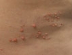
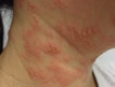
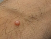
Plantar Molluscum Contagiosum: A Case Report of Molluscum Contagiosum Occurring on the Sole of the Foot and a Review of the World Literature
Omphalith-Associated Relapsing Umbilical Cellulitis: Recurrent Omphalitis Secondary to a Hair-Containing Belly Button Bezoar
Harpist's Finger: Case Report of a Trauma-Induced Blister in a Beginner Harpist and Review of String Instrument–Associated Skin Problems in Musicians
Musicians are at risk for developing instrument-associated dermatologic conditions. The cutaneous problems are frequently secondary to abnormalities of saliva production, contact dermatitis, hyperhidrosis, infection, or physical trauma.1-10 Harpist's finger is reported in a beginner harpist who developed a trauma-induced blister on the finger she repetitively used to play her instrument. Skin problems in musicians caused by string instruments are reviewed.
Case Report
A healthy 6-year-old Chinese girl presented with a blister on her left index finger. She did not have a history of skin fragility or bullous skin disorders. Clinical evaluation revealed an 8X6-mm tense, tender, fluid-filled vesicle on the distal ventral pad of her left index finger. Additional questioning revealed that she had recently begun playing the harp and was playing the notes by plucking the strings with her fingers (Figure). Her wooden harp (an Allegro) was made of solid Honduras mahogany with a birch-laminated soundboard and finished with a water-white precatalyzed nitrocellulose lacquer with a semigloss (50%) sheen (unicoat). The nylon strings were either monofilament or wound (nylon wrap over nylon). The red (C musical note) and blue strings (F musical note) were created by placing the nylon strings in boiling water that was used to dissolve scarlet or navy blue dye powder (Rit® dye), respectively.
The diagnosis of a blister secondary to repeated trauma between her distal digit and the taut harp strings was established based on the correlation of the patient's history and clinical findings. The blister subsequently flattened and its roof spontaneously shed. The girl continued to play the harp and eventually developed a callus at this location on her finger.
Comment
Dermatologic conditions in musicians that are caused by the instruments they play are frequently observed in orchestra members. Rimmer and Spielvogel1 distributed a survey regarding skin problems to 84 members of a professional symphony orchestra; 22 of 24 musicians who replied had instrument-associated dermatologic conditions. Intrigued by this study, Nethercott and Holness2 administered a health questionnaire to 41 orchestra members; 8 musicians reported a current work-related skin problem. More recently, Onder et al3 distributed a questionnaire to 97 orchestra members; 40 of 47 musicians who claimed to have either prior or current skin problems considered their dermatoses to be directly related to instrument use. Instrument-related skin conditions in musicians have been designated using several different classifications. Some of the conditions are described by their mechanism of pathogenesis and the resulting problem. For example, pizzicato paronychia is an infection of the nail fold in string players resulting from pizzicato playing whereby the musician plucks the instrument's strings instead of using a bow.4 Other dermatologic conditions in musicians are designated by combining the name of the instrument with either the anatomic location of the problem (eg, harpist's finger) or the description of the dermatosis (eg, piano paronychia). Alliteration often is incorporated into the nomenclature when these conditions are described, such as cellist's chest, clarinetist's cheilitis, drummer's digits, fiddler's fingers, guitarist's groin, piano paronychia, and pizzicato paronychia.1-6Harpists may develop calluses on the sides and tips of their fingers (harpist's finger), resulting from pressure and friction between the harpist's fingers and the strings of the instrument. The calluses may be painful or become irritated. Similar to the reported beginner harpist, neophyte harpists are especially susceptible to finger injury, such as sore fingertips and blister formation, prior to the development of calluses. Gradually increasing the time devoted to practicing the harp may enable the beginner harpist to avoid these problems.1,2,4,6,7,9,11 Harpists also are at risk for developing other instrument-associated dermatologic conditions, including not only intracorneal hemorrhage of the fingertips, similar to the friction-induced bleeding within the upper epidermis observed on the toes of runners (talon noir), but also nail dystrophies. Loosening of the nail plates and onycholysis may result from repetitive glissando playing in which the harpist rapidly glides one or more fingertips across multiple consecutive strings. In addition, paronychia, such as pizzicato paronychia from plucking the strings, and subungual hemorrhage with hematoma formation are other nail-related problems in harpists.1,4,6,7,9,11-13 Allergic contact dermatitis also has been observed, albeit rarely, in harpists. A 25-year-old woman who was a harpist developed an eczematous eruption of 5 months' duration on the fingertips of her right hand.2 Patch testing documented a 2+ reaction to potassium dichromate. Additional investigation revealed that this allergen was used as a tanning agent for the harp strings.2 Instrument-associated skin maladies also have been described in individuals playing other string instruments (Table).1-20 The main causes for these dermatoses are allergic contact dermatitis and physical trauma. The most common allergens associated with string instruments are chromium, nickel, colophony, paraphenylenediamine, propolis, and exotic woods.1-3,5-8,15,16,20
The 2 metallic substances, chromium and nickel, have been observed as allergens in musicians who play cello, guitar, harp, sitar, and violin. Allergic contact dermatitis to chromium has been described in electric guitarists, caused by contact with chromated steel components of their instruments, such as the strings, bridge, and frets, as well as the chromated leather accessories such as the guitar strap; a harpist for whom the allergen was used as a tanning agent for the harp strings; and a violinist whose E string was gold plated surrounding a chromated steel core.2,6,16 Nickel-related contact dermatitis has been described in a cellist whose wooden bow handle contained nickel, guitarists whose guitar strings contained the allergen, sitarists from the nickel mizrab covering the right index finger, and a violinist from the metal clamp of the violin's chin rest.1,3,6,8,20 Colophony (rosin) is a naturally occurring complex mixture of resin acids and neutral substances obtained from different species of coniferous trees (family Pinaceae). The principal sensitizer of colophony is abietic acid and its derivatives. Allergic contact dermatitis to colophony has been observed in cellists, violinists, and viola players who apply rosin to wax the strings on the bows of their instruments.1-8,15 Paraphenylenediamine is not only a skin sensitizer but also a skin irritant. Its principal use is in cosmetics as a black hair dye ingredient. Patch testing with a positive reaction to paraphenylenediamine was observed in an 11-year-old girl who had a cello with a black-string bow. She presented with scaling on her right thumb, index finger, and middle finger. A positive patch test result for paraphenylenediamine also was noted in a violinist who developed cutaneous eruptions located on his neck where it came into contact with the instrument's black chin rest.5-7 Propolis, also known as bee glue, has been reported to cause allergic contact dermatitis not only in musicians, such as cellists and violinists, but also in instrument makers. It can be found in many products of everyday use, such as chewing gum, facial creams, mouthwash preparations, and toothpastes. In addition, propolis is used as an ingredient in violin varnish.6,7 Exotic woods occasionally are used in the construction of the fingerboards of guitars and the body and/or chin rests of violas and violins. In string instruments, allergic contact dermatitis has been attributed to Brazilian rosewood (Dalbergia nigra), Indian rosewood (Dalbergiones species), East Indian rosewood (Dalbergia latifolia Roxb.), and ebony (Diospyros species) in chin rests and Makassar ebony in a violin. Because boxwood (Buxus sempervirens) does not contain any known allergens, it has been suggested as an alternative chin rest wood for musicians with allergic contact dermatitis to the wooden chin rest of their instrument.1,5-8 Physical trauma in musicians who play string instruments can result from the instrument or its strings. Dermatologic problems can be the sequelae of string instrument–induced physical trauma caused by the interaction of the fingers with the strings, which results in acro-osteolysis; callosities, such as harpist's fingers; Garrod pads; and nail dystrophies. Alternatively, mechanical trauma–associated skin conditions can occur secondary to pressure of the instrument against either the neck, chest, groin, knee, nipple, proximal arm, or scrotum.1,3-7,9,11-14,17-20
Conclusion
Musicians who play string instruments, such as the cello, guitar, harp, sarod, sitar, viola, and violin, can develop skin problems that usually result from either allergic contact dermatitis or physical trauma. The most common allergens are chromium, nickel, colophony, paraphenylenediamine, propolis, and exotic woods. Trauma-associated skin conditions are caused by either the interaction of the musician's fingers with the strings or pressure of the instrument against the musician's body. Harpist's finger is an example of a trauma-induced dermatologic problem, presenting as a callus on the affected finger, which may be preceded by soreness and blister formation that can be observed in beginner harpists.
- Rimmer S, Spielvogel RL. Dermatologic problems of musicians. J Am Acad Dermatol. 1990;22:657-663.
- Nethercott JR, Holness DL. Dermatologic problems of musicians [letter]. J Am Acad Dermatol. 1991;25 (5, pt 1):870.
- Onder M, Aksakal AB, Oztas MO, et al. Skin problems of musicians. Int J Dermatol. 1999;38:192-195.
- Fisher AA. Dermatitis in a musician. part III: injuries caused by specific musical instruments. Cutis. 1998;62: 261-262.
- Liu S, Hayden GF. Maladies in musicians. South Med J. 2002;95:727-734.
- Gambichler T, Boms S, Freitag M. Contact dermatitis and other skin conditions in instrumental musicians. BMC Dermatology. 2004;4:3.
- Adams RM. Skin conditions of musicians. Cutis. 2000;65:37-38.
- Fisher AA. Allergic contact dermatitis from musical instruments. Cutis. 1993;51:75-76.
- Fisher AA. Dermatitis in a musician. part II: injuries to skin, soft tissue, and bone from musical instruments. Cutis. 1998;62:214-215.
- Fisher AA. Dermatitis in a musician. part IV: physiologic, emotional, and infectious problems in musicians. Cutis. 1999;63:13-14.
- Kanerva L. Physical causes and radiation effects. In: Adams RM, ed. Occupational Skin Disease. 3rd ed. Philadelphia, PA: WB Saunders Co; 1999:35-68.
- Martinelli PT, Cohen PR, Schulze KE, et al. Intracorneal hemorrhage. Dermatol Nurs. 2006;18:373, 382.
- Cohen PR, Schulze KE, Nelson BR. Subungual hematoma. Dermatol Nurs. 2007;19:83-84.
- Bird HA. Development of Garrod's pads in the fingers of a professional violinist. Ann Rheum Dis. 1987;46: 169-170.
- Fisher AA. Allergic contact dermatitis in a violinist. the role of abietic acid—a sensitizer in rosin (colophony)—as the causative agent. Cutis. 1981;27:466-473.
- Buckley DA, Rogers S. 'Fiddler's fingers': violin-string dermatitis. Contact Dermatitis. 1995;32:46-47.
- Baran R, Tosti A. Occupational acroosteolysis in a guitar player. Acta Derm Venereol. 1993;73:64-65.
- Semple R, Gillingham J. Musical bumps [letter]. Br Med J. 1974;2:504.
- Curtis P. Guitar nipple [letter]. Br Med J. 1974;2:226.
- Kanwar AJ, Kaur S. More dermatologic problems of musicians [lettter]. J Am Acad Dermatol. 1991;24:321-322.
Musicians are at risk for developing instrument-associated dermatologic conditions. The cutaneous problems are frequently secondary to abnormalities of saliva production, contact dermatitis, hyperhidrosis, infection, or physical trauma.1-10 Harpist's finger is reported in a beginner harpist who developed a trauma-induced blister on the finger she repetitively used to play her instrument. Skin problems in musicians caused by string instruments are reviewed.
Case Report
A healthy 6-year-old Chinese girl presented with a blister on her left index finger. She did not have a history of skin fragility or bullous skin disorders. Clinical evaluation revealed an 8X6-mm tense, tender, fluid-filled vesicle on the distal ventral pad of her left index finger. Additional questioning revealed that she had recently begun playing the harp and was playing the notes by plucking the strings with her fingers (Figure). Her wooden harp (an Allegro) was made of solid Honduras mahogany with a birch-laminated soundboard and finished with a water-white precatalyzed nitrocellulose lacquer with a semigloss (50%) sheen (unicoat). The nylon strings were either monofilament or wound (nylon wrap over nylon). The red (C musical note) and blue strings (F musical note) were created by placing the nylon strings in boiling water that was used to dissolve scarlet or navy blue dye powder (Rit® dye), respectively.
The diagnosis of a blister secondary to repeated trauma between her distal digit and the taut harp strings was established based on the correlation of the patient's history and clinical findings. The blister subsequently flattened and its roof spontaneously shed. The girl continued to play the harp and eventually developed a callus at this location on her finger.
Comment
Dermatologic conditions in musicians that are caused by the instruments they play are frequently observed in orchestra members. Rimmer and Spielvogel1 distributed a survey regarding skin problems to 84 members of a professional symphony orchestra; 22 of 24 musicians who replied had instrument-associated dermatologic conditions. Intrigued by this study, Nethercott and Holness2 administered a health questionnaire to 41 orchestra members; 8 musicians reported a current work-related skin problem. More recently, Onder et al3 distributed a questionnaire to 97 orchestra members; 40 of 47 musicians who claimed to have either prior or current skin problems considered their dermatoses to be directly related to instrument use. Instrument-related skin conditions in musicians have been designated using several different classifications. Some of the conditions are described by their mechanism of pathogenesis and the resulting problem. For example, pizzicato paronychia is an infection of the nail fold in string players resulting from pizzicato playing whereby the musician plucks the instrument's strings instead of using a bow.4 Other dermatologic conditions in musicians are designated by combining the name of the instrument with either the anatomic location of the problem (eg, harpist's finger) or the description of the dermatosis (eg, piano paronychia). Alliteration often is incorporated into the nomenclature when these conditions are described, such as cellist's chest, clarinetist's cheilitis, drummer's digits, fiddler's fingers, guitarist's groin, piano paronychia, and pizzicato paronychia.1-6Harpists may develop calluses on the sides and tips of their fingers (harpist's finger), resulting from pressure and friction between the harpist's fingers and the strings of the instrument. The calluses may be painful or become irritated. Similar to the reported beginner harpist, neophyte harpists are especially susceptible to finger injury, such as sore fingertips and blister formation, prior to the development of calluses. Gradually increasing the time devoted to practicing the harp may enable the beginner harpist to avoid these problems.1,2,4,6,7,9,11 Harpists also are at risk for developing other instrument-associated dermatologic conditions, including not only intracorneal hemorrhage of the fingertips, similar to the friction-induced bleeding within the upper epidermis observed on the toes of runners (talon noir), but also nail dystrophies. Loosening of the nail plates and onycholysis may result from repetitive glissando playing in which the harpist rapidly glides one or more fingertips across multiple consecutive strings. In addition, paronychia, such as pizzicato paronychia from plucking the strings, and subungual hemorrhage with hematoma formation are other nail-related problems in harpists.1,4,6,7,9,11-13 Allergic contact dermatitis also has been observed, albeit rarely, in harpists. A 25-year-old woman who was a harpist developed an eczematous eruption of 5 months' duration on the fingertips of her right hand.2 Patch testing documented a 2+ reaction to potassium dichromate. Additional investigation revealed that this allergen was used as a tanning agent for the harp strings.2 Instrument-associated skin maladies also have been described in individuals playing other string instruments (Table).1-20 The main causes for these dermatoses are allergic contact dermatitis and physical trauma. The most common allergens associated with string instruments are chromium, nickel, colophony, paraphenylenediamine, propolis, and exotic woods.1-3,5-8,15,16,20
The 2 metallic substances, chromium and nickel, have been observed as allergens in musicians who play cello, guitar, harp, sitar, and violin. Allergic contact dermatitis to chromium has been described in electric guitarists, caused by contact with chromated steel components of their instruments, such as the strings, bridge, and frets, as well as the chromated leather accessories such as the guitar strap; a harpist for whom the allergen was used as a tanning agent for the harp strings; and a violinist whose E string was gold plated surrounding a chromated steel core.2,6,16 Nickel-related contact dermatitis has been described in a cellist whose wooden bow handle contained nickel, guitarists whose guitar strings contained the allergen, sitarists from the nickel mizrab covering the right index finger, and a violinist from the metal clamp of the violin's chin rest.1,3,6,8,20 Colophony (rosin) is a naturally occurring complex mixture of resin acids and neutral substances obtained from different species of coniferous trees (family Pinaceae). The principal sensitizer of colophony is abietic acid and its derivatives. Allergic contact dermatitis to colophony has been observed in cellists, violinists, and viola players who apply rosin to wax the strings on the bows of their instruments.1-8,15 Paraphenylenediamine is not only a skin sensitizer but also a skin irritant. Its principal use is in cosmetics as a black hair dye ingredient. Patch testing with a positive reaction to paraphenylenediamine was observed in an 11-year-old girl who had a cello with a black-string bow. She presented with scaling on her right thumb, index finger, and middle finger. A positive patch test result for paraphenylenediamine also was noted in a violinist who developed cutaneous eruptions located on his neck where it came into contact with the instrument's black chin rest.5-7 Propolis, also known as bee glue, has been reported to cause allergic contact dermatitis not only in musicians, such as cellists and violinists, but also in instrument makers. It can be found in many products of everyday use, such as chewing gum, facial creams, mouthwash preparations, and toothpastes. In addition, propolis is used as an ingredient in violin varnish.6,7 Exotic woods occasionally are used in the construction of the fingerboards of guitars and the body and/or chin rests of violas and violins. In string instruments, allergic contact dermatitis has been attributed to Brazilian rosewood (Dalbergia nigra), Indian rosewood (Dalbergiones species), East Indian rosewood (Dalbergia latifolia Roxb.), and ebony (Diospyros species) in chin rests and Makassar ebony in a violin. Because boxwood (Buxus sempervirens) does not contain any known allergens, it has been suggested as an alternative chin rest wood for musicians with allergic contact dermatitis to the wooden chin rest of their instrument.1,5-8 Physical trauma in musicians who play string instruments can result from the instrument or its strings. Dermatologic problems can be the sequelae of string instrument–induced physical trauma caused by the interaction of the fingers with the strings, which results in acro-osteolysis; callosities, such as harpist's fingers; Garrod pads; and nail dystrophies. Alternatively, mechanical trauma–associated skin conditions can occur secondary to pressure of the instrument against either the neck, chest, groin, knee, nipple, proximal arm, or scrotum.1,3-7,9,11-14,17-20
Conclusion
Musicians who play string instruments, such as the cello, guitar, harp, sarod, sitar, viola, and violin, can develop skin problems that usually result from either allergic contact dermatitis or physical trauma. The most common allergens are chromium, nickel, colophony, paraphenylenediamine, propolis, and exotic woods. Trauma-associated skin conditions are caused by either the interaction of the musician's fingers with the strings or pressure of the instrument against the musician's body. Harpist's finger is an example of a trauma-induced dermatologic problem, presenting as a callus on the affected finger, which may be preceded by soreness and blister formation that can be observed in beginner harpists.
- Rimmer S, Spielvogel RL. Dermatologic problems of musicians. J Am Acad Dermatol. 1990;22:657-663.
- Nethercott JR, Holness DL. Dermatologic problems of musicians [letter]. J Am Acad Dermatol. 1991;25 (5, pt 1):870.
- Onder M, Aksakal AB, Oztas MO, et al. Skin problems of musicians. Int J Dermatol. 1999;38:192-195.
- Fisher AA. Dermatitis in a musician. part III: injuries caused by specific musical instruments. Cutis. 1998;62: 261-262.
- Liu S, Hayden GF. Maladies in musicians. South Med J. 2002;95:727-734.
- Gambichler T, Boms S, Freitag M. Contact dermatitis and other skin conditions in instrumental musicians. BMC Dermatology. 2004;4:3.
- Adams RM. Skin conditions of musicians. Cutis. 2000;65:37-38.
- Fisher AA. Allergic contact dermatitis from musical instruments. Cutis. 1993;51:75-76.
- Fisher AA. Dermatitis in a musician. part II: injuries to skin, soft tissue, and bone from musical instruments. Cutis. 1998;62:214-215.
- Fisher AA. Dermatitis in a musician. part IV: physiologic, emotional, and infectious problems in musicians. Cutis. 1999;63:13-14.
- Kanerva L. Physical causes and radiation effects. In: Adams RM, ed. Occupational Skin Disease. 3rd ed. Philadelphia, PA: WB Saunders Co; 1999:35-68.
- Martinelli PT, Cohen PR, Schulze KE, et al. Intracorneal hemorrhage. Dermatol Nurs. 2006;18:373, 382.
- Cohen PR, Schulze KE, Nelson BR. Subungual hematoma. Dermatol Nurs. 2007;19:83-84.
- Bird HA. Development of Garrod's pads in the fingers of a professional violinist. Ann Rheum Dis. 1987;46: 169-170.
- Fisher AA. Allergic contact dermatitis in a violinist. the role of abietic acid—a sensitizer in rosin (colophony)—as the causative agent. Cutis. 1981;27:466-473.
- Buckley DA, Rogers S. 'Fiddler's fingers': violin-string dermatitis. Contact Dermatitis. 1995;32:46-47.
- Baran R, Tosti A. Occupational acroosteolysis in a guitar player. Acta Derm Venereol. 1993;73:64-65.
- Semple R, Gillingham J. Musical bumps [letter]. Br Med J. 1974;2:504.
- Curtis P. Guitar nipple [letter]. Br Med J. 1974;2:226.
- Kanwar AJ, Kaur S. More dermatologic problems of musicians [lettter]. J Am Acad Dermatol. 1991;24:321-322.
Successful Treatment of Auricular Pseudocyst Using a Surgical Bolster [letter]
Community-Acquired Methicillin-Resistant Staphylococcus Aureus Skin Infection Presenting as a Periumbilical Folliculitis
Familial Median Canaliform Nail Dystrophy
Median canaliform nail dystrophy is a nail abnormality that typically involves one or both thumbnails. The first case of this disorder was recorded by Heller1 in 1928. Median canaliform nail dystrophy presents as a central longitudinal groove of the nail plate, extending proximally from the end of the nail.2 This condition is usually not inherited. However, it may be acquired following trauma to the nail. We describe a man with familial median canaliform nail dystrophy and discuss the differential diagnosis.
Case Report
A 68-year-old man presented with bilateral dystrophy of his thumbnails. The nail abnormality initially appeared at 34 years of age with no preceding trauma to the digits. His older brother and his mother also had developed the same nail changes as young adults. Neither the patient nor his brother or mother rubbed their proximal thumbnail fold with the tip of their second finger; the absence of this behavioral activity was repeatedly confirmed by both the patient and his wife during several subsequent office visits.
Examination of both thumbnails showed an asymptomatic distal fissure with a fir tree–like pattern (Figure). Proximally, the nail plates showed transverse grooves. In addition, the lunula was red and enlarged.
Please refer to the PDF to view the figure
Comment
Median canaliform nail dystrophy appears as a long longitudinal groove extending from either the proximal nail fold or a more distal portion of the nail plate to the end of the nail. Lateral extensions of this fissure create a conspicuous inverted fir tree–like pattern. In severe cases, the nail can split along the groove.3 Thickening of the proximal nail fold, enlargement of the lunula, and redness of the lunula also may occur.4-9
The diagnosis of this condition is usually established based on clinical features because pathologic correlation is rarely available. However, specimens for microscopic evaluation have occasionally been provided. Parakeratosis, as well as an accumulation of melanin within and between the nail bed keratinocytes, was demonstrated in the evaluation of an affected nail by Heller10 in 1927. Subsequently, parakeratosis and intranuclear pigmentation were found within the longitudinal canal of the affected nail plate of a 12-year-old girl with median canaliform nail dystrophy who was described by Robinson and Weidman11 in 1948.
Median canaliform nail dystrophy may present following trauma to the nail plate or nail matrix.3-7,12-15 In addition, coexisting conditions such as either soft tissue in the nail defect or dental caries have been observed in some patients with median canaliform nail dystrophy. In one case, a 19-year-old woman presented with a flabby filament of fleshy tissue that was observed within the dystrophic nail canal.14 The tissue was extracted, and the nail abnormality resolved. Subsequently, the nail dystrophy, including the associated tissue, reappeared.14 Tooth decay associated with median canaliform nail dystrophy was reported in a 23-year-old woman with a deformity that involved many of the nails on both of her hands. Her nail condition spontaneously cleared after 3 carious teeth were extracted.16
Medication was postulated as the causative factor for the development of median canaliform nail dystrophy in 3 patients who were receiving isotretinoin. The first, reported by Bottomley and Cunliffe17 in 1992, was a 38-year-old woman who developed median canaliform nail dystrophy 6 weeks after beginning treatment with isotretinoin. Her thumbnail returned to normal 4 weeks after she discontinued the drug.17 The second patient, described by Griego et al4 in 1995, was an 18-year-old man who developed median canaliform nail dystrophy of both thumbnails after starting therapy with isotretinoin for his acne. The nail disfigurement became distinct after 4 months of treatment; his new thumbnail dystrophy resolved 5 months after he discontinued the medication.4 A third patient was reported by Dharmagunawardena and Charles-Holmes12 in 1997. They described a 19-year-old man who developed median canaliform nail dystrophy in both thumbnails within 4 weeks after starting treatment with isotretinoin for his acne. His nails returned to normal 3 months after completing a 5-month course of isotretinoin therapy.12
Familial median canaliform nail dystrophy has not been associated with any systemic syndromes. In our patient and his family, the nail dystrophy was not congenital but rather appeared as an acquired abnormality of the nails in adulthood.
The etiology of median canaliform nail dystrophy is unknown.5,7,13,16-18 It usually is an acquired condition. Nail matrix trauma may precede the onset; however, an associated nail injury has often not occurred.3-7,12-16,19-21 This nail dystrophy is not considered to be inherited. The familial occurrence of median canaliform nail dystrophy has rarely been described. Indeed, to the best of our knowledge, in addition to our patient, only 3 families with median canaliform nail dystrophy have been described.20,22,23 In the first such family, a 16-year-old girl with bilateral median canaliform nail dystrophy of her thumbnails since the age of 13 years had a mother with similar-appearing thumbnails.20 A second such family also included a mother and daughter.22 Long longitudinal grooves were present in the daughter's left thumbnail since the age of 11 years; her mother had a similar dystrophy involving her right thumbnail that began when she was 12 years old. Her mother, currently 34 years old, still has recurrent episodes of spontaneously resolving median canaliform nail dystrophy. The family had no history of other hereditary diseases.22 The third family in which median canaliform nail dystrophy occurred was reported by Bossi23 in the Italian literature in 1965. Our patient and his brother and mother represent the fourth such family.
The differential diagnosis of median canaliform nail dystrophy includes habit tic deformity (Table). It also includes other causes of longitudinal splits in the nail plate such as direct trauma to the nail unit. In addition, digital mucous cyst (synovial cyst), lichen striatus, nail-patella syndrome, pterygium, Raynaud disease, and trachyonychia are other conditions in which a longitudinal nail defect has been described.5,7,30,31
Please refer to the PDF to view the table
Habit tic deformity is usually present in one or both thumbnails and results in alteration of the normal nail growth. It is caused by the constant or habitual rubbing of the thumb's proximal nail fold by the tip of the second digit. The subsequent damage to the nail matrix causes clinical changes in the nail plate that appear different than those of median canaliform nail dystrophy. The habit tic deformity produces transverse ridges along the central nail plate depression instead of a longitudinal groove with lateral projections. The depth of the central nail plate canal depends on the intensity of the inflicted trauma by the index finger to the matrix of the thumbnail. In addition, the lunula may appear red and enlarged.9,29 Also, the proximal nail fold may be swollen.5,13
Median canaliform nail dystrophy has occasionally been described to periodically disappear; often, the nail defect reappears in these individuals.4-7,13,15,17,24 In some patients, the central nail defect is replaced by a longitudinal ridge5,6; however, in most patients, such as ours, the condition does not resolve spontaneously. Keeping the nail length short and buffing the surface of the nail can prevent the edge of the nail plate from catching on clothing and other objects.5 Covering the nail plate with tape or a nail wrap also can aid in ensuring that jagged edges are not present.4,7
Conclusion
Familial median canaliform nail dystrophy has rarely been described. Our patient had adult onset of his condition involving both thumbnails with associated red macrolunula. His brother and his mother also experienced the same nail dystrophy. Including our patient and his family, median canaliform nail dystrophy has only been reported in 4 families. The mode of inheritance for median canaliform nail dystrophy in these families remains to be determined.
- Heller J. Zur kasuistik seltener nagelkrankheiten: dystrophia unguium mediana canaliformis. Dermat Ztschr. 1928;51:416-419.
- Ronchese F. Peculiar nail anomalies. AMA Arch Derm Syphilol. 1951;63:565-580.
- Baran R. Modifications of the nail surface. In: Pierre M, ed. The Nail. Edinburgh, Scotland: Churchill Livingstone; 1981:26-29.
- Griego RD, Orengo IF, Scher RK. Median nail dystrophy and habit tic deformity: are they different forms of the same disorder? Int J Dermatol. 1995;34:799-800.
- Samman PD, Fenton DA. Miscellaneous acquired nail disorders. In: Samman PD, Fenton DA, eds. Samman's The Nails in Disease. 5th ed. Oxford, England: Butterworth-Heinemann; 1995:97-110.
- Samman PD. The nails. In: Rook A, Wilkinson DS, Ebling FJG, eds. Textbook of Dermatology. 3rd ed. Oxford, England: Blackwell Scientific; 1979:1825-1855.
- Baran R, Dawber RPR, Richert B, et al. Physical signs. In: Baran R, Dawber RPR, de Berker DAR, et al, eds. Diseases of the Nails and Their Management. 3rd ed. Oxford, England: Blackwell Science; 2001:48-103.
- Zelger J, Wohlfarth B, Putz R. Dystrophia unguium mediana canaliformis Heller. Hautarzt. 1974;25:629-631.
- Cohen PR. The lunula. J Am Acad Dermatol. 1996;34:943-955.
- Heller J. In: Jadassohn J. Handbuch der Haut-und Geschlechtskrankheiten. Berlin, Germany: Springer; 1927. Cited by: De Nicola P, Morsiani M, Zavagli G. Nail symptoms. In: Nail Diseases in Internal Medicine. Springfield, Ill: Charles C. Thomas; 1974:29-57.
- Robinson MM, Weidman FD. Dystrophia unguium mediana canaliformis. AMA Arch Derm Syphilol. 1948;57:328-331.
- Dharmagunawardena B, Charles-Holmes R. Median canaliform dystrophy following isotretinoin therapy [letter]. Br J Dermatol. 1997;137:658-659.
- Van Dijk E. Dystrophia unguium mediana canaliformis. Dermatologica. 1978;156:358-366.
- Sutton RL Jr. Solenonychia: canaliform dystrophy of the nails. South Med J. 1965;58:1143-1146.
- Sweet RD. Dystrophia unguium mediana canaliformis. AMA Arch Derm Syphilol. 1951;64:61-62.
- Fowle LP, Wiggall RH. Dystrophia unguium mediana canaliformis: report of a case. AMA Arch Derm Syphilol. 1944;50:267-268.
- Bottomley WW, Cunliffe WJ. Median nail dystrophy associated with isotretinoin therapy. [letter]. Br J Dermatol. 1992;127:447-448.
- Costa OG. Median canal-like dystrophy of the nails. Arch Dermatol. 1943;49:406-407.
- Oliver EA, Bluefarb SM. Nevus striatus symmetricus unguis. AMA Arch Derm Syphilol. 1944;49:190.
- Rehtijarvi K. Dystrophia unguis mediana canaliformis. Acta Derm Venereol. 1971;51:316-317.
- Krause ME, Cole HN, Driver JR. Dystrophia mediana canaliformis. AMA Arch Derm Syphilol. 1945;52:418.
- Seller H. Dystrophia unguis mediana canaliformis. Familial occurrence [in German]. Hautarzt. 1974;25:456.
- Bossi G. Heller’s dystrophia unguium mediana canaliformis [in Italian]. Minerva Dermatol.1965;40:303-304. Cited by:Van Dijk E. Dystrophia unguium mediana canaliformis. Dermatologica.1978;156:358-366.
- De Nicola P, Morsiani M, Zavagli G. Nail symptoms. In:Nail Diseases in Internal Medicine. Springfield, Ill: Charles C.Thomas; 1974:29-57.
- Samman PD. A traumatic nail dystrophy produced by ahabit tic. Arch Dermatol. 1963;88:895-896.
- Samman PD. Nail deformities due to trauma. In: Samman PD, Fenton DA, eds. The Nails in Disease. 5th ed. Oxford, England: Butterworth-Heinemann; 1995:148-168.
- Oppenheim M, Cohen D. Naevus striatus symmetricus of the thumbs. AMA Arch Derm Syphilol.1942;45:253.
- Macaulay WL. Transverse ridging of the thumbnails. “washboard thumbnails.” Arch Dermatol. 1966;93:421-423.
- Vittorio CC, Phillips KA. Treatment of habit-tic deformity with fluoxetine. Arch Dermatol. 1997;133:1203-1204.
- Anderson CR. Longitudinal grooving of the nails caused by synovial lesions. AMA Arch Derm Syph. 1947;55:828-830.
- Smith EB, Skipworth GB, Van der Ploeg DE. Longitudinal grooving of nails due to synovial cysts. Arch Dermatol.1964;89:364-366.
Median canaliform nail dystrophy is a nail abnormality that typically involves one or both thumbnails. The first case of this disorder was recorded by Heller1 in 1928. Median canaliform nail dystrophy presents as a central longitudinal groove of the nail plate, extending proximally from the end of the nail.2 This condition is usually not inherited. However, it may be acquired following trauma to the nail. We describe a man with familial median canaliform nail dystrophy and discuss the differential diagnosis.
Case Report
A 68-year-old man presented with bilateral dystrophy of his thumbnails. The nail abnormality initially appeared at 34 years of age with no preceding trauma to the digits. His older brother and his mother also had developed the same nail changes as young adults. Neither the patient nor his brother or mother rubbed their proximal thumbnail fold with the tip of their second finger; the absence of this behavioral activity was repeatedly confirmed by both the patient and his wife during several subsequent office visits.
Examination of both thumbnails showed an asymptomatic distal fissure with a fir tree–like pattern (Figure). Proximally, the nail plates showed transverse grooves. In addition, the lunula was red and enlarged.
Please refer to the PDF to view the figure
Comment
Median canaliform nail dystrophy appears as a long longitudinal groove extending from either the proximal nail fold or a more distal portion of the nail plate to the end of the nail. Lateral extensions of this fissure create a conspicuous inverted fir tree–like pattern. In severe cases, the nail can split along the groove.3 Thickening of the proximal nail fold, enlargement of the lunula, and redness of the lunula also may occur.4-9
The diagnosis of this condition is usually established based on clinical features because pathologic correlation is rarely available. However, specimens for microscopic evaluation have occasionally been provided. Parakeratosis, as well as an accumulation of melanin within and between the nail bed keratinocytes, was demonstrated in the evaluation of an affected nail by Heller10 in 1927. Subsequently, parakeratosis and intranuclear pigmentation were found within the longitudinal canal of the affected nail plate of a 12-year-old girl with median canaliform nail dystrophy who was described by Robinson and Weidman11 in 1948.
Median canaliform nail dystrophy may present following trauma to the nail plate or nail matrix.3-7,12-15 In addition, coexisting conditions such as either soft tissue in the nail defect or dental caries have been observed in some patients with median canaliform nail dystrophy. In one case, a 19-year-old woman presented with a flabby filament of fleshy tissue that was observed within the dystrophic nail canal.14 The tissue was extracted, and the nail abnormality resolved. Subsequently, the nail dystrophy, including the associated tissue, reappeared.14 Tooth decay associated with median canaliform nail dystrophy was reported in a 23-year-old woman with a deformity that involved many of the nails on both of her hands. Her nail condition spontaneously cleared after 3 carious teeth were extracted.16
Medication was postulated as the causative factor for the development of median canaliform nail dystrophy in 3 patients who were receiving isotretinoin. The first, reported by Bottomley and Cunliffe17 in 1992, was a 38-year-old woman who developed median canaliform nail dystrophy 6 weeks after beginning treatment with isotretinoin. Her thumbnail returned to normal 4 weeks after she discontinued the drug.17 The second patient, described by Griego et al4 in 1995, was an 18-year-old man who developed median canaliform nail dystrophy of both thumbnails after starting therapy with isotretinoin for his acne. The nail disfigurement became distinct after 4 months of treatment; his new thumbnail dystrophy resolved 5 months after he discontinued the medication.4 A third patient was reported by Dharmagunawardena and Charles-Holmes12 in 1997. They described a 19-year-old man who developed median canaliform nail dystrophy in both thumbnails within 4 weeks after starting treatment with isotretinoin for his acne. His nails returned to normal 3 months after completing a 5-month course of isotretinoin therapy.12
Familial median canaliform nail dystrophy has not been associated with any systemic syndromes. In our patient and his family, the nail dystrophy was not congenital but rather appeared as an acquired abnormality of the nails in adulthood.
The etiology of median canaliform nail dystrophy is unknown.5,7,13,16-18 It usually is an acquired condition. Nail matrix trauma may precede the onset; however, an associated nail injury has often not occurred.3-7,12-16,19-21 This nail dystrophy is not considered to be inherited. The familial occurrence of median canaliform nail dystrophy has rarely been described. Indeed, to the best of our knowledge, in addition to our patient, only 3 families with median canaliform nail dystrophy have been described.20,22,23 In the first such family, a 16-year-old girl with bilateral median canaliform nail dystrophy of her thumbnails since the age of 13 years had a mother with similar-appearing thumbnails.20 A second such family also included a mother and daughter.22 Long longitudinal grooves were present in the daughter's left thumbnail since the age of 11 years; her mother had a similar dystrophy involving her right thumbnail that began when she was 12 years old. Her mother, currently 34 years old, still has recurrent episodes of spontaneously resolving median canaliform nail dystrophy. The family had no history of other hereditary diseases.22 The third family in which median canaliform nail dystrophy occurred was reported by Bossi23 in the Italian literature in 1965. Our patient and his brother and mother represent the fourth such family.
The differential diagnosis of median canaliform nail dystrophy includes habit tic deformity (Table). It also includes other causes of longitudinal splits in the nail plate such as direct trauma to the nail unit. In addition, digital mucous cyst (synovial cyst), lichen striatus, nail-patella syndrome, pterygium, Raynaud disease, and trachyonychia are other conditions in which a longitudinal nail defect has been described.5,7,30,31
Please refer to the PDF to view the table
Habit tic deformity is usually present in one or both thumbnails and results in alteration of the normal nail growth. It is caused by the constant or habitual rubbing of the thumb's proximal nail fold by the tip of the second digit. The subsequent damage to the nail matrix causes clinical changes in the nail plate that appear different than those of median canaliform nail dystrophy. The habit tic deformity produces transverse ridges along the central nail plate depression instead of a longitudinal groove with lateral projections. The depth of the central nail plate canal depends on the intensity of the inflicted trauma by the index finger to the matrix of the thumbnail. In addition, the lunula may appear red and enlarged.9,29 Also, the proximal nail fold may be swollen.5,13
Median canaliform nail dystrophy has occasionally been described to periodically disappear; often, the nail defect reappears in these individuals.4-7,13,15,17,24 In some patients, the central nail defect is replaced by a longitudinal ridge5,6; however, in most patients, such as ours, the condition does not resolve spontaneously. Keeping the nail length short and buffing the surface of the nail can prevent the edge of the nail plate from catching on clothing and other objects.5 Covering the nail plate with tape or a nail wrap also can aid in ensuring that jagged edges are not present.4,7
Conclusion
Familial median canaliform nail dystrophy has rarely been described. Our patient had adult onset of his condition involving both thumbnails with associated red macrolunula. His brother and his mother also experienced the same nail dystrophy. Including our patient and his family, median canaliform nail dystrophy has only been reported in 4 families. The mode of inheritance for median canaliform nail dystrophy in these families remains to be determined.
- Heller J. Zur kasuistik seltener nagelkrankheiten: dystrophia unguium mediana canaliformis. Dermat Ztschr. 1928;51:416-419.
- Ronchese F. Peculiar nail anomalies. AMA Arch Derm Syphilol. 1951;63:565-580.
- Baran R. Modifications of the nail surface. In: Pierre M, ed. The Nail. Edinburgh, Scotland: Churchill Livingstone; 1981:26-29.
- Griego RD, Orengo IF, Scher RK. Median nail dystrophy and habit tic deformity: are they different forms of the same disorder? Int J Dermatol. 1995;34:799-800.
- Samman PD, Fenton DA. Miscellaneous acquired nail disorders. In: Samman PD, Fenton DA, eds. Samman's The Nails in Disease. 5th ed. Oxford, England: Butterworth-Heinemann; 1995:97-110.
- Samman PD. The nails. In: Rook A, Wilkinson DS, Ebling FJG, eds. Textbook of Dermatology. 3rd ed. Oxford, England: Blackwell Scientific; 1979:1825-1855.
- Baran R, Dawber RPR, Richert B, et al. Physical signs. In: Baran R, Dawber RPR, de Berker DAR, et al, eds. Diseases of the Nails and Their Management. 3rd ed. Oxford, England: Blackwell Science; 2001:48-103.
- Zelger J, Wohlfarth B, Putz R. Dystrophia unguium mediana canaliformis Heller. Hautarzt. 1974;25:629-631.
- Cohen PR. The lunula. J Am Acad Dermatol. 1996;34:943-955.
- Heller J. In: Jadassohn J. Handbuch der Haut-und Geschlechtskrankheiten. Berlin, Germany: Springer; 1927. Cited by: De Nicola P, Morsiani M, Zavagli G. Nail symptoms. In: Nail Diseases in Internal Medicine. Springfield, Ill: Charles C. Thomas; 1974:29-57.
- Robinson MM, Weidman FD. Dystrophia unguium mediana canaliformis. AMA Arch Derm Syphilol. 1948;57:328-331.
- Dharmagunawardena B, Charles-Holmes R. Median canaliform dystrophy following isotretinoin therapy [letter]. Br J Dermatol. 1997;137:658-659.
- Van Dijk E. Dystrophia unguium mediana canaliformis. Dermatologica. 1978;156:358-366.
- Sutton RL Jr. Solenonychia: canaliform dystrophy of the nails. South Med J. 1965;58:1143-1146.
- Sweet RD. Dystrophia unguium mediana canaliformis. AMA Arch Derm Syphilol. 1951;64:61-62.
- Fowle LP, Wiggall RH. Dystrophia unguium mediana canaliformis: report of a case. AMA Arch Derm Syphilol. 1944;50:267-268.
- Bottomley WW, Cunliffe WJ. Median nail dystrophy associated with isotretinoin therapy. [letter]. Br J Dermatol. 1992;127:447-448.
- Costa OG. Median canal-like dystrophy of the nails. Arch Dermatol. 1943;49:406-407.
- Oliver EA, Bluefarb SM. Nevus striatus symmetricus unguis. AMA Arch Derm Syphilol. 1944;49:190.
- Rehtijarvi K. Dystrophia unguis mediana canaliformis. Acta Derm Venereol. 1971;51:316-317.
- Krause ME, Cole HN, Driver JR. Dystrophia mediana canaliformis. AMA Arch Derm Syphilol. 1945;52:418.
- Seller H. Dystrophia unguis mediana canaliformis. Familial occurrence [in German]. Hautarzt. 1974;25:456.
- Bossi G. Heller’s dystrophia unguium mediana canaliformis [in Italian]. Minerva Dermatol.1965;40:303-304. Cited by:Van Dijk E. Dystrophia unguium mediana canaliformis. Dermatologica.1978;156:358-366.
- De Nicola P, Morsiani M, Zavagli G. Nail symptoms. In:Nail Diseases in Internal Medicine. Springfield, Ill: Charles C.Thomas; 1974:29-57.
- Samman PD. A traumatic nail dystrophy produced by ahabit tic. Arch Dermatol. 1963;88:895-896.
- Samman PD. Nail deformities due to trauma. In: Samman PD, Fenton DA, eds. The Nails in Disease. 5th ed. Oxford, England: Butterworth-Heinemann; 1995:148-168.
- Oppenheim M, Cohen D. Naevus striatus symmetricus of the thumbs. AMA Arch Derm Syphilol.1942;45:253.
- Macaulay WL. Transverse ridging of the thumbnails. “washboard thumbnails.” Arch Dermatol. 1966;93:421-423.
- Vittorio CC, Phillips KA. Treatment of habit-tic deformity with fluoxetine. Arch Dermatol. 1997;133:1203-1204.
- Anderson CR. Longitudinal grooving of the nails caused by synovial lesions. AMA Arch Derm Syph. 1947;55:828-830.
- Smith EB, Skipworth GB, Van der Ploeg DE. Longitudinal grooving of nails due to synovial cysts. Arch Dermatol.1964;89:364-366.