User login
Can acid suppression prevent recurrent idiopathic gastroduodenal ulcer bleeding?
SAN DIEGO – However, these patients face a considerable risk of overall mortality, despite a low incidence of recurrent GI bleeding.
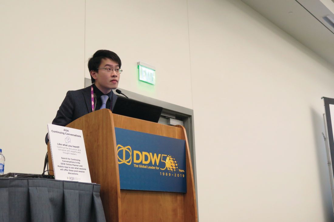
“Peptic ulcer disease is a commonly encountered condition in our daily clinical practice,” one of the study authors, Louis Lau, MD, said at the annual Digestive Disease Week. “Most of the ulcers are due to H. pylori and NSAIDs or aspirin. However, idiopathic peptic ulcers remain an area to be investigated.”
According to Dr. Lau of the division of gastroenterology and hepatology at Prince of Wales Hospital, Hong Kong, H. pylori–negative idiopathic ulcers have increased in prevalence in the Asian Pacific region, from 1%-4% in 2000 to up to 20% in recent data. These ulcers have been shown to have a bleeding rate of up to 13% within 1 year (Gastroenterology. 2005;128:1845-50) and have been associated with a mortality rate of up to 88% in 7 years of follow-up (Gastroenterology. 2009;137:525-31).
“There is no consensus for the optimal management of this particular ulcer,” he continued. “One proposed management was maintenance PPI [proton pump inhibitors]. However, we would like to look for the best optimal prevention strategy.”
Dr. Lau and associates conducted a double-blind, randomized trial at Prince of Wales Hospital to determine if there were any differences between the PPI lansoprazole and the histamine2 receptor antagonist famotidine for the prevention of recurrent bleeding in patients with H. pylori–negative idiopathic peptic ulcers. It involved a 2-year follow-up period and included a screening and end-of-study esophagogastroduodenoscopy. The drugs were repackaged into identical green capsules.
The researchers limited the analysis to 228 patients over the age 18 with H. pylori–negative idiopathic ulcers, defined as having endoscopic biopsies negative for both rapid urease test and histology for H. pylori; no exposure to aspirin, NSAIDs, or drugs of unknown nature including traditional Chinese medicine within 4 weeks; and no other causes of ulceration identified. Exclusion criteria included patients with a strong indication for maintenance therapy with PPIs, those with prior gastric surgery, pregnant patients, and those with advanced malignancy or comorbidity.
The primary endpoint was clinical GI bleeding or reduction in hemoglobin level of 2 g/dL or more and confirmation of peptic ulcers by endoscopy. The secondary endpoint was recurrent ulcer detected by end-of-study endoscopy, with or without clinical symptoms.
Of the 228 patients, 114 received lansoprazole 30 mg daily and 114 received famotidine 40 mg daily. Recurrent upper GI bleeding was suspected in 10 patients in each of the treatment groups. Ulcer confirmation via endoscopy detected one ulcer in the lansoprazole group and three in the famotidine group (0.9% vs. 2.6%, respectively; P = .62). For the secondary endpoint, there were five recurrent ulcers in the lansoprazole group and nine in the famotidine group, (4.4% vs. 7.9%, respectively; P = .27).
Dr. Lau and colleagues observed eight deaths in the lansoprazole group (7%), compared with five (4.4%) in the famotidine group (P = .39), mainly caused by pneumonia, renal failure, and other chronic medical conditions. Intention-to-treat analysis at 2 years found that both groups were comparable in terms of time to analysis of recurrent upper GI bleeding, adjusted for death as a competing risk (Gray’s test, P = .313).
“In this randomized, controlled trial, we found that both a PPI and an H2 blocker are comparable in the prevention of idiopathic ulcer recurrent bleeding,” Dr. Lau concluded. “The rebleeding rate was five times lower than a previous cohort reported with no or irregular acid suppression therapy [Gastroenterology. 2005;128[7]:1845-50]. With all this, there is still a considerable risk of recurrent ulcers and death. We postulate that there is an underlying mechanism for this type of idiopathic ulcer.”
The study was funded in part by the General Research Fund–Research Grant Council. Dr. Lau reported having no financial disclosures. Many of his coauthors reported having numerous financial ties to the pharmaceutical industry.
SOURCE: Lau L et al. DDW 2019, Abstract 322.
SAN DIEGO – However, these patients face a considerable risk of overall mortality, despite a low incidence of recurrent GI bleeding.
“Peptic ulcer disease is a commonly encountered condition in our daily clinical practice,” one of the study authors, Louis Lau, MD, said at the annual Digestive Disease Week. “Most of the ulcers are due to H. pylori and NSAIDs or aspirin. However, idiopathic peptic ulcers remain an area to be investigated.”
According to Dr. Lau of the division of gastroenterology and hepatology at Prince of Wales Hospital, Hong Kong, H. pylori–negative idiopathic ulcers have increased in prevalence in the Asian Pacific region, from 1%-4% in 2000 to up to 20% in recent data. These ulcers have been shown to have a bleeding rate of up to 13% within 1 year (Gastroenterology. 2005;128:1845-50) and have been associated with a mortality rate of up to 88% in 7 years of follow-up (Gastroenterology. 2009;137:525-31).
“There is no consensus for the optimal management of this particular ulcer,” he continued. “One proposed management was maintenance PPI [proton pump inhibitors]. However, we would like to look for the best optimal prevention strategy.”
Dr. Lau and associates conducted a double-blind, randomized trial at Prince of Wales Hospital to determine if there were any differences between the PPI lansoprazole and the histamine2 receptor antagonist famotidine for the prevention of recurrent bleeding in patients with H. pylori–negative idiopathic peptic ulcers. It involved a 2-year follow-up period and included a screening and end-of-study esophagogastroduodenoscopy. The drugs were repackaged into identical green capsules.
The researchers limited the analysis to 228 patients over the age 18 with H. pylori–negative idiopathic ulcers, defined as having endoscopic biopsies negative for both rapid urease test and histology for H. pylori; no exposure to aspirin, NSAIDs, or drugs of unknown nature including traditional Chinese medicine within 4 weeks; and no other causes of ulceration identified. Exclusion criteria included patients with a strong indication for maintenance therapy with PPIs, those with prior gastric surgery, pregnant patients, and those with advanced malignancy or comorbidity.
The primary endpoint was clinical GI bleeding or reduction in hemoglobin level of 2 g/dL or more and confirmation of peptic ulcers by endoscopy. The secondary endpoint was recurrent ulcer detected by end-of-study endoscopy, with or without clinical symptoms.
Of the 228 patients, 114 received lansoprazole 30 mg daily and 114 received famotidine 40 mg daily. Recurrent upper GI bleeding was suspected in 10 patients in each of the treatment groups. Ulcer confirmation via endoscopy detected one ulcer in the lansoprazole group and three in the famotidine group (0.9% vs. 2.6%, respectively; P = .62). For the secondary endpoint, there were five recurrent ulcers in the lansoprazole group and nine in the famotidine group, (4.4% vs. 7.9%, respectively; P = .27).
Dr. Lau and colleagues observed eight deaths in the lansoprazole group (7%), compared with five (4.4%) in the famotidine group (P = .39), mainly caused by pneumonia, renal failure, and other chronic medical conditions. Intention-to-treat analysis at 2 years found that both groups were comparable in terms of time to analysis of recurrent upper GI bleeding, adjusted for death as a competing risk (Gray’s test, P = .313).
“In this randomized, controlled trial, we found that both a PPI and an H2 blocker are comparable in the prevention of idiopathic ulcer recurrent bleeding,” Dr. Lau concluded. “The rebleeding rate was five times lower than a previous cohort reported with no or irregular acid suppression therapy [Gastroenterology. 2005;128[7]:1845-50]. With all this, there is still a considerable risk of recurrent ulcers and death. We postulate that there is an underlying mechanism for this type of idiopathic ulcer.”
The study was funded in part by the General Research Fund–Research Grant Council. Dr. Lau reported having no financial disclosures. Many of his coauthors reported having numerous financial ties to the pharmaceutical industry.
SOURCE: Lau L et al. DDW 2019, Abstract 322.
REPORTING FROM DDW 2019
Artificial intelligence advances optical biopsy
SAN DIEGO – Artificial intelligence is improving the accuracy of optical biopsies, a development that may ultimately avoid the need for tissue biopsies of many low-risk colonic polyps, Michael Byrne, MD, said at the annual Digestive Disease Week.

Dr. Byrne, chief executive officer of Satisfai Health, founder of ai4gi, and gastroenterologist at Vancouver General Hospital; Nicolas Guizard, medical imaging researcher at Imagia; and their colleagues at ai4gi developed a “full clinical workflow” for detecting colonic polyps and performing optical biopsies of the polyps.”
Using narrow band imaging (NBI) enhanced with artificial intelligence, the system was used to review 21,804 colonoscopy frames and it achieved a “near-perfect” diagnostic accuracy of 99.9%. In an assessment of colonoscopy videos that included 125 polyps, the system had 95.9% sensitivity, with a specificity of 91.6% and a negative predictive value of 93.6%, Dr. Byrne said.
The speed of the system’s decision-making is rapid, with a typical reaction time of 360 milliseconds. The system was able to make diagnostic inferences at a rate of 26 milliseconds per frame.
With exposure to more learning experiences, the artificial intelligence system improved and committed to a prediction for 97.6% of the polyps it visualized. Dr. Byrne said this result represented a 12.8% improvement from previously published data on the model’s performance.
Dr. Byrne and his colleagues found the system had a tracking accuracy of 92.8%, meaning that this percentage of polyps was both correctly detected and assigned to a unique identifier for follow-up of the site of each excised polyp over time. The interface worked even when multiple polyps were seen on the same screen.
In a video interview, Dr. Byrne discussed the implications for gastroenterology and plans for a clinical trial for rigorous testing of the model.
ai4gi is developing the AI colonoscopy technology. Dr. Byrne is founder of the ai4gi joint venture, which holds a technology codevelopment agreement with Olympus US.
SAN DIEGO – Artificial intelligence is improving the accuracy of optical biopsies, a development that may ultimately avoid the need for tissue biopsies of many low-risk colonic polyps, Michael Byrne, MD, said at the annual Digestive Disease Week.
Dr. Byrne, chief executive officer of Satisfai Health, founder of ai4gi, and gastroenterologist at Vancouver General Hospital; Nicolas Guizard, medical imaging researcher at Imagia; and their colleagues at ai4gi developed a “full clinical workflow” for detecting colonic polyps and performing optical biopsies of the polyps.”
Using narrow band imaging (NBI) enhanced with artificial intelligence, the system was used to review 21,804 colonoscopy frames and it achieved a “near-perfect” diagnostic accuracy of 99.9%. In an assessment of colonoscopy videos that included 125 polyps, the system had 95.9% sensitivity, with a specificity of 91.6% and a negative predictive value of 93.6%, Dr. Byrne said.
The speed of the system’s decision-making is rapid, with a typical reaction time of 360 milliseconds. The system was able to make diagnostic inferences at a rate of 26 milliseconds per frame.
With exposure to more learning experiences, the artificial intelligence system improved and committed to a prediction for 97.6% of the polyps it visualized. Dr. Byrne said this result represented a 12.8% improvement from previously published data on the model’s performance.
Dr. Byrne and his colleagues found the system had a tracking accuracy of 92.8%, meaning that this percentage of polyps was both correctly detected and assigned to a unique identifier for follow-up of the site of each excised polyp over time. The interface worked even when multiple polyps were seen on the same screen.
In a video interview, Dr. Byrne discussed the implications for gastroenterology and plans for a clinical trial for rigorous testing of the model.
ai4gi is developing the AI colonoscopy technology. Dr. Byrne is founder of the ai4gi joint venture, which holds a technology codevelopment agreement with Olympus US.
REPORTING FROM DDW 2019

SEER data show that pancreatic cancer mortality rates differ by ethnicity
SAN DIEGO – .

However, between 2013 and 2015, mortality rates began to fall among all ethnicities, for reasons that remain unclear. “There is still a significant disparity between the [white] and African American populations,” lead study author Mohamed M. Gad, MD, said at the annual Digestive Disease Week. “Trends for the Asian/American Indian populations were similar to that of the [white] population.”
While most patients diagnosed with pancreatic cancer are white men, the disparities among racial and ethnic groups have not been addressed on a large scale, said Dr. Gad, an internal medicine resident at the Cleveland Clinic. In an effort to investigate the racial disparities in mortality rates in patients with pancreatic cancer, he and colleagues collected data from the National Cancer Institute’s Surveillance, Epidemiology and End-Results Program (SEER) 9 database during 1973 to 2015. The researchers calculated mortality rates for all pancreatic cancer cases by race. Next, they calculated the Observed/Expected (O/E) ratio and the excess risk per 10,000 person-years to estimate the change of risk following the diagnosis in different ethnicities, when compared to the general population, using Joinpoint Regression software.
Dr. Gad reported results from 65,985 patients who died from pancreatic cancer between 1973 and 2015. The overall mortality rate was highest for African Americans (9.4%), followed by whites (6.4%), and Asians/American Indians (5.4%). The researchers observed that mortality rates of all racial groups continued to increase from 1973 until 2013. However, between 2013 and 2015, the mortality rates decreased by 20.5% for African Americans, by 27.1% for whites, and by 25.1% for Asians/American Indians (P less than .001 for all decreases). Socioeconomic status and access to health care have been hypothesized to play a role in these observed outcomes, Dr. Gad said, but more research is needed to understand the observed disparities and assess the need for intervention. For example, he said, one line of research would be to evaluate the difference between access to health care and the quality of health care delivery for African American patients compared with their white counterparts. “Once we identify that, how can we improve outcomes for the African American population?” Dr. Gad asked.
He reported having no financial disclosures.
SAN DIEGO – .
However, between 2013 and 2015, mortality rates began to fall among all ethnicities, for reasons that remain unclear. “There is still a significant disparity between the [white] and African American populations,” lead study author Mohamed M. Gad, MD, said at the annual Digestive Disease Week. “Trends for the Asian/American Indian populations were similar to that of the [white] population.”
While most patients diagnosed with pancreatic cancer are white men, the disparities among racial and ethnic groups have not been addressed on a large scale, said Dr. Gad, an internal medicine resident at the Cleveland Clinic. In an effort to investigate the racial disparities in mortality rates in patients with pancreatic cancer, he and colleagues collected data from the National Cancer Institute’s Surveillance, Epidemiology and End-Results Program (SEER) 9 database during 1973 to 2015. The researchers calculated mortality rates for all pancreatic cancer cases by race. Next, they calculated the Observed/Expected (O/E) ratio and the excess risk per 10,000 person-years to estimate the change of risk following the diagnosis in different ethnicities, when compared to the general population, using Joinpoint Regression software.
Dr. Gad reported results from 65,985 patients who died from pancreatic cancer between 1973 and 2015. The overall mortality rate was highest for African Americans (9.4%), followed by whites (6.4%), and Asians/American Indians (5.4%). The researchers observed that mortality rates of all racial groups continued to increase from 1973 until 2013. However, between 2013 and 2015, the mortality rates decreased by 20.5% for African Americans, by 27.1% for whites, and by 25.1% for Asians/American Indians (P less than .001 for all decreases). Socioeconomic status and access to health care have been hypothesized to play a role in these observed outcomes, Dr. Gad said, but more research is needed to understand the observed disparities and assess the need for intervention. For example, he said, one line of research would be to evaluate the difference between access to health care and the quality of health care delivery for African American patients compared with their white counterparts. “Once we identify that, how can we improve outcomes for the African American population?” Dr. Gad asked.
He reported having no financial disclosures.
REPORTING FROM DDW 2019
Coffee, tea, and soda all up GERD risk
SAN DIEGO – .
In an interview following the oral presentation, Raaj S. Mehta, MD, said that patients in his primary care panel at Massachusetts General Hospital, Boston, where he’s a senior resident, frequently came to him with GERD. In addition to questions about diet, patients frequently wanted to know which beverages might provoke or exacerbate their GERD.
In trying to help his patients, Dr. Mehta said he realized that there wasn’t a prospective evidence base to answer their questions about beverages and GERD, so he and his colleagues used data from the Nurses’ Health Study II (NHS II), a prospective cohort study, to look at the association between various beverages and the incidence of GERD.
“What’s exciting is that we were able to find that coffee, tea, and soda – all three – increase your risk for gastroesophageal reflux disease,” Dr. Mehta said in a video interview. “At the highest quintile level, so looking at people who consume six or more cups per day, you’re looking at maybe a 25%-35% increase in risk of reflux disease.”
There was a dose-response relationship as well: “You do see a slight increase as you go from one cup, to two, to three, and so on, all the way up to six cups” of the offending beverages, said Dr. Mehta.
Overall, the risk for GERD rose from 1.17 to 1.34 with coffee consumption as servings per day increased from less than one to six or more (P for trend less than .0001). Tea consumption was associated with increased GERD risk ranging from 1.08 to 1.26 as consumption rose (P for trend .001). For soda, the increased risk went from 1.12 at less than one serving daily, to 1.41 at four to five servings daily, and then fell to 1.29 at six or more daily servings (P for trend less than .0001).
Whether the beverages were caffeinated or not, said Dr. Mehta, only made a “minimal difference” in GERD risk.
“In contrast, we didn’t see an association for beverages like water, juice, and milk,” he said – reassuring findings in light of fruit juice’s anecdotal status as a GERD culprit.
The NHS II collected data every 2 years from 48,308 female nurses aged 42-62 years at the beginning of the study. Every 4 years dietary information was collected, and on the opposite 4-year cycle, participants answered questions about GERD. Medication use, including the incident use of proton pump inhibitors, was collected every 2 years.
Patients with baseline GERD or use of PPIs or H2 receptor antagonists were excluded from participation.
The quantity and type of beverages were assessed by food frequency questionnaires; other demographic, dietary, and medication variables were also gathered and used to adjust the statistical analysis.
A substitution analysis answered the “what-if” question of the effect of substituting two glasses of plain water daily for either coffee, tea, or soda. Dr. Mehta and colleagues saw a modest reduction in risk for GERD with this strategy.
In addition to the prospective nature of the study (abstract 514, doi: 10.1016/S0016-5085(19)37044-1), the large sample size, high follow-up rates, and well validated dietary data were all strengths, said Dr. Mehta. However, the study’s population is relatively homogeneous, and residual confounding couldn’t be excluded. Also, GERD was defined by self-report, though participants were asked to respond to clear, validated criteria.
For Dr. Mehta, he’s glad to have a clear answer to a common clinic question. “I think that this is one additional thing that I can recommend as a primary care provider to my patients when they come into my office,” he said.
Dr. Mehta reported no conflicts of interest.
REPORTING FROM DDW 2019

Early cholecystectomy prevents recurrent biliary events
SAN DIEGO – In a retrospective study of 234 patients admitted for gallstone pancreatitis, almost 90% of recurrent biliary events occurred in patients who did not receive a cholecystectomy within 60 days of hospital discharge. The overall rate of recurrence was 19%, and over half of patients (59%) did not receive a cholecystectomy during their index hospitalization.

Additionally, none of the recurrent biliary events occurred in those patients who did receive a cholecystectomy during the index hospitalization or within the first 30 days after discharge. “It really is the case that, ‘if you snooze, you lose,’ ” said Vijay Dalapathi, MD, presenting the findings during an oral presentation at the annual Digestive Disease Week.
Dr. Dalapathi and colleagues had observed that cholecystectomy during an index hospitalization for mild biliary pancreatitis was a far from universal practice, despite guidelines recommending early cholecystectomy.
To delve further into practice patterns, Dr. Dalapathi, first author Mohammed Ullah, MD, and their coauthors at the University of Rochester (N.Y.) conducted a single-site retrospective study of patients who were admitted with gallstone pancreatitis over a 5-year period ending December 2017. Dr. Dalapathi and Dr. Ullah are both second-year gastroenterology fellows.
The study had twin primary outcome measures: cholecystectomy rates performed during an index hospitalization for gallstone pancreatitis and recurrent biliary events after hospitalization. Adult patients were included if they had a diagnosis of acute gallstone pancreatitis, with or without recurrent cholangitis, choledocholithiasis, or acute cholecystitis. Pediatric patients and those with prior cholecystectomy were excluded.
A total of 234 patients were included in the study. Their mean age was 58.3 years, and patients were mostly female (57.3%) and white (91.5%). Mean body mass index was 29.1 kg/m2. A total of 175 patients (74.8%) had endoscopic retrograde cholangiopancreatography.
Out of the entire cohort of patients, 138 (59%) did not have a cholecystectomy during the index hospitalization. Among the patients who did not receive a cholecystectomy, 33 (24%) were deemed unsuitable candidates for the procedure, either because they were critically ill or because they were poor candidates for surgery for other reasons. No reason was provided for the nonperformance of cholecystectomy for an additional 28 patients (20%).
The remaining 75 patients (54%) were deferred to outpatient management. Looking at this subgroup of patients, Dr. Dalapathi and his coinvestigators tracked the amount of time that passed before cholecystectomy.
The researchers found that 19 patients (25%) had not had a cholecystectomy within the study period. A total of 21 patients (28%) had the procedure more than 60 days from hospitalization, and another 23 (31%) had the procedure between 30 and 60 days after hospitalization. Just 12 patients (16%) of this subgroup had their cholecystectomy within 30 days of hospitalization.
Among patients who were discharged without a cholecystectomy, Dr. Dalapathi and his coauthors found 26 recurrent biliary events (19%): 15 were gallstone pancreatitis and 10 were cholecystitis; 1 patient developed cholangitis.
The crux of the study’s findings came when the investigators looked at the association between recurrent events and cholecystectomy timing. They found no recurrent biliary events among those who received cholecystectomy while hospitalized or within the first 30 days after discharge. Of the 26 events, 3 (12%) occurred in those whose cholecystectomies came 30-60 days after discharge. The remaining 23 events (88%) were seen in those receiving a cholecystectomy more than 60 days after discharge, or not at all.
Guidelines from the American Gastroenterological Association, the Society of American Gastrointestinal and Endoscopic Surgeons, and the American College of Gastroenterology all recommend early cholecystectomy after mild acute gallstone pancreatitis, said Dr. Dalapathi.
However, two separate systematic reviews including a total of 22 studies and over 3,000 patients showed that about half (48% and 51%) of patients admitted with mild acute biliary pancreatitis received a cholecystectomy during the index hospitalization or within 14 days of the hospitalization.
Further, he said, previous work had shown recurrent biliary event rates approaching 20% for patients whose biliary pancreatitis bout was not followed by cholecystectomy, a figure in line with the rate seen in the present study.
“Cholecystectomy should be performed during index hospitalization or as soon as possible within 30 days of mild biliary pancreatitis to minimize risk of recurrent biliary events,” said Dr. Dalapathi.
The authors reported no outside sources of funding and no conflicts of interest.
SOURCE: Ullah M. et al. DDW 2019, Abstract 24.
SAN DIEGO – In a retrospective study of 234 patients admitted for gallstone pancreatitis, almost 90% of recurrent biliary events occurred in patients who did not receive a cholecystectomy within 60 days of hospital discharge. The overall rate of recurrence was 19%, and over half of patients (59%) did not receive a cholecystectomy during their index hospitalization.
Additionally, none of the recurrent biliary events occurred in those patients who did receive a cholecystectomy during the index hospitalization or within the first 30 days after discharge. “It really is the case that, ‘if you snooze, you lose,’ ” said Vijay Dalapathi, MD, presenting the findings during an oral presentation at the annual Digestive Disease Week.
Dr. Dalapathi and colleagues had observed that cholecystectomy during an index hospitalization for mild biliary pancreatitis was a far from universal practice, despite guidelines recommending early cholecystectomy.
To delve further into practice patterns, Dr. Dalapathi, first author Mohammed Ullah, MD, and their coauthors at the University of Rochester (N.Y.) conducted a single-site retrospective study of patients who were admitted with gallstone pancreatitis over a 5-year period ending December 2017. Dr. Dalapathi and Dr. Ullah are both second-year gastroenterology fellows.
The study had twin primary outcome measures: cholecystectomy rates performed during an index hospitalization for gallstone pancreatitis and recurrent biliary events after hospitalization. Adult patients were included if they had a diagnosis of acute gallstone pancreatitis, with or without recurrent cholangitis, choledocholithiasis, or acute cholecystitis. Pediatric patients and those with prior cholecystectomy were excluded.
A total of 234 patients were included in the study. Their mean age was 58.3 years, and patients were mostly female (57.3%) and white (91.5%). Mean body mass index was 29.1 kg/m2. A total of 175 patients (74.8%) had endoscopic retrograde cholangiopancreatography.
Out of the entire cohort of patients, 138 (59%) did not have a cholecystectomy during the index hospitalization. Among the patients who did not receive a cholecystectomy, 33 (24%) were deemed unsuitable candidates for the procedure, either because they were critically ill or because they were poor candidates for surgery for other reasons. No reason was provided for the nonperformance of cholecystectomy for an additional 28 patients (20%).
The remaining 75 patients (54%) were deferred to outpatient management. Looking at this subgroup of patients, Dr. Dalapathi and his coinvestigators tracked the amount of time that passed before cholecystectomy.
The researchers found that 19 patients (25%) had not had a cholecystectomy within the study period. A total of 21 patients (28%) had the procedure more than 60 days from hospitalization, and another 23 (31%) had the procedure between 30 and 60 days after hospitalization. Just 12 patients (16%) of this subgroup had their cholecystectomy within 30 days of hospitalization.
Among patients who were discharged without a cholecystectomy, Dr. Dalapathi and his coauthors found 26 recurrent biliary events (19%): 15 were gallstone pancreatitis and 10 were cholecystitis; 1 patient developed cholangitis.
The crux of the study’s findings came when the investigators looked at the association between recurrent events and cholecystectomy timing. They found no recurrent biliary events among those who received cholecystectomy while hospitalized or within the first 30 days after discharge. Of the 26 events, 3 (12%) occurred in those whose cholecystectomies came 30-60 days after discharge. The remaining 23 events (88%) were seen in those receiving a cholecystectomy more than 60 days after discharge, or not at all.
Guidelines from the American Gastroenterological Association, the Society of American Gastrointestinal and Endoscopic Surgeons, and the American College of Gastroenterology all recommend early cholecystectomy after mild acute gallstone pancreatitis, said Dr. Dalapathi.
However, two separate systematic reviews including a total of 22 studies and over 3,000 patients showed that about half (48% and 51%) of patients admitted with mild acute biliary pancreatitis received a cholecystectomy during the index hospitalization or within 14 days of the hospitalization.
Further, he said, previous work had shown recurrent biliary event rates approaching 20% for patients whose biliary pancreatitis bout was not followed by cholecystectomy, a figure in line with the rate seen in the present study.
“Cholecystectomy should be performed during index hospitalization or as soon as possible within 30 days of mild biliary pancreatitis to minimize risk of recurrent biliary events,” said Dr. Dalapathi.
The authors reported no outside sources of funding and no conflicts of interest.
SOURCE: Ullah M. et al. DDW 2019, Abstract 24.
REPORTING FROM DDW 2019
Adding drugs to gastric balloons increases weight loss
SAN DIEGO – In a multicenter study involving four academic medical centers, the addition of weight loss drugs to intragastric balloons resulted in better weight loss 12 months after balloon placement.
In a video interview at the annual Digestive Disease Week, study investigator Reem Sharaiha, MD, explained that one of the drawbacks of intragastric balloons is that, although they produce weight loss for the 6 or 12 months that they are in place, patients tend to regain that weight after they are removed. The study, involving 111 patients, was designed to determine whether the addition of weight loss drugs could mitigate this effect and improve weight loss, said Dr. Sharaiha of Weill Cornell Medical Center, New York.
Adding drugs such as metformin or weight loss drugs tailored to patients’ particular weight issues (cravings, anxiety, or fast gastric emptying) at the 3- or 6-month mark while the intragastric balloon was in place helped patients continue losing weight after balloon removal. At 12 months, the percentage of total body weight lost was significantly greater in the intragastric balloon group with concurrent pharmacotherapy (21.4% vs. 13.1%).
SOURCE: Shah SL et al. DDW 2019, Abstract 1105.
REPORTING FROM DDW 2019

Ethnicity seems to affect predisposition to components of metabolic syndrome
SAN DIEGO – .

“It appears that not all components of metabolic syndrome directly correlate with visceral fat,” lead study author Patrick Chen, DO, said at the annual Digestive Disease Week.
According to Dr. Chen, a gastroenterology fellow at Wright State University, Dayton, Ohio, the prevalence of obesity in the United States increased from 30.5% in 2000 to 39.6% in 2015, a condition that costs the U.S. medical system $150 billion each year and is associated with 19% of all deaths. “We also know that abdominal visceral fat is more associated with mortality and cardiac events than overall adiposity, while visceral fat contributes to nonalcoholic fatty liver disease and metabolic syndrome,” he said.
For Asian populations, the World Health Organization has set body mass indexes of 23 kg/m2 for overweight and 27.5 kg/m2 for obesity. “This has led to an ongoing debate as to whether we should be adopting regional anthropometric criteria based on race-unique risk factors,” Dr. Chen said. “Despite all of these association studies, the mechanisms are still undergoing research and still unknown to this day.”
For the current analysis, he and his colleagues assessed the prevalence of type 2 diabetes, hypertension, and hyperlipidemia and the impact of increasing BMI among racial groups in the United States. They drew from the National Ambulatory Medical Care Survey from 2011 to 2015 to collect and compare data on patient race, BMI, and the prevalence of type 2 diabetes, hypertension, and dyslipidemia, and used SPSS software for chi-square analysis. They excluded patients under the age of 18, those with type 1 diabetes, those with no listed race, and Native American populations, “since there were too few patients for meaningful analysis,” he said.
The 69,949 patients in the analysis included 57,448 whites, 9,281 African Americans, and 2,142 Asians. The majority (64,091) were listed as non-Hispanic, while 5,858 were listed as Hispanic. The mean age of the study population ranged from 49 to 55 years. African Americans had the highest mean BMI (31 kg/m2), followed by whites (29 kg/m2) and Asians (25 kg/m2). Meanwhile, both Hispanics and non-Hispanics had a mean BMI of 29 kg/m2.
Dr. Chen reported that African Americans (19.3%) and Asians (18.5%) had a higher prevalence of type 2 diabetes compared with whites (13.4%; P less than .001), while Hispanics had a higher prevalence of type 2 diabetes compared with non-Hispanics (18.9% vs. 13.9%; P less than .001). At the same time, African Americans had a higher prevalence of hypertension (49.6%) compared with whites (38.2%) and Asians (37.9%; P less than .001 for both associations), while non-Hispanics had a higher prevalence of hypertension compared with Hispanics (40.4% vs. 33.1%; P less than .001). Asians had a higher prevalence of hyperlipidemia (28.4%) compared with whites (25.6%; P = .004); both groups had a higher prevalence compared with African Americans (21.9%, P less than .001). Hispanics had a lower prevalence of hyperlipidemia compared with non-Hispanics (23.6% vs. 25.1%; P = .005).
“Diabetes, hypertension, and hyperlipidemia likely have different mechanisms that lead to different race’s predisposition to these diseases,” Dr. Chen concluded. “This study supports that regional anthropometric criteria should be done based on ethnicity-specific risk factors. Some food for thought is whether we need to change our screening guidelines for conditions like diabetes for Asian patients and hypertension in African American patients. We should also consider ethnicity when we look for NAFLD. Overall, continued research is needed to explain these correlations.”
The researchers reported having no financial disclosures.
SOURCE: Chen P et al. DDW 2019, Abstract 447.
SAN DIEGO – .
“It appears that not all components of metabolic syndrome directly correlate with visceral fat,” lead study author Patrick Chen, DO, said at the annual Digestive Disease Week.
According to Dr. Chen, a gastroenterology fellow at Wright State University, Dayton, Ohio, the prevalence of obesity in the United States increased from 30.5% in 2000 to 39.6% in 2015, a condition that costs the U.S. medical system $150 billion each year and is associated with 19% of all deaths. “We also know that abdominal visceral fat is more associated with mortality and cardiac events than overall adiposity, while visceral fat contributes to nonalcoholic fatty liver disease and metabolic syndrome,” he said.
For Asian populations, the World Health Organization has set body mass indexes of 23 kg/m2 for overweight and 27.5 kg/m2 for obesity. “This has led to an ongoing debate as to whether we should be adopting regional anthropometric criteria based on race-unique risk factors,” Dr. Chen said. “Despite all of these association studies, the mechanisms are still undergoing research and still unknown to this day.”
For the current analysis, he and his colleagues assessed the prevalence of type 2 diabetes, hypertension, and hyperlipidemia and the impact of increasing BMI among racial groups in the United States. They drew from the National Ambulatory Medical Care Survey from 2011 to 2015 to collect and compare data on patient race, BMI, and the prevalence of type 2 diabetes, hypertension, and dyslipidemia, and used SPSS software for chi-square analysis. They excluded patients under the age of 18, those with type 1 diabetes, those with no listed race, and Native American populations, “since there were too few patients for meaningful analysis,” he said.
The 69,949 patients in the analysis included 57,448 whites, 9,281 African Americans, and 2,142 Asians. The majority (64,091) were listed as non-Hispanic, while 5,858 were listed as Hispanic. The mean age of the study population ranged from 49 to 55 years. African Americans had the highest mean BMI (31 kg/m2), followed by whites (29 kg/m2) and Asians (25 kg/m2). Meanwhile, both Hispanics and non-Hispanics had a mean BMI of 29 kg/m2.
Dr. Chen reported that African Americans (19.3%) and Asians (18.5%) had a higher prevalence of type 2 diabetes compared with whites (13.4%; P less than .001), while Hispanics had a higher prevalence of type 2 diabetes compared with non-Hispanics (18.9% vs. 13.9%; P less than .001). At the same time, African Americans had a higher prevalence of hypertension (49.6%) compared with whites (38.2%) and Asians (37.9%; P less than .001 for both associations), while non-Hispanics had a higher prevalence of hypertension compared with Hispanics (40.4% vs. 33.1%; P less than .001). Asians had a higher prevalence of hyperlipidemia (28.4%) compared with whites (25.6%; P = .004); both groups had a higher prevalence compared with African Americans (21.9%, P less than .001). Hispanics had a lower prevalence of hyperlipidemia compared with non-Hispanics (23.6% vs. 25.1%; P = .005).
“Diabetes, hypertension, and hyperlipidemia likely have different mechanisms that lead to different race’s predisposition to these diseases,” Dr. Chen concluded. “This study supports that regional anthropometric criteria should be done based on ethnicity-specific risk factors. Some food for thought is whether we need to change our screening guidelines for conditions like diabetes for Asian patients and hypertension in African American patients. We should also consider ethnicity when we look for NAFLD. Overall, continued research is needed to explain these correlations.”
The researchers reported having no financial disclosures.
SOURCE: Chen P et al. DDW 2019, Abstract 447.
REPORTING FROM DDW 2019
Treatment-resistant GERD reported by more than half of patients
SAN DIEGO – Gastroesophageal reflux disease refractory to proton pump inhibitors may affect nearly half of those treated, according to the findings of a population-based sample of more than 70,000 Americans.
As part of the National Institutes of Health GI Patient Reported Outcomes Measurement Information System (NIH GI-PROMIS) questionnaire, respondents could download a free app called “My GI Health,” which led them through a series of questions about GI diseases. Sean Delshad, MD, MBA, of Cedars-Sinai Medical Center Los Angeles, and his colleagues examined data on symptom responses about GERD and heartburn.
Their somewhat surprising findings were that 44% of respondents had ever had GERD and that 70% of those respondents had symptoms in the past week. GERD seemed to be more common in women than in men, and in non-Hispanic whites more than other demographic groups. The rate of proton pump inhibitor–refractory GERD was reported at 54%.
Dr. Delshad discussed the implications of the study results for treatment and research in a video interview at the annual Digestive Disease Week.
REPORTING FROM DDW 2019

Bariatric surgery found to be effective in IBD patients
SAN DIEGO – In carefully selected patients with well-controlled inflammatory bowel disease (IBD), bariatric surgery results in sustained weight loss over a 2-year period, results from a retrospective study suggest.
“Obesity is increasing in patients with inflammatory bowel disease at a rate similar to that seen in the general population,” the study’s primary author, Nicholas P. McKenna, MD, said in an interview in advance of the annual Digestive Disease Week. “While bariatric surgery is a well-accepted therapy for obesity in patients without IBD, its use in patients with IBD is less well studied.”
For the current study, Dr. McKenna, a resident in the department of surgery at the Mayo Clinic in Rochester, Minn., and colleagues collected data on 33 patients who underwent bariatric surgery with a pre- or postoperative diagnosis of IBD across three academic centers between August 2006 and December 2017. They evaluated IBD characteristics and medications; postoperative complications; the need for future IBD-related surgery; and weight loss at 6, 12, and 24 months.
The patients underwent 34 bariatric operations. Their median age was 51 years and their median duration of IBD was 13 years. Of the 33 patients, 16 underwent a Roux-en-Y gastric bypass procedure: 9 who had ulcerative colitis, 6 who had Crohn’s disease, and 1 who had indeterminate colitis. A total of 14 patients underwent sleeve gastrectomy: 7 who had ulcerative colitis and 7 who had Crohn’s disease. Four patients underwent a gastric band procedure, all of whom had ulcerative colitis. The mean body mass index of patients prior to their bariatric procedures was 42.7 kg/m2. A total of 31 patients had an existing diagnosis of IBD, and 2 were diagnosed with Crohn’s disease after Roux-en-Y gastric bypass. In addition, 9 patients were on preoperative immunosuppression for IBD, and 11 had undergone prior intestinal resection for IBD.
Dr. McKenna reported that the average hospitalization for all patients was 3.6 days and that only four 30-day infectious complications occurred: two superficial surgical site infections, one infected intra-abdominal hematoma, and one hepatic abscess. In the long term, seven patients required reoperation: three for failed gastric band, two for reduction of internal hernia, and two for cholelithiasis. The researchers found that the mean percentage of overall excess weight loss was 57.5% at 6 months, 63.3% at 12 months, and 58.6% at 24 months. During a mean follow-up of 3.4 years, no IBD flares requiring surgery were observed.
“Our hypothesis based on the existing literature was that bariatric surgery would be safe in carefully selected patients with IBD and result in sustained weight loss, so we were not surprised with these results,” Dr. McKenna said. “We were not sure if medication requirements would change after surgery as the literature is conflicted on this. We observed that most patients continued to require no immunosuppression for control of their IBD after surgery. Further, we did not observe that any patients required future surgery at the time of last follow-up for an IBD flare.”
He acknowledged certain limitations of the study, including its retrospective design. “Additionally, though it is a relatively large sample, compared to the existing literature on bariatric surgery in IBD, it is still only 33 patients. This limits the comparisons that can be performed between patients with ulcerative colitis and Crohn’s disease and between the operation choices.”
The study’s secondary author, Alaa Sada, MD, a surgery resident at Mayo, presented the findings at the meeting. The researchers reported having no financial disclosures.
REPORTING FROM DDW 2019
In duodenal neuroendocrine tumors, resection technique matters
SAN DIEGO – according to a study presented at the annual Digestive Disease Week.
In a retrospective case series of 20 patients, local recurrence was seen primarily in patients who had cold forceps, rather than deeper, excision techniques. However, most patients who had cold forceps resections also remained recurrence-free, said Jonathan Ragheb, MD, a resident physician at the Cleveland Clinic.
Duodenal neuroendocrine tumors are becoming increasingly prevalent, so Dr. Ragheb and colleagues were interested in “seeing what we should do with them when we encounter them in clinical practice – whether it be surgery or endoscopic intervention,” he said.
In an interview, Dr. Ragheb said that he and his colleagues structured the study to answer the question: “What is the impact of the margin status on the recurrence of the tumor?” This relationship is important in guiding neuroendocrine tumor (NET) management, he said. The technique used for NET removal may also have effects on recurrence rates, so Dr. Ragheb and his collaborators were also interested in answering that question.
The investigators looked at patients at two facilities with a histopathologic diagnosis of duodenal NET who had endoscopic tumor resection during 2004-2018. They excluded patients who had cold forceps endoscopic resection (ER) and clear margins, patients who had further surgical therapy, and those who were lost to endoscopic follow-up.
Assessment of resection margin status was performed independently by pathologists at each study center.
“We found that people with clear margins tend not to have any recurrence, and this is over the course of a year to a year and a half of follow-up,” said Dr. Ragheb, adding, “Those patients who did have some positive margins – whether lateral margins or vertical margins – the majority of them did not have recurrence over that time period.” However, 4 of the patients in the 20-patient cohort did have some tumor recurrence, and all of these patients had an incomplete initial resection.
The investigators took a closer look at which resection techniques were most likely to result in clear margins and no recurrences, and they found that deeper techniques were associated with fewer recurrences. These included endoscopic submucosal or mucosal resection and en bloc snare polypectomy; all were associated with fewer recurrences than resections performed with cold forceps biopsy.
In all, 7 patients had clear (R0) margins, while 13 patients had an incomplete (R1) resection from the biopsy. Of the patients who had R1 margins with local recurrence, three had received a cold forceps biopsy. The other recurrence was in a patient who had endoscopic mucosal resection.
“Margin status is not the sole contributor to recurrence rates of these duodenal neuroendocrine tumors,” said Dr. Ragheb, noting that previous work has identified other possible factors, including tumor grade and biology, that can affect recurrence.
Knowledge gaps still exist regarding best practices for biopsy and decision of duodenal NETs, acknowledged Dr. Ragheb. The present study only followed patients for about a year and a half, so longer-term recurrence patterns and their relationship with various resection techniques aren’t known.
“Larger studies considering tumor grading and ER [endoscopic resection] technique are needed to fully elucidate the risk of local recurrences after ER,” wrote Dr. Ragheb and colleagues.
Dr. Ragheb reported no outside sources of funding and no conflicts of interest.
REPORTING FROM DDW 2019