User login
Benefits of lebrikizumab in asthma inconsistent
LONDON – Two large phase III trials with lebrikizumab, a monoclonal antibody that blocks the activity of interleukin-13 (IL-13), produced inconsistent evidence of benefit in patients with uncontrolled asthma, according to a presentation of both sets of data at the annual congress of the European Respiratory Society.
The two phase III studies compared lebrikizumab in doses of 37.5 mg and 125 mg to placebo, each administered subcutaneously every 4 weeks. The results were mixed not only for the primary endpoint, which was a reduction in the rate of exacerbations, but also for several secondary outcomes.
Evenly randomized into three treatment arms, 1,081 patients were enrolled in LAVOLTA I and 1,081 in LAVOLTA II. For entry, patients were required to have a forced expiratory volume in 1 second (FEV1) between 40% and 80% of predicted. Lebrikizumab was added on top of stable background therapy. For the efficacy analyses, patients were divided into those who were biomarker high, defined as having levels of periostin greater than 50 ng/mL or blood eosinophils greater than 300 mcg/L, or biomarker low, defined as having lower levels of periostin and blood eosinophils.
Over the 52-week study period in the biomarker high patients enrolled in LAVOLTA I, the rate of exacerbations was 51% lower among those randomized to the 37.5 mg dose (P less than .001) and 30% lower (P = .0232) among those randomized to the 125-mg dose relative to placebo. In the biomarker low group, a statistically significant 45% reduction in exacerbations was achieved with the 37.5 mg dose relative to placebo (P = .0318), but a 28% reduction in exacerbations, which was achieved in patients taking the 125-mg dose, was not statistically significant.
In the biomarker high patients enrolled in LAVOLTA II, both the 37.5-mg and the 125-mg doses reduced the rate of exacerbations by 26% relative to placebo, but neither reduction reached statistical significance. In the biomarker low patients, the 37.5-mg dose was associated with a 46% increase in exacerbations and the 125-mg dose was associated with a 6% increase in exacerbations relative to placebo.
In LAVOLTA I, lung function (as measured by FEV1) improved in biomarker high patients in both active treatment arms relative to placebo. The relative advantage of taking lebrikizumab was seen only in the biomarker low patients who took the 125-mg dose.
In LAVOLTA II, a similar advantage was observed in biomarker high patients who took both doses of lebrikizumab over biomarker high patients who took the placebo. There were no significant differences in outcomes observed between any treatment arms in the biomarker low group of LAVOLTA II.
Changes in other secondary endpoints were also inconsistent between the two studies. For example, the time to first exacerbation was significantly increased by lebrikizumab in LAVOLTA I but not in LAVOLTA II. Although use of rescue medications was significantly lower in patients receiving lebrikizumab in both studies, the rate of urgent health care utilization, such as emergency department visits, was lower in patients who took lebrikizumab in LAVOLTA 1, but not in those patients who too the drug in LAVOLTA II.
One consistency between the two studies’ outcomes was a lack of “meaningful improvements” having occurred in patients’ quality of life, as measured by the Asthma Quality of Life Questionnaire and the 5-item Asthma Control Questionnaire, according to Dr. Hanania.
The only difference in the occurrences of events associated with impaired immune function observed between any of the groups was a slight increase in the rate of herpes zoster among those treated with lebrikizumab relative to placebo (1.1% vs. 0.3%), Dr. Hanania reported. The only death that occurred in either study involved a patient in a placebo group. The rates of other side effects were comparable among patients in the active treatment and placebo groups.
An improvement in exacerbations would be consistent with the purported mechanism of lebrikizumab, because IL-13 is believed to induce expression of periostin, which, in turn, is thought to mediate bronchial hyper-responsiveness. In previously completed phase II trials, lebrikizumab was associated with a significant reduction in the rate of exacerbations over 24 weeks among patients with moderate to severe asthma (Thorax. 2015;80:748-56).
Asked in the discussion period whether the results of LAVOLTA I and II signaled the end of lebrikizumab studies in asthma control, Dr. Hanania deferred the question to Roche, which is developing this agent.
He noted that additional studies in a more select group of patients may be warranted and that lebrikizumab is still being evaluated for use in other lung diseases, such as idiopathic pulmonary fibrosis.
Dr. Hanania reports financial relationships with Forest, GlaxoSmithKline, Novartis, Pfizer, and Roche.
AT THE ERS CONGRESS 2016
Key clinical point: The primary efficacy endpoint was achieved in only one of two phase III trials of lebrikizumab for treatment of uncontrolled asthma.
Major finding: For lebrikizumab, which targets IL-13, the inconsistency of significant benefit clouds its future as an asthma treatment.
Data source: A placebo-controlled multicenter phase III program.
Disclosures: Dr. Hanania reports financial relationships with Forest, GlaxoSmithKline, Novartis, Pfizer, and Roche.
CT evidence of emphysema in ever smokers predicts activity limitation
LONDON – Recent evidence of ever smokers having measurable deficits in activity even when lung function is preserved has been further expanded by findings suggesting a different phenotype for patients in this group with radiologic evidence of emphysema, according to data from SPIROMICS (Subpopulations and Intermediate Outcome Measures In COPD Study).
The most recent data were presented at the annual congress of the European Respiratory Society.
Earlier this year, published data drawn from SPIROMICS demonstrated that current and former smokers with preserved lung function (forced expiratory volume in 1 second:forced vital capacity greater than or equal to 0.70) had activity limitations, respiratory symptoms, and exacerbations even though they did not meet the current definition of chronic obstructive pulmonary disease (COPD) (N Engl J Med. 2016;374:1811-21). In the new analysis, further distinctions could be made for patients in this subgroup who also had emphysema on computed tomography (CT).
“Among smokers with preserved lung function, emphysema on CT was associated with reduced activity levels and desaturation on the 6-minute walk test (6MWT),” reported Christian M. Lo Cascio, MD, Columbia University, New York. Compared with smokers with preserved lung function without CT evidence of emphysema, the patients with emphysema on CT did not have more symptoms or exacerbations.
“These and prior findings suggest two distinct but overlapping phenotypes in ever smokers with preserved lung function: an airway disease with respiratory symptoms and frequent exacerbations and emphysema characterized by activity limitation and increased mortality,” said Dr. Lo Cascio.
SPIROMICS, an observational study supported by the National Heart, Lung, and Blood Institute, has enrolled current and former smokers at six centers in the United States to prospectively analyze biomarker, genetic, and clinical data. In the previously published analysis, the focus was on the difference between 849 ever smokers with preserved lung function and 963 patients with mild to moderate COPD. In the data presented by Dr. Lo Cascio, 901 SPIROMICS patients with preserved lung function were analyzed with the focus on the difference between the 66 (7%) who had radiologic evidence of emphysema and the 835 (93%) who had undergone CT indicating that they did not have emphysema.
“Our hypothesis was that the subgroup of patients with emphysema on CT would have lower activity levels, greater desaturation of at least 4% on the 6MWT, more respiratory symptoms, and more exacerbations,” Dr. Lo Cascio reported.
The hypothesis was only half correct. As measured on the activity component of the St. Georges Respiratory Questionnaire, there was about a 10-point (P less than .001) greater reduction in reported activity levels among those with emphysema on CT relative to those without. In addition, the odds ratio (OR) for significant oxygen desaturation, defined as a 4% fall in oxygen saturation of hemoglobin during the 6MWD, was approximately two times greater (P less than .001) for the patients with CT evidence of emphysema.
There were no significant differences between the two groups in walking distance on the 6MWD. Both sets of patients had the same rate of exacerbations and scores on the symptom component of the St. Georges Respiratory Questionnaire, which captures cough, phlegm, wheezing, shortness of breath, and symptom-free days.
Overall, when ever smokers with preserved lung function were subdivided into those with and without CT evidence of emphysema, several similarities were found between the two groups. For those with CT evidence of emphysema and those with no CT evidence of emphysema, mean pack-years of smoking were 43 years and 40 years, respectively, and average body mass indexes were 28.1 and 29.1, respectively. The percentage of patients who had a COPD Assessment Test score of greater than or equal to 10 was 61% among those with CT evidence of emphysema, vs. 50% among those patients without CT evidence of emphysema.
When asked if looking for emphysema on CT in ever smokers with preserved lung function might have clinical utility, Dr. Lo Cascio suggested that it is not yet clear how this information might change management. These data strengthen other evidence from SPIROMICS that ever smokers who do not meet the definition of COPD already have a significant disease burden, he said.
Dr. Lo Cascio reports no relevant financial relationships.
AT THE ERS CONGRESS 2016
Key clinical point: Among ever smokers with preserved lung function, CT evidence of emphysema identifies a group with even greater functional impairment.
Major finding: Despite preserved lung function in both groups, physical activity and desaturation on exercise was greater (P less than .001) in patients with emphysema.
Data source: Subpopulation analysis of prospective registry study.
Disclosures: Dr. Lo Cascio reports no relevant financial relationships.
Inhaled antibiotic regimen reduces bronchiectasis exacerbations
LONDON – In patients with non–cystic fibrosis bronchiectasis (NCFB), use of inhaled ciprofloxacin on a schedule of 14 days on and 14 days off significantly reduced the rate of exacerbations, in a multicenter placebo-controlled trial. The results were presented at the annual congress of the European Respiratory Society.
“At last, an antibiotic intervention trial [in NCFB] with a positive outcome,” reported Anthony De Soyza, MBChB, PhD, senior lecturer in respiratory medicine, Newcastle (England) University. Dr. De Soyza was the study’s principal investigator.
In this study, called RESPIRE 1, the researchers conducted two placebo-controlled arms simultaneously. In one, patients randomized to inhaled ciprofloxacin or placebo took their assigned treatment twice daily for 14 days on and then 14 days off. In the other, patients took their assigned treatment of ciprofloxacin or placebo twice daily, but for 28 days on and then 28 days off. The on-off schedules were maintained for 48 weeks. The ratio of randomization of active therapy to placebo was 2:1.
There were two co-primary endpoints. One was the number of exacerbations over the 48 weeks of follow-up. Exacerbations were strictly defined as the worsening of at least three respiratory symptoms (dyspnea, wheezing, cough, increased sputum volume over 24 hours, or increased sputum purulence) plus fever with malaise or fatigue, and systemic antibiotic use. The other endpoint was the time to first exacerbation.
When the 111 patients randomized to the 14-day on-off regimen of inhaled ciprofloxacin were compared to 49 placebo patients, the mean number of exacerbations over 48 weeks of follow-up was 0.6 in the active treatment group, versus 1.0 in the placebo group (adjusted hazard ratio of 0.61, P = .0061), according to Dr. De Soyza.
The mean time to first exacerbation approached 1 year in those randomized to the 14-day on-off schedule of inhaled ciprofloxacin. In the two placebo arms (pooled for this analysis), the mean time to first exacerbation was 186 days. This difference was highly significant (HR = 0.53; P = .0005).
On the 28-day schedule, for 174 patients, there was a trend for a delay in the time to first exacerbation (HR 0.73; P = .0650) for those randomized to inhaled ciprofloxacin relative to placebo. The difference in the mean number of exacerbations between patients in the experimental group and the placebo group did not approach significance, which Dr. De Soyza said may have been caused by a lack of protection from antibiotics during the off period.
Ciprofloxacin, which was delivered in a dose of 32.5 mg in a proprietary dry powder inhalation device, was well tolerated. The overall rate of adverse events was similar across the study arms. The rates of discontinuation for adverse events in the 14-day and 28-day arms of ciprofloxacin were 12.5% and 10.6%, respectively. These rates were numerically lower than the 13.9% rate of adverse events that occurred among the patients receiving placebos.
According to previously published data cited by Dr. De Soyza, the proportion of NCFB patients with chronic lung infections is 70%, and approximately 40% of these patients have at least two exacerbations per year. In patients with at least two exacerbations per year, the 4-year mortality rate is 15%, Dr. De Soyza reported.
“There is an urgent medical need for better therapeutic strategies to improve outcome in patients with this disease,” he emphasized.
In this particular study, enrolled patients had relatively advanced NCFB. About 30% had a baseline forced expiratory volume in one second of less than 50% of predicted, about 55% had experienced two or more exacerbations in the past 12 months, 20% had been hospitalized in the last year, and more than 15% were on long-term oral macrolides, Dr. De Soyza reported.
These data will be rendered even more compelling if the similarly designed and nearly completed RESPIRE 2 trial produces similar results, he said. If both studies demonstrate protection from exacerbations, they might lead to “a paradigm shift” in the treatment of NCFB, he added.
In the context of the limited strategies currently available for NCFB, “the punch line is that this is a really exciting outcome,” he said.
Dr. De Soyza has financial relationships with AstraZeneca, Bayer, Chiesi, Forrest Labs, GlaxoSmithKline, Novartis, and Teva.
AT THE ERS CONGRESS 2016
Key clinical point: An every-14-day regimen of inhaled ciprofloxacin reduced the number of and the rate of exacerbations in non–cystic fibrosis bronchiectasis patients.
Major finding: The rate of exacerbations was reduced by 39% over a 48-week period in those randomized to the inhaled antibiotic relative to placebo.
Data source: A multicenter double-blind, placebo-controlled trial.
Disclosures: Dr. De Soyza has financial relationships with AstraZeneca, Bayer, Chiesi, Forrest Labs, GlaxoSmithKline, Novartis, and Teva.
Periostin level may indicate upper-airway disease in asthmatics
LONDON – Measuring serum levels of the extracellular protein periostin could help identify patients with asthma who have comorbid upper-airway disease, according to research from a late-breaking oral abstract presented at the annual congress of the European Respiratory Society.
Periostin is known to be involved in the pathophysiology of upper-airway disease, linked to both airway remodeling and inflammation. Levels of periostin have been shown to be higher in patients with asthma, allergic rhinitis, and chronic rhinosinusitis than in healthy individuals, and there are data suggesting that it could be linked to severity. However, little is known about whether serum periostin can be used to detect and perhaps monitor upper-airway disease in patients with asthma.
Serum periostin levels were found to be higher in asthma patients with comorbid chronic rhinosinusitis, compared with those with no comorbid upper-airway disease (119.8 vs. 86.3 ng/mL; P = .04).
Levels of periostin in the serum were also significantly higher in asthma patients with chronic rhinosinusitis who also had nasal polyps than in those who did not have nasal polyps (143.3 vs. 94.0 ng/mL; P = .01).
“Serum periostin is a sensitive biomarker for detecting comorbid chronic rhinosinusitis, especially in patients with chronic rhinosinusitis with nasal polyps,” said Dr. Takamitsu Asano, who reported the findings at the meeting. Dr. Asano of Nagoya (Japan) City University Graduate School of Medical Sciences noted that the serum periostin could also reflect the severity of chronic rhinosinusitis in patients with asthma.
“Upper-airway diseases are considered risk factors for the exacerbation and poor control of asthma,” Dr. Asano explained. It is thought, he said, that 40%-50% of patients with asthma have comorbid chronic rhinosinusitis, and 57%-70% have comorbid allergic rhinitis.
Dr. Asano and colleagues at Nagoya City University and Saga (Japan) Medical School recruited 65 patients with stable asthma, with or without comorbid upper-airway disease, between July 2014 and December 2015. Of these patients, 20 had no comorbid upper-airway disease, 22 had rhinitis, 21 had chronic rhinosinusitis, and two had confirmed nasal polyps. Of the 21 patients with chronic rhinosinusitis, 10 had and 11 did not have nasal polyps. Patients’ diagnoses were checked by otorhinolaryngology specialists.
The recruited patients, who were a mean age of 56 years, with an asthma disease duration of 9 years, underwent the following tests: fractional exhaled nitric oxide testing, spirometry, sputum induction, and sinus computed tomography scanning. They also had their blood tested for serum periostin, eosinophils, eotaxin, and immunoglobulin E levels.
Serum periostin levels were found to correlate with the results of fractional exhaled nitric oxide testing, and with blood and sputum eosinophil counts, with respective correlation coefficients of 0.36 (P = .003), 0.35 (P = .005), and 0.44 (P = .004).
In a separate study presented by Dr. Cecile Holweg of Genentech, several patient factors were found to influence the level of serum periostin. Using data from trials of the experimental asthma therapy lebrikizumab and the asthma therapy omalizumab (Xolair), the biologic variability in serum periostin and blood eosinophils was characterized according to patient demographics, asthma severity, concomitant medication use, and comorbidities, including upper-airway diseases.
Multivariate analysis showed that serum periostin levels were higher in patients with nasal polyps and in patients from South America and Asia. Conversely, serum periostin levels were lower in patients with high body mass index and in patients who were current smokers.
Raised eosinophil counts were also seen in patients with nasal polyps and lower eosinophil counts were observed in current smokers.
“These factors should be taken into account when making treatment decisions based on these biomarkers,” Dr. Holweg concluded.
Dr. Asano has received financial support from Novartis, AstraZeneca, Merck Sharp & Dohme, and Eisai Co. Dr. Holweg is an employee of Genentech.
AT THE ERS CONGRESS 2016
Key clinical point: Measuring serum periostin could be useful for chronic rhinosinusitis detection in asthma patients.
Major finding: Serum periostin levels were higher in patients with comorbid chronic rhinosinusitis, compared with those with no comorbid upper-airway disease (119.8 vs. 86.3 ng/mL, P = .04).
Data source: Prospective study of comorbid upper-airway disease, rhinitis, and chronic rhinosinusitis in 65 patients with asthma.
Disclosures: Dr. Asano has received financial support from Novartis, AstraZeneca, Merck Sharp & Dohme, and Eisai Co.
Lung cryobiopsies could reduce need for surgical biopsy
LONDON – The vast majority of surgical lung biopsies currently used to diagnose interstitial lung diseases (ILDs) could be avoided, suggests research presented at the annual congress of the European Respiratory Society.
During an oral presentation, Benjamin Bondue, MD, of Hopital Erasme, Brussels, presented the preliminary results of a Belgian prospective study evaluating the role of transbronchial lung cryobiopsies in 24 patients with undefined ILD treated at three participating centers.
Cryobiopsies were found to have a diagnostic yield of 79%, meaning that patients might be able to avoid undergoing a more invasive surgical removal of tissue in many cases. Compared with surgical biopsy, cryobiopsies offered the potential advantage of lower morbidity and shorter hospitalization time, Dr. Bondue said. He reported that patients needed to stay in hospital just 1.2 days after the procedure in the study.
“Our data also show that there is some benefit of surgical lung biopsy after cryobiopsy if we identify an NSIP [nonspecific interstitial pneumonia] pattern or idiopathic conditions, or if we cannot obtain a clear pathological diagnosis,” he reported. Acknowledging the study was small and conducted in a single center, he said the use of cryobiopsies following surgical biopsy might be worth further study.
Transbronchial lung cryobiopsy is a relatively new technique that uses a cryoprobe inserted down through a bronchoscope about 1-2 cm from the thoracic wall. Once in place, the probe is cooled for between 3 and 6 seconds, lung tissue freezes to the probe, and the probe and bronchoscope are removed together. This method allows for larger samples of tissue to be taken than does traditional transbronchial biopsy, which involves using large forceps to obtain tissue samples (Respirology. 2014;19:645-54).
In the Belgian study, Dr. Bondue noted that a Fogarty balloon was used to control any bleeding and that four transbronchial lung cryobiopsies were obtained from two different segments of the same lobe of a patient’s lungs. All biopsies were then analyzed by an expert pathologist in ILDs, and reviewed by two other expert pathologists when needed. The mean sample size obtained was 16 mm2.
The patients included in the study had undergone chest X-ray and had inconclusive findings in the majority (84%) of cases. They then had the option to undergo cryobiopsy or surgical lung biopsy, with the latter performed following discussion among a multidisciplinary team’s members.
Following cryobiopsy, 16 of the 24 patients – who were a mean age of 62 years, and over half of whom were past (56%) or current (12%) smokers – were diagnosed with a specific pattern of ILD not due to NSIP. Of the 16 cases, 6 were due to hypersensitive pneumonitis, 4 were due to interstitial pulmonary fibrosis, and 2 were due to sarcoidosis. The other four cases included patients with one of the following conditions: adenocarcinoma, desquamative interstitial pneumonia, eosinophilic pneumonia, and amyloidosis.
Six of the 24 cases were defined as NSIP, with 2 reclassified as definite and 1 as probable hypersensitive pneumonitis, after discussion within the multidisciplinary team.
Five patients – three who had been diagnosed with NSIP and two who had been given no pathological diagnosis after cryobiopsy – underwent surgical lung biopsy. Of these, following the surgical biopsies, only one patient was considered to have NSIP and the other four were eventually diagnosed with interstitial pulmonary fibrosis.
In terms of safety, five patients experienced pneumothorax, two patients required chest drainage, two needed simple aspiration and one underwent observation. In the majority of cases, patients experienced mild bleeding, with only one patient having experienced severe bleeding. During this study, none of the participants experienced significant chest pain, acute exacerbations, or infections, and none of them died.
Dr. Bondue has received research grants and fees for consulting from Boehringer Ingelheim and Roche.
AT THE ERS CONGRESS 2016 LONDON
Key clinical point: Transbronchial lung cryobiopsies are useful for the diagnosis of interstitial lung diseases and could help avoid surgical lung biopsies.
Major finding: Transbronchial lung cryobiopsy had a diagnostic yield of 79%.
Data source: Single-center study of 24 patients with interstitial lung diseases who underwent transbronchial lung cryobiopsies, surgical lung biopsies, or both.
Disclosures: Dr. Bondue has received research grants and fees for consulting from Boehringer Ingelheim and Roche.
Artificial intelligence beats standard algorithms for diagnosing lung disease
LONDON – In a proof-of-principle study, artificial intelligence (AI) led more frequently to the correct diagnosis of underlying lung disease than did physicians’ use of standard algorithms, such as those recommended by the American Thoracic Society and the European Respiratory Society, according to late-breaker data presented at the annual congress of the European Respiratory Society.
“The beauty of this approach is that artificial intelligence can simulate the complex reasoning that clinicians use to reach their diagnosis but in a more standardized and objective fashion, so it removes any bias,” reported Wim Janssens, MD, PhD, of the division of medicine and respiratory rehabilitation at University of Leuven (Belgium).
The AI employed in this study was based on a subfield of computer science that relies on patterns within statistics to build decision trees. Often called machine learning, this type of AI grows smarter as it learns from the patterns it finds in the data provided.
In this case, the AI was designed to provide diagnoses for lung diseases based on patterns drawn from clinical and lung function data. The computer-based choices were compared to diagnoses reached by clinicians. The final diagnoses were validated by a consensus of expert clinicians.
“The computer-based choices were in almost all cases better than the choices made by standard diagnostic algorithms,” reported Marko Topalovic, PhD, a researcher in AI who is affiliated with the University of Leuven. Dr. Topalovic presented the data at the ERS.
The study involved 968 patients presenting with lung symptoms to a pulmonary clinic for the first time. Standard clinical data, such as smoking history, body mass index, and age, were collected. Lung function studies conducted in all patients included spirometry, body plethysmography, and airway diffusion, although participating clinicians were permitted to order additional tests at their own discretion. Clinical diagnoses were separated into 10 predefined disease groups.
The average accuracy of clinicians’ diagnoses was 38%. The clinicians were best at identifying chronic obstructive pulmonary disease (COPD), having accurately diagnosed 74% of the cases of this disease. For other disease groups, the clinician’s accuracy rarely exceeded 50%.
The diagnoses made by AI, on the other hand, on average, were 68% accurate. For diagnosing COPD, the AI achieved a positive predictive value of 83% and a sensitivity of 78%. The positive predictive value and sensitivity of AI for asthma (66% and 82%, respectively) and interstitial lung disease (52% and 59%) were both significantly greater than those achieved by the clinicians.
When findings are ambiguous or there are anomalies in the clinical picture, a final clinical diagnosis can be challenging, according to Dr. Janssens. He suggested that automation eliminates the potential for bias, which often occurs when clinicians inadvertently give more weight to one clinical variable relative to another.
The decision-making system tested in this study was characterized as “a first step to automated interpretation of lung function,” Dr. Topalovic said. He added that he expects the AI to improve as it receives more data.
“Not only do we think this system can help nonexperienced clinicians, but it will help experts reach a diagnosis more quickly,” Dr. Topalovic said. Noting that this same approach is being pursued in other fields of medicine, he said he thinks adding AI to respiratory medicine will reduce the number of redundant tests and, in other ways, introduce opportunities for efficiencies and reduced costs.
Dr. Topalovic and Dr. Janssens reported no relevant financial relationships.
AT THE ERS CONGRESS 2016
Key clinical point: When given the same clinical information, artificial intelligence is more likely than are clinicians to diagnose lung diseases correctly.
Major finding: The diagnostic label was correct in 38% of cases with standard algorithms, versus 68% with artificial intelligence.
Data source: Prospective study.
Disclosures: Dr. Marko Topalovic and Dr. Wim Janssens reported no relevant financial relationships.
ICS/LABA exacerbation benefit outweighs pneumonia risk
LONDON – The benefit of a fixed-dose inhaled corticosteroid (ICS) and long-acting beta-agonist (LABA) combination in reducing exacerbations of chronic obstructive pulmonary disease (COPD) far outweighed any risk for pneumonia in a post hoc analysis of the 48-week FORWARD study.
Although there were 13 extra pneumonia events when a fixed-dose combination of beclometasone diproprionate and formoterol fumarate (Foster, Chiesi Farmaceutici SpA) was used as compared to formoterol fumarate alone, there were 123 fewer moderate to severe COPD exacerbations over a 342-day analysis period.
“Analysis of pneumonia and exacerbation cumulative number of events shows that the number of incident pneumonia remains very small relative to that of moderate to severe exacerbations,” Massimo Corradi, MD, of the University of Parma (Italy), reported at the annual congress of the European Respiratory Society.
Dr. Corradi added that the new analysis confirms that the ICS/LABA combination has a “positive risk-benefit balance over LABA monotherapy, supporting [the argument that] the benefits of adding an ICS to a bronchodilator significantly outweigh potential risks.”

The FORWARD study was a two-arm trial designed to compare the efficacy and safety of fixed-dose treatment with beclometasone diproprionate and formoterol fumarate versus formoterol fumarate alone in 1,199 patients with severe COPD.
For inclusion in the study, patients had to have a post-bronchodilator forced expiratory volume in 1 second below 50% of predicted and a forced vital capacity ratio of less than 0.7. They also had to have a smoking history of 10 pack-years or more, and a history of at least one COPD exacerbation in the previous 12 months that had required treatment or hospitalization (Eur Respir J. 2013;41[1]:12-7).
After a 2-week run-in period, where all patients received a 24-mcg dose of formoterol fumarate, patients were randomized to continue treatment with formoterol fumarate or to receive the fixed-dose combination of beclometasone diproprionate 400 mcg and beclometasone diproprionate 24 mcg for 48 weeks.
A total of 1,186 patients, most of whom were male (69%) with a mean age of 64 years, formed the intention-to-treat population.
Published results (Respir Med. 2014;108[8]:1153-62) showed that the combination of the ICS beclometasone diproprionate and the LABA formoterol fumarate (Chiesi Farmaceutici SpA) was associated with a 28% reduction in the annual rate of moderate to severe exacerbations versus the LABA alone.
The adjusted rate of exacerbations per patient per year was 0.80 in patients treated with the ICS/LABA combination versus 1.12 for those treated with just the LABA, with an adjusted rate ratio of 0.72 (P less than .001).
The published data also showed that pneumonia was reported by 23 patients (3.8%) treated with the ICS/LABA and by 11 (1.8%) treated with the LABA only.
For the new analysis, Dr. Corradi and his coinvestigators looked at the cases of pneumonia and COPD exacerbations in more detail, plotting out the cumulative number of events over time and also characterizing the types of pneumonia in more detail.
All patients had a chest x-ray to confirm the presence of pneumonia, he said, noting that overall there were 35 cases of pneumonia, 24 occurring in patients treated with the fixed-dose beclometasone diproprionate and formoterol fumarate combination and 11 in patients treated only with formoterol fumarate.
Of these cases, 25 required in-hospital treatment – 16 patients in the ICS/LABA arm and 9 in the LABA-only arm. There were three instances of patients acquiring pneumonia in hospital – two in the ICS/LABA and one in the LABA-only arm.
There were also two fatal cases of pneumonia – one in each treatment group. Neither were thought to be related to either of the treatments.
These findings are in line with a recent review of the use of ICS for COPD by the European Medicines Agency (EMA/488280/2016), which noted that “overall the benefits of inhaled corticosteroid medicines in treating COPD continue to outweigh their risks and there should be no change to the way in which these medicines are used.”
The European Medicines Agency advised that patients and clinicians need to “be alert for signs and symptoms of pneumonia, bearing in mind that the clinical features of pneumonia overlap with those of a worsening (exacerbation) of the underlying disease.”
Dr. Corradi has received speaker fees from Chiesi Farmaceutici SpA, which funded the FORWARD study, and his coauthors are employees of the company.
LONDON – The benefit of a fixed-dose inhaled corticosteroid (ICS) and long-acting beta-agonist (LABA) combination in reducing exacerbations of chronic obstructive pulmonary disease (COPD) far outweighed any risk for pneumonia in a post hoc analysis of the 48-week FORWARD study.
Although there were 13 extra pneumonia events when a fixed-dose combination of beclometasone diproprionate and formoterol fumarate (Foster, Chiesi Farmaceutici SpA) was used as compared to formoterol fumarate alone, there were 123 fewer moderate to severe COPD exacerbations over a 342-day analysis period.
“Analysis of pneumonia and exacerbation cumulative number of events shows that the number of incident pneumonia remains very small relative to that of moderate to severe exacerbations,” Massimo Corradi, MD, of the University of Parma (Italy), reported at the annual congress of the European Respiratory Society.
Dr. Corradi added that the new analysis confirms that the ICS/LABA combination has a “positive risk-benefit balance over LABA monotherapy, supporting [the argument that] the benefits of adding an ICS to a bronchodilator significantly outweigh potential risks.”
The FORWARD study was a two-arm trial designed to compare the efficacy and safety of fixed-dose treatment with beclometasone diproprionate and formoterol fumarate versus formoterol fumarate alone in 1,199 patients with severe COPD.
For inclusion in the study, patients had to have a post-bronchodilator forced expiratory volume in 1 second below 50% of predicted and a forced vital capacity ratio of less than 0.7. They also had to have a smoking history of 10 pack-years or more, and a history of at least one COPD exacerbation in the previous 12 months that had required treatment or hospitalization (Eur Respir J. 2013;41[1]:12-7).
After a 2-week run-in period, where all patients received a 24-mcg dose of formoterol fumarate, patients were randomized to continue treatment with formoterol fumarate or to receive the fixed-dose combination of beclometasone diproprionate 400 mcg and beclometasone diproprionate 24 mcg for 48 weeks.
A total of 1,186 patients, most of whom were male (69%) with a mean age of 64 years, formed the intention-to-treat population.
Published results (Respir Med. 2014;108[8]:1153-62) showed that the combination of the ICS beclometasone diproprionate and the LABA formoterol fumarate (Chiesi Farmaceutici SpA) was associated with a 28% reduction in the annual rate of moderate to severe exacerbations versus the LABA alone.
The adjusted rate of exacerbations per patient per year was 0.80 in patients treated with the ICS/LABA combination versus 1.12 for those treated with just the LABA, with an adjusted rate ratio of 0.72 (P less than .001).
The published data also showed that pneumonia was reported by 23 patients (3.8%) treated with the ICS/LABA and by 11 (1.8%) treated with the LABA only.
For the new analysis, Dr. Corradi and his coinvestigators looked at the cases of pneumonia and COPD exacerbations in more detail, plotting out the cumulative number of events over time and also characterizing the types of pneumonia in more detail.
All patients had a chest x-ray to confirm the presence of pneumonia, he said, noting that overall there were 35 cases of pneumonia, 24 occurring in patients treated with the fixed-dose beclometasone diproprionate and formoterol fumarate combination and 11 in patients treated only with formoterol fumarate.
Of these cases, 25 required in-hospital treatment – 16 patients in the ICS/LABA arm and 9 in the LABA-only arm. There were three instances of patients acquiring pneumonia in hospital – two in the ICS/LABA and one in the LABA-only arm.
There were also two fatal cases of pneumonia – one in each treatment group. Neither were thought to be related to either of the treatments.
These findings are in line with a recent review of the use of ICS for COPD by the European Medicines Agency (EMA/488280/2016), which noted that “overall the benefits of inhaled corticosteroid medicines in treating COPD continue to outweigh their risks and there should be no change to the way in which these medicines are used.”
The European Medicines Agency advised that patients and clinicians need to “be alert for signs and symptoms of pneumonia, bearing in mind that the clinical features of pneumonia overlap with those of a worsening (exacerbation) of the underlying disease.”
Dr. Corradi has received speaker fees from Chiesi Farmaceutici SpA, which funded the FORWARD study, and his coauthors are employees of the company.
AT THE ERS CONGRESS 2016
Key clinical point: The risk for pneumonia associated with drugs containing inhaled corticosteroids (ICS) is outweighed by the reduction in chronic obstructive pulmonary disease (COPD) exacerbations that can be achieved.
Major finding: There were 13 extra pneumonia events but 123 fewer COPD exacerbations when a fixed dose ICS/long-acting beta-agonist (LABA) combination was used versus a LABA alone.
Data source: Post hoc analysis of the FORWARD study, a multicenter, randomized, double-blind, active-controlled 48-week trial of a fixed-dose ICS/LABA combination versus LABA for reducing exacerbations in 1,186 patients with COPD.
Disclosures: Dr. Corradi has received speaker fees from Chiesi Farmaceutici SpA, which funded the FORWARD study, and his coauthors are employees of the company.
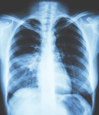
ACOS definitions under fire
LONDON – A study comparing patient data with six definitions of the Asthma-COPD Overlap Syndrome (ACOS) found only one of the patients analyzed met all definitions. This provoked an animated discussion at the annual congress of the European Respiratory Society about the utility of ACOS as a clinical entity.
Of 864 patients diagnosed with chronic obstructive pulmonary disease (COPD) or asthma drawn from the Netherlands Epidemiology of Obesity cohort (a population-based study with 5,784 patients), 39.1% (338 patients) met at least one of the definitions of ACOS, while 0.1% (one patient) met the criteria for all six definitions.
When this finding was presented, the ERS audience first laughed and then applauded. At the end of the presentation, long lines formed at the microphones. Every comment made was hostile to the concept of ACOS.
“Let us bring ACOS to an honorable death,” said one audience member. His point, reiterated by all who commented subsequently, was that ACOS confuses efforts to treat the underlying respiratory symptoms. Even in those who have both asthma and COPD, the speaker, like other members of the audience, said he considered the diagnosis of ACOS unhelpful.
The six definitions in the study included the latest and just published consensus definition from the ERS (Eur Respir J. 2016;48[3]:664-73). According to the ERS definition, the key features of ACOS are age greater than 40 years, long-term history of asthma (since childhood or early adulthood), and significant exposure to cigarette or biomass smoke.
The other definitions analyzed included a medical history of both asthma and COPD, a self-reported history of both asthma and COPD, and a record of the proportion of a person’s vital capacity that he/she is able to expire in 1 second of forced expiration of less than 0.7 plus a record of fractionated nitric oxide concentration in exhaled breath of greater than or equal to 45 parts per billion.
Although attempted, a Venn diagram that would show overlapping subsets of patients that fell into these definitions “was not possible,” according to Tobias Bonten, MD, University of Leiden, the Netherlands.
Asthma duration was just over 10 years in those identified as having ACOS by medical history alone (registry-based definitions), just over 20 years in those with a medical history and objective evidence of impaired lung function, but about 40 years in those with a self-report of both asthma and COPD.
One area that all groups created by the ACOS definitions did have in common was demographic variables, such as median age, proportion of patients defined as overweight or obese by body mass index, and proportion who were current smokers.
Members of the audience acknowledged the importance of considering the coexistence of asthma and COPD, but expressed skepticism about the value of ACOS as a separate entity in the clinic.
“ACOS is something like the emperor’s new clothes,” one audience member said during the discussion. “It is important to identify asthma patients with obstruction because they have reduced lung function that should be treated more actively, but I find the definition [of ACOS] unnecessary,” he said.
A similar conclusion was drawn in a review article devoted to ACOS published last year (N Engl J Med. 2015;373[13]:1241-9). “It is premature to recommend the designation of ACOS as a disease entity,” the authors wrote.
This is a position widely shared by clinicians, judging from audience comments provoked by this demonstration.
For the sake of time, the moderators were forced to end the discussion with significant lines of clinicians at the microphone.
“It is quite clear that ACOS should die,” said one of the last speakers given a chance to voice an opinion. He suggested that the coexistence of asthma and COPD is something that “quite clearly can happen,” but he objected to definitions he said are unhelpful for clinical care.
Dr. Bonten reported no relevant financial relationships.
AT THE ERS CONGRESS 2016
Key clinical point: A study comparing current definitions of Asthma-COPD Overlap Syndrome (ACOS) provoked criticism of the very concept.
Major finding: When six definitions of ACOS were compared in a population-based study, only 1 (0.1%) of 864 possible candidates met all criteria of all definitions.
Data source: Population-based cohort study.
Disclosures: Dr. Bonten reported no relevant financial relationships.
New method proposed for phenotyping COPD patients
LONDON – Based on readily available clinical data, patients with chronic obstructive pulmonary disease (COPD) can be placed into five phenotypes with different characteristics and risk profiles, according to data generated by a cluster analysis and presented at the European Respiratory Society International Congress 2016.
The algorithm that places patients into these phenotypes was developed from a cluster analysis, reported Pierre-Regis Burgel, MD, PhD, professor of respiratory medicine, Université Réné Descartes, Paris. Mortality rates at 3 years for these phenotypes ranged from 2.6% to 49.5%

“We think that this could be the basis for recognizing clinically distinct COPD phenotypes and designing better tailored management,” Dr. Burgel explained. “We also think it has potential use in routine clinical assessment.”
To create these phenotypes, data from 2,049 COPD patients enrolled in a French-Belgian collaborative cohort were evaluated with Classification And Regression Tree (CART) analyses. The five phenotypes were derived from symptom burden, respiratory function, relative age, and presence of comorbidities.
Based on these characteristics, “a set of clinical rules” to phenotype patients was developed, according to Dr. Burgel. This algorithm was then further validated with the 3,651 COPD patients enrolled in the prospective COPD Cohorts Collaborative International Assessment (3CIA) initiative.
The two initial branches in the algorithm are created by dividing patients into those with and without cardiovascular comorbidities or diabetes. In those without cardiovascular disease, the phenotypes are defined by relative symptom severity, using cut-off scores from the modified Medical Research Council (mMRC) dyspnea assessment tool and relative degrees of lung function impairment as measured with forced expiratory volume in 1 second (FEV1).
In those with cardiovascular disease or diabetes, mMRC-defined symptoms and FEV1-defined lung function impairment also create decision points in the algorithm, but age and body mass index (BMI) are additional variables that direct patients to a specific phenotype. Class 4 and 5 are reached in the absence of cardiovascular disease or diabetes only, while cardiovascular disease is a prerequisite to reach Classes 4 and 5. Class 2 is the only phenotype that can be reached through the algorithm irrespective of the presence or absence of cardiovascular disease.
Using this algorithm, each of the phenotypes was associated with similar relative hierarchy in mortality in the two cohorts, even though mortality rates for each phenotype were consistently lower in the 3CIA group.
For class 1, which was reached by patients with cardiovascular disease or diabetes, the greatest symptom burden, and the lowest lung function, the mortality rates at 3 years were 49.5% and 23.2% for the French-Belgian and 3CIA cohorts, respectively.
For class 4, which was also defined by the greatest symptom burden and the lowest lung function without cardiovascular disease or diabetes the mortality rates were 45.3% and 27.3%, respectively. The lowest mortality rates, which were 2.6% and 4.0%, respectively, were found in the class 5 phenotype, which contained patients with a low symptom burden (mMRC less than or equal to 1), relatively good lung function (FEV1 greater than or equal to 60%), and no history of cardiovascular disease or diabetes.
In classes 2 and 3, the mortality rates fell in between those of the lowest- and highest-risk phenotypes. Specifically, these 3-year mortality rates were 22.9% and 24%, respectively, in the French-Belgian cohort, and 11.1% and 14.1%, respectively, in the 3CIA cohort.
The consistency of the hierarchy of outcomes in the two cohorts provides the basis for suggesting that these phenotypes are effective for categorizing relative risk, according to Dr. Burgel. He believes that the phenotypes are clinically important, and he emphasized that the algorithm relies on clinical information that is already routinely collected and readily accessible.
“There is growing awareness that COPD phenotypes are important and are likely to be valuable in managing patients,” Dr. Burgel explained. “We feel that we have created simple rules for allocating patients that we think will be useful for research and for clinical application.”
Dr. Burgel reported financial relationships with AstraZeneca, Boehringer Ingelheim, Chiesi, GlaxoSmithKline, Novartis, Pfizer, and Vertex.
LONDON – Based on readily available clinical data, patients with chronic obstructive pulmonary disease (COPD) can be placed into five phenotypes with different characteristics and risk profiles, according to data generated by a cluster analysis and presented at the European Respiratory Society International Congress 2016.
The algorithm that places patients into these phenotypes was developed from a cluster analysis, reported Pierre-Regis Burgel, MD, PhD, professor of respiratory medicine, Université Réné Descartes, Paris. Mortality rates at 3 years for these phenotypes ranged from 2.6% to 49.5%
“We think that this could be the basis for recognizing clinically distinct COPD phenotypes and designing better tailored management,” Dr. Burgel explained. “We also think it has potential use in routine clinical assessment.”
To create these phenotypes, data from 2,049 COPD patients enrolled in a French-Belgian collaborative cohort were evaluated with Classification And Regression Tree (CART) analyses. The five phenotypes were derived from symptom burden, respiratory function, relative age, and presence of comorbidities.
Based on these characteristics, “a set of clinical rules” to phenotype patients was developed, according to Dr. Burgel. This algorithm was then further validated with the 3,651 COPD patients enrolled in the prospective COPD Cohorts Collaborative International Assessment (3CIA) initiative.
The two initial branches in the algorithm are created by dividing patients into those with and without cardiovascular comorbidities or diabetes. In those without cardiovascular disease, the phenotypes are defined by relative symptom severity, using cut-off scores from the modified Medical Research Council (mMRC) dyspnea assessment tool and relative degrees of lung function impairment as measured with forced expiratory volume in 1 second (FEV1).
In those with cardiovascular disease or diabetes, mMRC-defined symptoms and FEV1-defined lung function impairment also create decision points in the algorithm, but age and body mass index (BMI) are additional variables that direct patients to a specific phenotype. Class 4 and 5 are reached in the absence of cardiovascular disease or diabetes only, while cardiovascular disease is a prerequisite to reach Classes 4 and 5. Class 2 is the only phenotype that can be reached through the algorithm irrespective of the presence or absence of cardiovascular disease.
Using this algorithm, each of the phenotypes was associated with similar relative hierarchy in mortality in the two cohorts, even though mortality rates for each phenotype were consistently lower in the 3CIA group.
For class 1, which was reached by patients with cardiovascular disease or diabetes, the greatest symptom burden, and the lowest lung function, the mortality rates at 3 years were 49.5% and 23.2% for the French-Belgian and 3CIA cohorts, respectively.
For class 4, which was also defined by the greatest symptom burden and the lowest lung function without cardiovascular disease or diabetes the mortality rates were 45.3% and 27.3%, respectively. The lowest mortality rates, which were 2.6% and 4.0%, respectively, were found in the class 5 phenotype, which contained patients with a low symptom burden (mMRC less than or equal to 1), relatively good lung function (FEV1 greater than or equal to 60%), and no history of cardiovascular disease or diabetes.
In classes 2 and 3, the mortality rates fell in between those of the lowest- and highest-risk phenotypes. Specifically, these 3-year mortality rates were 22.9% and 24%, respectively, in the French-Belgian cohort, and 11.1% and 14.1%, respectively, in the 3CIA cohort.
The consistency of the hierarchy of outcomes in the two cohorts provides the basis for suggesting that these phenotypes are effective for categorizing relative risk, according to Dr. Burgel. He believes that the phenotypes are clinically important, and he emphasized that the algorithm relies on clinical information that is already routinely collected and readily accessible.
“There is growing awareness that COPD phenotypes are important and are likely to be valuable in managing patients,” Dr. Burgel explained. “We feel that we have created simple rules for allocating patients that we think will be useful for research and for clinical application.”
Dr. Burgel reported financial relationships with AstraZeneca, Boehringer Ingelheim, Chiesi, GlaxoSmithKline, Novartis, Pfizer, and Vertex.
AT THE ERS CONGRESS 2016
Key clinical point: A relatively simple method proposed for identifying COPD phenotypes has implications for clinical care as well as research.
Major finding: Five phenotypes were identified with mortality rates at 3 years ranging from 2.6% to 49.5%
Data source: Cohort analyses.
Disclosures: Dr. Burgel reported financial relationships with AstraZeneca, Boehringer Ingelheim, Chiesi, GlaxoSmithKline, Novartis, Pfizer, and Vertex.

Cytokine shows promise as biomarker for sepsis outcomes
LONDON – A cytokine known as tumor necrosis factor–related apoptosis-inducing ligand (TRAIL) is showing potential as a biomarker for evaluating the severity of sepsis and septic shock, according to results of a prospective study presented at the annual congress of the European Respiratory Society.
In a prospective study undertaken in patients administered to an intensive care unit, “lower levels of plasma TRAIL correlated with both organ system dysfunction and mortality,” reported Thomas Nicholson, MD, of Cornell University, New York.
A series of studies associating low levels of TRAIL with increased sepsis severity have attracted attention to this potential biomarker, according to Dr. Nicholson. In a mouse model of sepsis, for example, exogenous administration of TRAIL was associated with improved survival. In a clinical study conducted in CHINA that was cited by Dr. Nicholson, low levels of TRAIL correlated with lower rates of survival.
In the data presented at the ERS Congress, plasma TRAIL was collected from 108 patients on the first day of ICU admission. Of these patients 59 (54%) had sepsis and 23 (21%) had septic shock. Those with a noninfectious critical illness served as controls.
All patients with sepsis or septic shock were required to meet diagnostic criteria from the recently published Third International Consensus Definitions for Sepsis and Septic Shock (JAMA. 2016;315[8]:801-10). This is important because the newer criteria, relative to previous criteria, have “moved conceptually away from a condition defined by inflammatory biomarkers toward one that emphasizes signs of organ dysfunction,” Dr. Nicholson reported.
Although a dysregulated host response to infection is still a fundamental concept to the newer definition of sepsis, the importance of biomarkers of inflammation has been deemphasized, a change that would not be expected to favor an inflammatory cytokine as a biomarker.
Despite this, plasma TRAIL levels, which were measured with commercially available ELISA kits, were significantly lower for those with sepsis (P = .038) and for those with septic shock (P less than .001) relative to those with a noninfectious critical illness. There was a trend (P = .077) for lower plasma levels of TRAIL in patients with septic shock relative to sepsis.
In addition, there was a positive and significant correlation (r = –0.1983; P = .0397) between plasma TRAIL and degree of organ dysfunction as measured with the Sequential Organ Failure Assessment (SOFA) score. Higher plasma TRAIL levels also predicted survival at 28 days (P = .016). Although the overall mortality for patients with sepsis or septic shock in this series was 23%, there were no deaths among sepsis or septic shock patients with a TRAIL above the mean, which was 26.8 pg/mL.
“For every 10 mg/mL increase in TRAIL the odds ratio for survival increased by 1.9-fold,” Dr. Nicholson reported.
TRAIL is implicated in several processes that may explain these observations, according to Dr. Nicholson. For example, he reported that there is evidence that TRAIL induces apoptosis in neutrophils, a suspected mediator of sepsis-related injury.
“The observations in our study are consistent with a growing literature suggesting that TRAIL is an important mediator of inflammatory cells, such as neutrophils, tempering the degree of inflammation,” Dr. Nicholson explained.
More data are needed to verify that TRAIL is a clinically useful biomarker, but Dr. Nicholson emphasized that this is a promising area of research. He said the biomarker is being developed as a potential tool for evaluating sepsis severity.
Dr. Nicholson reported no relevant financial relationships.
AT THE ERS CONGRESS 2016
Key clinical point: A biomarker – TRAIL – is demonstrating promise as a tool to evaluate the presence and severity of sepsis and septic shock.
Major finding: In a prospective evaluation, low levels of TRAIL correlated with sepsis and organ dysfunction and higher levels of TRAIL were associated with survival.
Data source: Ongoing prospective cohort study.
Disclosures: Dr. Nicholson reported no relevant financial relationships.