User login
American Neurological Association (ANA): Annual Meeting 2017
APOE4 may drive tau deposition in Alzheimer’s
SAN DIEGO – The apolipoprotein E e4 allele is well known for its association with amyloid deposition in Alzheimer’s disease, but now it also appears to help drive the other key pathological process in the disease: deposition of hyperphosphorylated tau protein.
That’s according to the authors of a PET neuroimaging study presented at the annual meeting of the American Neurological Association.
The finding suggests a pathophysiologic mechanism for Alzheimer’s disease cases that predominantly affect memory.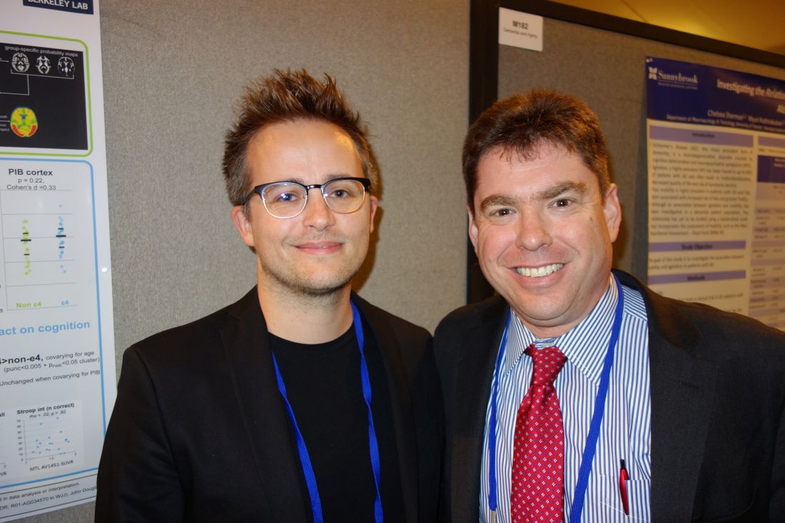
The team then compared the results with uptake of the radiotracers in 71 cognitively normal control subjects who were a mean age of 79 years, 23 of whom (32%) were APOE4 carriers.
APOE4 was associated with higher cortical amyloid in the controls, but not tau deposition. Although AV1451 uptake in the temporal lobe was increased in e4 carriers, the effect was not statistically significant after controlling for PiB uptake.
“We saw an APOE4 effect on amyloid but not on tau in normal people,” said senior investigator Gil Rabinovici, MD, a neurologist and professor of memory and aging at the University of California, San Francisco.
All the AD patients had PiB uptake, with no difference in uptake between e4 carriers and noncarriers. However, APOE4 carriers had higher AV1451 uptake in their anterior medial temporal lobes (MTL), a difference that remained unchanged after controlling for PiB.
Carriers of the e4 allele who had AV1451 uptake in their MTLs had a harder time than other AD patients on a test in which they were asked to recall a series of words after a 10-minute break (California Verbal Learning Test), but they did not perform worse on other cognitive measures.
“In cognitively normal individuals, the effect of APOE4 on tau pathology seems to be mediated by the effect of e4 on Ab [amyloid-beta] deposition,” the investigators noted. “However, when assessing amyloid-positive symptomatic AD patients, APOE4 was associated with increased AV1451 binding in the MTL.
“This suggests that, in addition to its effect on Ab pathology, APOE4 might influence the topographical distribution of tau pathology and, potentially, the cognitive symptoms in patients,” the researchers concluded.
“We are very interested in heterogeneity in Alzheimer’s disease – age of onset, rate of progression, which brain areas are affected,” Dr. Rabinovici said. “This study is beginning to dig into some of the factors that might explain that; APOE4 is one potential modifier. It may have a direct effect on tau phosphorylation, and it changes where tau is located in the setting of Alzheimer’s disease.”
The finding of increased MTL tau in APOE4-positive Alzheimer’s patients helps explain why patients who carry the allele tend to have more memory problems, he said, while AD patients who don’t carry the allele tend to have more of a cortical-predominant presentation, with more visual-spatial and language problems.
“I think it’s very likely that APOE4-related disease may be driven by a different mechanism than APOE4-negative disease,” Dr. Rabinovici said. “In the future, APOE4-positive or APOE4-negative might be used to stratify therapy and the measurements used for disease progression. APOE4 itself may be an interesting therapeutic target because it has downstream effects on amyloid and tau.”
Meanwhile, there’s just not a lot of tau pathology in cognitively normal people, which is likely why the effect didn’t show up in the controls, noted lead investigator Renaud La Joie, PhD, a neuroimaging researcher at UCSF.
The work was funded in part by the National Institutes of Health. Avid Pharmaceuticals provided the AV1451. Dr. Rabinovici is an advisor for Genentech, Merck, and Roche, and has research support from Avid Radiopharmaceuticals and Eli Lilly, among others.
SOURCE: La Joie R, et al. Abstract M183, American Neurological Association 2017 annual meeting.
REPORTING FROM ANA 2017
Key clinical point:
Major finding: APOE4 carriers had higher AV1451-uptake in their anterior medial temporal lobes, a difference that remained unchanged after controlling for Pittsburgh compound B.
Study details: An analysis of radiotracer PET imaging in 67 Alzheimer’s disease patients and 71 controls.
Disclosures: The work was funded in part by the National Institutes of Health. Avid Pharmaceuticals provided the AV1451. The senior investigator is an advisor for Genentech, Merck, and Roche, and has research support from Avid Radiopharmaceuticals and Eli Lilly, among others.
Source: La Joie R, et al. Abstract M183, American Neurological Association 2017 annual meeting.
Cars that recognize hypoglycemia? Maybe soon
SAN DIEGO – When researchers at the University of Nebraska placed sensors in the cars of patients with type 1 diabetes, they found something interesting: About 3.4% of the time, the patients were driving with a blood glucose below 70 mg/dL.
Almost 10% of the time, it was above 300 mg/dL, and both hyper and hypoglycemia, but especially hypoglycemia, corresponded with erratic driving, especially at highway speeds.
The finding explains why patients taking insulin for type 1 diabetes have a 12%-19% higher risk of crashing their cars, compared with the general population. But in a larger sense, the study speaks to a new possibility as cars become smarter: monitoring drivers’ mental states and pulling over to the side of the road or otherwise taking control if there’s a problem.
The “results show that vehicle sensor and physiologic data can be successfully linked to quantify individual driver performance and behavior in drivers with metabolic disorders that affect brain function. The work we are doing could be used to tune the algorithm that drive these automated vehicles. I think this is a very important area of study,” said senior investigator Matthew Rizzo, MD, chair of the university’s department of neurological sciences in Omaha.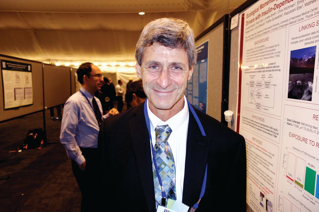
Participants had the devices in their cars for a month, during which time the diabetes patients were also on continuous, 24-hour blood glucose monitoring. The investigators then synched the car data with the glucose readings, and compared it with the data from the controls’ cars. In all, the system recorded more than 1,000 hours of road time across 3,687 drives and 21,232 miles.
“What we found was that the drivers with diabetes had trouble,” Dr. Rizzo said at the American Neurological Association annual meeting.
Glucose was dangerously high or low about 13% of the time when people with diabetes were behind the wheel. Their accelerometer profiles revealed more risky maneuvering and variability in pedal control even during periods of euglycemia and moderate hyperglycemia, but particularly when hypoglycemia occurred at highway speeds.
One driver almost blacked out behind the wheel when his blood glucose fell below 40 mg/dL. “He might have been driving because he was not aware he had a problem,” Dr. Rizzo said. He is now; he was shown the video.
The team reviewed their subjects’ department of motor vehicles records for the 2 years before the study. All three car crashes in the study population were among drivers with diabetes, and they received 11 of the 13 citations (85%).
The technology has many implications. In the short term, it’s a feedback tool to help people with diabetes stay safer on the road. But the work is also “a model for us to be able to approach all kinds of medical disorders in the real world. We want generalizable models that go beyond type 1 diabetes to type 2 diabetes and other forms of encephalopathy, of which there are many in neurology.” Those models could one day lead to “automated in-vehicle technology responsive to driver’s momentary neurocognitive state. You could have [systems] that alert the car that the driver is in no state to drive; the car could even take over. We are very excited about” the possibilities, Dr. Rizzo said.
Meanwhile, “just the diagnosis of diabetes itself is not enough to restrict a person from driving. But if you record their sugars over long periods of time, and you see the kind of changes we saw in some of the drivers, it means the license might need to be adjusted slightly,” he said.
Dr. Rizzo had no relevant disclosures. One of the investigators was an employee of the Toyota Collaborative Safety Research Center.
REPORTING FROM ANA 2017
Key clinical point:
Major finding: Glucose was dangerously high or low about 13% of the time when people with diabetes were behind the wheel.
Study details: Investigators paired real-time driving data with continuous glucose monitoring in patients with type 1 diabetes to asses how blood sugar levels affected driving.
Disclosures: Toyota funded the work. The senior investigator had no relevant disclosures.
Source: Rizzo M, et al. ANA 2017 abstract number S131.
Corynebacterium in the gut can trigger Parkinson’s disease
SAN DIEGO – The presence of Corynebacterium in the gut microbiome of people with two G alleles at the rs356219 single nucleotide polymorphism locus of the alpha-synuclein gene was associated with 100% probability of having Parkinson’s disease in a study conducted by the NeuroGenetics Research Consortium.
If the finding is replicated, it means that Corynebacterium is the trigger for Parkinson’s disease (PD) in people with the GG genotype. The GG signature at rs356219 is the strongest genetic risk factor for PD identified to date, but it’s not necessarily strong enough to cause the disease on its own. “It definitely needs a trigger,” and there’s a good chance that Corynebacterium is it, said senior investigator Haydeh Payami, PhD, professor of neurology and genomics at the University of Alabama, Birmingham.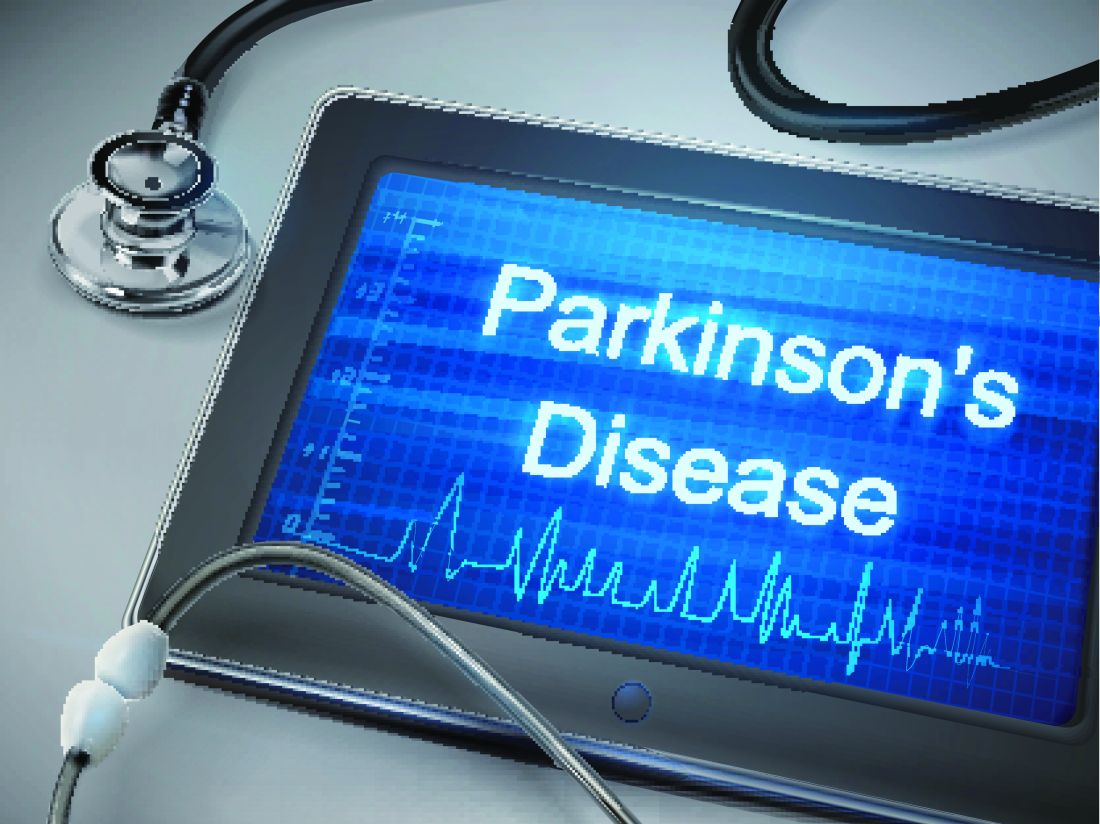
Her team genotyped SNCA rs356219 from blood samples in 197 middle-aged PD patients and 115 age-matched controls. They also extracted DNA from stool samples to see what bacteria were in their gut and then looked for interactions between rs356219 genotype, gut microbiome, and PD risk.
The medical literature has been full of hints for a while now that PD might be set off by something going wrong in the gastrointestinal tract. Colonic inflammation, alpha-synuclein pathology in the gut, and dysbiosis of the gut microbiome in PD are among the many clues. The goal of the work was to find the link between PD and its GI aberrations.
Ninety genera were identified in the stool samples, but “no matter how you looked at the data, whichever method you used, one [genus] kept coming up” for interaction with the rs356219 genotype, “and that was Corynebacterium,” Dr. Payami said.
As in past studies, the rs356219 AA genotype did not increase the odds of PD, and there was no difference in microbiome abundance between PD patients and controls. The GA genotype increased the odds slightly without Corynebacterium, but it increased the odds more than fivefold when Corynebacterium was in the gut (odds ratio, 5.9; P = .04). If people had GG plus Corynebacterium, however, “you nailed it,” Dr. Payami said: The odds of developing PD were infinite (P = .0003).
Corynebacterium was more abundant in GA subjects with PD than GA subjects without PD, but it was by far the most abundant in GG subjects, and every person who had the GG genotype and gut Corynebacterium also had PD.
Corynebacterium are gram-positive, aerobic bacilli commonly found on the skin. Some members of the genus are opportunistic pathogens. It’s not clear how they get incorporated into the gut microbiome, or if they can be wiped out selectively in the gut with antibiotics or probiotics.
Perhaps Corynebacterium in the GI tract induces expression of alpha-synuclein protein, a major component of PD Lewy bodies that’s known to travel from the gut to the brain. Maybe the amount expressed depends on how many Gs people have in rs356219. Perhaps “if you have two Gs, you get so much alpha-synuclein that’s there’s no turning back, and it’s enough to cause PD,” Dr. Payami said.
The study was led by Zachary Wallen, a PhD candidate in Dr. Payami’s lab, and presented by him at the annual meeting of the American Neurological Association. The work was supported by the National Institutes of Health. Dr. Payami and Mr. Wallen had no industry disclosures.
SOURCE: Wallen Z et al. ANA 2017 abstract number S268
REPORTING FROM ANA 2017
Key clinical point: in genetically susceptible people.
Major finding: Those who have Corynebacterium in their gut microbiome plus two G alleles at the rs356219 locus of the alpha-synuclein gene have infinite odds of developing PD (P = .0003).
Study details: A case-control study involving 197 middle-aged PD patients and 115 age-matched controls.
Disclosures: The work was supported by the National Institutes of Health. The lead and senior investigators had no industry disclosures.
Source: Wallen Z et al. ANA 2017 abstract number S268
Head injury linked to amyloid deposition decades later
SAN DIEGO – , according to PET imaging in 329 older adults without dementia.
Previous studies have found an association between head injury and later dementia, but the mechanism isn’t understood. The importance of the new investigation is that it “hints at pathophysiology. We were very excited” to find amyloid in the frontal cortex, “which is also associated with Alzheimer’s disease,” said lead investigator Andrea Schneider, MD, PhD, a neurology resident at Johns Hopkins University, Baltimore.
Increased amyloid deposition was not found in other areas associated with the disease, but frontal cortex involvement “does suggest perhaps a common” denominator, she noted.
The older adults in the study were all participants in the Atherosclerosis Risk in Communities (ARIC) cohort, a group of over 15,000 community-dwelling adults who have been followed since the late 1980s. All 329 had brain amyloid deposition assessed by florbetapir (Amyvid) PET scans in 2011-2013; mean age was 76 years.
Sixty-six (20%) reported a history of head trauma at a median of about 25 years before the scan, with self-reports correlating well with hospitalization billing codes collected as part of ARIC. The cause of the head trauma was not known.
Head injury was associated with elevated standardized uptake value ratios (SUVRs, greater than 1.2). Head injury patients had a 1.31-fold increased prevalence of elevated global amyloid deposition (95% confidence interval, 1.07-1.60) as well as a 1.24-fold increased prevalence of elevated deposition in the orbitofrontal cortex (95% CI, 1.02-1.50) and prefrontal cortex (95% CI, 1.03-1.49), and 1.29-fold increased prevalence in the superior frontal cortex (95% CI, 1.06-1.56).
There were no differences in the prevalence of elevated SUVRs in the anterior cingulate, posterior cingulate, precuneus, or the lateral temporal, parietal, or occipital lobes (all P greater than .05) Dr. Schneider reported.
The model was adjusted for age, sex, race, total intracranial volume, and cardiovascular disease, among other things.
Thirty percent of the participants had either one or two APOE4 alleles, and 27% had mild cognitive impairment; neither correlated with increased amyloid deposition.
Head injuries were more common among participants who were men (43%) or white (57%). There was a trend for a slightly stronger link between head injury and increased amyloid deposition among black individuals (P for interaction 0.169).
Dr. Schneider is continuing her work to illuminate the connection between head trauma and dementia. She plans to integrate more detailed cognitive data from ARIC into the analysis, and perhaps emergency department and outpatient data. She also wants to look at more acute imaging after head trauma.
The work was funded by the National Institutes of Health. Avid Radiopharmaceuticals provided the florbetapir. Dr. Schneider did not have any relevant disclosures.
SOURCE: Schneider A et al. ANA 2017 abstract M336WIP
REPORTING FROM ANA 2017
Key clinical point: Head injury is associated with increased brain amyloid deposition globally and in the frontal cortex, which may help explain the association between head trauma and dementia.
Major finding: Head injury patients had a 1.31-fold increased prevalence of elevated global amyloid deposition (95% CI, 1.07-1.60) as well as a 1.24-fold increased prevalence of elevated deposition in the orbitofrontal cortex (95% CI, 1.02-1.50) and prefrontal cortex (95% CI, 1.03-1.49) and 1.29-fold increased prevalence in the superior frontal cortex (95% CI, 1.06-1.56).
Data source: PET imaging in 329 older adults without dementia.
Disclosures: The work was funded by the National Institutes of Health. Avid Radiopharmaceuticals provided the florbetapir. The lead author did not have any relevant disclosures.
Source: Schneider A et al. ANA 2017 abstract M336WIP.
Faciobrachial dystonic seizures require urgent immunotherapy
SAN DIEGO – The sooner immunotherapy is started for faciobrachial dystonic seizures, the better, according to a retrospective review presented at the American Neurological Association annual meeting.
Only recently described, patients with faciobrachial dystonic seizures (FBDS) have frequent episodes – sometimes hundreds a day – of abrupt, involuntary, stereotypical movements lasting 1-5 seconds and typically involving one half of their face and the arm on the same side, such as a left-sided grimace and arm flex. It’s closely associated with antibodies against leucine-rich glioma, inactivated 1 (LGI-1), a protein that plays a role in synaptic transmission and myelination.
“This is an autoimmune condition. It’s important to get onto it early; you’ve got to treat these seizures with immunotherapy and treat them fast,” she said.
A lot of the time, FBDS is probably mistaken for a movement disorder or tic, but tics aren’t typically dystonic, plus FBDS patients can’t suppress their movements and they don’t feel the buildup of tension, then release typical of tics. “If something doesn’t add up, test for the antibodies,” Dr. Thompson said.
Her team reviewed 103 cases culled from South Korea, Great Britain, Australia, and the United States, the largest cohort assembled to date. The median age was 64 years old; most patients were white, and 62% were men. There were no associations with tumors, and none of the seasonal variation typical of viral causes.
FBDS showed “a striking time-dependent response to immunotherapy. Prompt immunotherapy” stops the seizures and prevents brain damage, “maybe via inhibiting IgG1-mediated complement deposition,” Dr. Thompson said.
Antiepileptic drugs didn’t help much, but immunotherapy did; among patients who had immunotherapy, seizures stopped within 30 days in over half, and in 88% after 3 months. Only one patient with seizure cessation went on to develop cognitive impairment. For every day that immunotherapy was delayed, there was a 0.69% reduction in the probability of stopping the seizures. The window of opportunity appears to be short: among patients whose seizures weren’t terminated within 3 months, 56% progressed to cognitive impairment.
Immunotherapy varied greatly across the study centers, and included intravenous immunoglobulin, azathioprine, and mycophenolate, among other options that were generally given on a background of steroids.
“It became quite obvious that immunotherapy was the key,” but there weren’t enough patients to determine the optimal approach. Dr. Thompson and her colleagues are working to figure that out, as well as what triggers FBDS and its pathophysiology, she said.
Patients who just had seizures had almost exclusively LGI-1 IgG4 antibodies; those who had progressed to cognitive impairment had LGI-1 IgG1 antibodies. It’s unclear at this point what causes the subclass switch as patients progress.
Medial temporal lobe T2-hyperintensities and temporal and frontal lobe ictal EEG changes, as well as serum hyponatremia, were also found almost exclusively in patients with cognitive impairment.
Much remains to be learned about the condition.
There was no industry funding for the work, and Dr. Thompson didn’t have any disclosures.
AT ANA 2017
Key clinical point:
Major finding: For every day that immunotherapy was delayed, there was a 0.69% reduction in the probability of stopping the seizures and progression to limbic encephalitis and permanent disability.
Data source: Review of 103 patients, the largest cohort assembled to date.
Disclosures: There was no industry funding for the work, and the lead investigator had no disclosures.
Salivary biomarker identified for Huntington’s disease
SAN DIEGO – Huntingtin protein – the key biomarker for Huntington’s disease – can be detected in saliva, which might prove to be an easy and inexpensive way to diagnose and monitor Huntington’s, and perhaps even predict clinical onset, according to investigators at the University of California, San Diego.
In a study of 178 subjects, they found that salivary total huntingtin protein (Htt) was significantly increased in saliva from individuals with Huntington’s disease (HD), compared with controls without HD (mean, 0.775 ng/mL vs. 0.359 ng/mL; P = .0012). Levels remained consistent throughout the day and from day to day, and were not affected by age or sex.
Meanwhile, salivary mutant Htt levels were higher in gene-positive, premanifest HD subjects than in normal controls (P less than .05). Salivary C-reactive protein level was also significantly elevated in premanifest HD subjects (9,548 pg/mL vs. 3,399 pg/mL, P = .025), indicating a pathologic inflammatory or metabolic state. When considered together, the two measurements might herald the onset of symptoms.
There’s an acute need for a convenient, inexpensive HD biomarker. Htt isn’t often measured in clinical practice, and when it is, it’s assessed from blood or cerebrospinal fluid. With salivary Htt, “you don’t need any specialized personnel, [and samples] are easy to obtain and process. They keep well and are very stable. We don’t have to rush to get them somewhere,” said lead investigator Jody Corey-Bloom, MD, PhD, professor emeritus of neurosciences at the university.
“We are really excited about the potential of salivary Htt. We think this is going to be an easy way to follow patients,” she said at the annual meeting of the American Neurological Association.
The next step is to see how salivary Htt correlates with cerebrospinal fluid and blood levels. The team will also investigate it as a diagnostic tool; perhaps there’s a cut point that diagnoses HD. “It’s an intriguing idea,” Dr. Corey-Bloom said.
Perhaps the greatest potential is for predicting disease onset so treatments can be started before symptoms emerge. There’s nothing on the market yet that can delay or prevent progression, but trials are in the works for therapeutics that lower levels of mutant Htt in the brain. “If we can use something simple like salivary Htt [to start preemptive treatment] that would be phenomenal. That’s the hope,” she said.
Subjects refrained from smoking, eating, drinking, and brushing their teeth for at least an hour before saliva samples were taken. Testing was done by Western blot and enzyme-linked immunosorbent assay.
The work was funded by the university. Dr. Corey-Bloom said she had no relevant disclosures.
AT ANA 2017
Key clinical point:
Major finding: Salivary total huntingtin protein was significantly increased in saliva from individuals with Huntington’s disease, compared with controls (mean, 0.775 ng/mL vs. 0.359 ng/mL, P = .0012).
Data source: Measurement of salivary Htt and other analytes in 178 subjects.
Disclosures: The work was funded by the University of California, San Diego. The lead investigator had no relevant financial disclosures.

In Lewy body disease, CSF may reveal dementia risk
SAN DIEGO – In patients with Lewy body disease, cerebrospinal fluid biomarkers may predict the extent of Alzheimer’s disease pathology, according to a new postmortem analysis.
In the small prospective study, David Irwin, MD, and his colleagues at the University of Pennsylvania, Philadelphia, found that higher antemortem cerebrospinal fluid (CSF) levels of total tau (t-tau) protein and lower amyloid-beta 1-42 (Abeta 1-42) levels were associated with postmortem evidence of cerebral Alzheimer’s disease and synuclein pathology.
The findings could have prognostic indications because about 40% of LBD patients have enough Abeta 1-42 and tau tangle pathology to have a secondary diagnosis of Alzheimer’s disease, and greater Alzheimer’s disease pathology has been linked to shorter survival. It could also help individualize therapy, as LBD patients with more Alzheimer’s disease character could be expected to respond better to antisynuclein and Alzheimer’s therapies, according to Dr. Irwin, who presented the study at a poster session at the annual meeting of the American Neurological Association.
In fact, the CSF ratio of CSF t-tau/Abeta 1-42 predicted the presence or absence of Alzheimer’s disease pathology with 90% sensitivity and 100% specificity.
If the results from the small study hold up, “we can identify who has Alzheimer’s copathology and who has a more aggressive form of Lewy body disease where it’s reaching the cortex rather than being in the brain stem, and that correlates with memory loss, dementia, and other things that impair the patient’s functioning,” Dr. Irwin said in an interview.
The study was possible because the Udall Center for Parkinson’s Research at the University of Pennsylvania has an active autopsy program and a significant number of Parkinson’s disease patients who have donated their brain for research. “It’s very rare to have autopsy-confirmed samples where we can validate what we’re measuring in the spinal fluid to what is actually accumulating in the brain,” Dr. Irwin said.
The study included 24 patients with autopsy-confirmed LBD who had undergone CSF testing while they were still alive. The researchers calculated ordinal pathology scores of tau tangles, Abeta 1-42 plaques, and alpha-synuclein pathology across seven regions of the brain, producing a global pathology score for each variable. The subjects were then divided into two groups: Lewy body synuclein stages with medium to high Alzheimer’s copathology (SYN+AD, n = 10) and those with low to no Alzheimer’s copathology (SYN-AD, n = 14).
In the CSF, the researchers analyzed associations with t-tau, phosphorylated tau (p-tau), and Abeta 1-42. They found that Abeta 1-42 levels were significantly different between SYN+AD and SYN-AD patients (147.2 pg/mL vs. 231.1 pg/mL, respectively; P = .001), as were CSF t-tau levels (63.9 pg/mL vs. 36.9 pg/mL; P = .02). P-tau was not significantly different (20.2 pg/mL vs. 15.5 pg/mL; P = 0.3), which was a surprise since it “is usually the strongest correlate of tangles in the brain,” Dr. Irwin said.
A ratio of CSF t-tau/Abeta 1-42 higher than 0.30 distinguished between SYN+AD and SYN-AD (area under the curve, 0.92; 95% confidence interval, 0.7-1.0; 90% sensitivity, 100% specificity), while CSF Abeta 1-42 less than 185 pg/mL distinguished neocortical synuclein stage (AUC, 0.92; 95% CI, 0.54-0.94; 77% sensitivity, 82% specificity).
The study was funded by the National Institute of Neurological Disorders and Stroke. Dr. Irwin reported having no financial disclosures.
AT ANA 2017
Key clinical point:
Major finding: A CSF ratio of t-tau/Abeta 1-42 greater than 0.30 predicted the presence or absence of Alzheimer’s disease pathology with 90% sensitivity and 100% specificity.
Data source: Prospective study of 24 patients with Lewy body disease.
Disclosures: The study was funded by the National Institute of Neurological Disorders and Stroke. Dr. Irwin reported having no financial disclosures.
VIDEO: Smartphones could ‘democratize’ EEG
SAN DIEGO – Epilepsy is a common condition, but many people live in areas too poor or underserved to benefit from electroencephalogram (EEG) testing to confirm a diagnosis. Modern technology may be changing that. Smartphones wirelessly linked to headsets can collect EEG data and potentially serve as an alternate platform for diagnosis of epilepsy.
To test the efficacy of one such system, Farrah Mateen, MD, PhD, a neurologist at Massachusetts General Hospital, Boston, conducted a study in the remote, mountainous Kingdom of Bhutan, which lies between India and China. Her group compared the signals between a smartphone EEG and standard EEG.
The sensitivity was low, at around 40%, but the device’s specificity was 90%-95%. For now, the device performs better as a confirmatory test than as a screening device, but the researchers hope to adjust the lead location to improve sensitivity.
The headset used was designed for use by video gamers, and at a cost of less than $300, it represents a low-cost entry into portable EEG.
Dr. Mateen discussed the smartphone EEG study in a video interview at the annual meeting of the American Neurological Association.
AT ANA 2017
New narcolepsy drug passes phase 3 test
SAN DIEGO – The selective dopamine and norepinephrine reuptake inhibitor solriamfetol is effective in reducing sleepiness in patients with narcolepsy, according to results of a phase 3 study.
At 150-mg and 300-mg doses, the drug had statistically significant effects on objective and subjective measures.
There are wake-promoting drugs available, such as amphetamine-related drugs that are often used off label, but addiction liability is a concern. The nonamphetamine modafinil has been approved by the Food and Drug Administration since 1998.
Jazz Pharmaceuticals is in the process of submitting solriamfetol for FDA evaluation. If approved, the drug will add to the options available for narcolepsy patients. “All of the available drugs have some limitations. Some have more abuse liability than others. Some have more robust wake-promoting properties than others. We haven’t done any head-to-head comparisons, so I can’t tell you how we will stack up,” Philip Jochelson, MD, said in an interview. Dr. Jochelson is vice president of clinical development at Jazz Pharmaceuticals and presented the results of the study at a poster session at the annual meeting of the American Neurological Association.
An earlier study showed the drug had less abuse potential than the schedule IV stimulant phentermine. That’s not surprising given the drug’s mechanism of action, Dr. Jochelson said. Amphetamine-based drugs stimulate dopamine release, which can prompt a dopamine surge that people equate with a high, he said. Solriamfetol also affects dopamine, but it is a reuptake inhibitor, so it doesn’t produce a surge.
If the drug gains approval, it remains to be seen how it will be classified on the Drug Enforcement Agency Controlled Substance scale. “Where it will fall in that spectrum is speculative at this point,” said Dr. Jochelson.
In the current study, 236 adults (aged 18-75 years) with type 1 narcolepsy were randomized to once-daily placebo, 75 mg solriamfetol, 150 mg solriamfetol, or 300 mg solriamfetol; 27.1% of patients in the 300-mg group discontinued, compared with 7.3% in the 150-mg group, 16.9% in the 75-mg group, and 10.3% in the placebo group. The mean change from baseline on the Maintenance of Wakefulness Test was statistically significant in the 300-mg group (12.3 minutes vs. 2.1 minutes for placebo, P less than .0001) and the 150-mg group (9.8 minutes vs. 2.1 minutes, P less than .0001) but not the 75-mg group (4.7 minutes vs. 2.1 minutes).
The drug also outperformed placebo at week 12 on the Epworth Sleepiness Scale. The mean change in the 300-mg group was –6.4 vs. –1.6 for placebo (P less than .001), –5.4 in the 150-mg group (P less than .0001), and –3.8 in the 75-mg group (P less than .05).
By both Maintenance of Wakefulness Test and Epworth Sleepiness Scale measures, the 150-mg and 300-mg solriamfetol groups had statistically significant differences as early as week 1.
The drug had some adverse effects, which were expected based on its pharmacologic profile. These included increases in headache (5.1% with placebo, 10.2% with 75 mg, 23.7% with 150 mg, 30.5% with 300 mg), nausea (1.7% for placebo, 5.1% for 75 mg, 10.2% for 150 mg, 16.9% for 300 mg), anxiety (1.7% with placebo, 1.7% with 75 mg, 5.1% with 150 mg, 8.5% with 300 mg), and insomnia (0% for placebo, 3.4% for 75 mg, 0% for 150 mg, 5.1% for 300 mg). Other adverse events occurring in at least 5% of patients were decreased appetite, nasopharyngitis, and dry mouth.
The study was funded by Jazz Pharmaceuticals. Dr. Jochelson is an employee of Jazz.
AT ANA 2017
Key clinical point: Solriamfetol outperformed placebo on both objective and subjective sleep measures.
Major finding: At 12 weeks, 150 mg increased Maintenance of Wakefulness Test score to 9.8 minutes, compared with 2.1 minutes in the placebo group.
Data source: A randomized, controlled trial (n = 236).
Disclosures: The study was funded by Jazz Pharmaceuticals. Dr. Jochelson is an employee of Jazz.
Stroke cognitive outcomes found worse in Mexican Americans
SAN DIEGO – A new analysis shows that Mexican Americans (MAs) have worse cognitive outcomes a year after having a stroke than do non-Hispanic whites (NHWs).
The findings, which show that MAs had a mean score of 86 on the Modified Mini-Mental State Examination (3MSE; range: 0-100), compared with 92 for NHWs, come from a prospective study of MAs and NHWs in Corpus Christi, Texas, where both populations have long-established residencies.
After controlling for all factors, the researchers found a difference of –6.73 (95% confidence interval, –3.88 to –9.57; P less than .001).
“The Mexican-American population is growing quickly and aging. The cost of stroke-related cognitive impairment is high for patient, family, and society. Efforts to combat stroke-related cognitive decline are critical,” said Dr. Morgenstern, professor of neurology and epidemiology at the University of Michigan, Ann Arbor.
The study grew out of the Brain Attack Surveillance in Corpus Christi (BASIC) Project, which began in 1999 and is funded until 2019. It is the only ongoing stroke surveillance program that focuses on Mexican Americans, who comprise the largest segment of Hispanic Americans.
The researchers analyzed data encompassing all stroke patients in the BASIC Project from October 2014 through January 2016 (n = 227). They analyzed cognitive outcome data from 3 months, 6 months, and 12 months. MAs were younger on average than NHWs (median age 66 vs. 70; P = .018), and were more likely to have diabetes (54% vs. 36%; P less than .001). They were less likely to have atrial fibrillation (13% vs. 20%; P = .025).
At 12 months, MAs had a lower median 3MSE score of 86 (interquartile range, 73-93, compared with 92 in NHWs (IQR, 83-96; P less than .001). As the researchers adjusted for additional factors, the discrepancy became larger. Adjustment for age and sex revealed a difference of 6.88 (95% confidence interval, 4.15-9.60). Additional adjustment for prestroke condition showed a difference of 7.04. Additional adjustment for insurance led to the same differential of 7.04. Adjustment for diabetes and comorbidities pushed the difference to 7.11. Adjustment for stroke severity (National Institutes of Health Stroke Scale) revealed a difference of 6.73 (P less than .001).
Asked if the results were surprising, Dr. Morgenstern replied: “I think it’s always surprising to see one population of U.S. citizens who have more disease or a worse outcome than another when it’s not explained by the many possible factors we considered.” He also called for additional studies of cognitive dysfunction in Hispanic communities in other forms of dementia, such as Alzheimer’s disease and vascular dementia. “There’s very little of that,” he said.
The National Institutes of Health funded the study. Dr. Morgenstern reported having no financial disclosures.
AT ANA 2017
Key clinical point: In an analysis, cognitive outcomes were worse in Mexican-American stroke survivors despite researchers’ controlling for many factors.
Major finding: At 12 months, Mexican Americans scored 6 points lower on the Modified Mini-Mental State Examination compared with non-Hispanic whites.
Data source: Prospective analysis of 227 stroke patients in Corpus Christi, Texas.
Disclosures: The National Institutes of Health funded the study. Dr. Morgenstern reported having no financial disclosures.