User login
What is the most effective management of acute fractures of the base of the fifth metatarsal?
For acute Jones’ fractures in recreationally active patients, early intramedullary screw fixation results in lower failure rates and shorter times to both clinical union and return to sports than non-weightbearing short leg casting (strength of recommendation [SOR]: A, based on 2 randomized controlled trials (RCT)]. Non-weightbearing short leg casting achieves union in 56% to 100% of patients but can require prolonged casting (SOR: B, based on 2 prospective cohorts and multiple retrospective, follow-up studies). Stress fractures were not included in this review.
For avulsion fractures of the fifth metatarsal tuberosity, a soft Jones–dressing allows earlier return to pre-injury levels of activity than rigid short leg casting (SOR: B, based on a lower-quality RCT).
For athletes, surgical correction of all Jones-type fractures usually preferred
Douglas F. Aukerman, MD
Family and Community Medicine, The Milton S. Hershey, Medical Center, Penn State University
Fifth metatarsal fractures are frequently seen in clinical practice. When faced with a fifth metatarsal fracture, determine its exact location, which influences treatment. Acute fractures to the proximal end of the bone within the cancellous bone area, if nondisplaced, do very well with closed treatment.
Fractures between the insertion of the peroneus brevis and tertius tendons, which marks a transition from mostly cancellous to relatively avascular cortical bone, can be problematic. This injury, often called a Jones fracture, needs to be identified as a chronic stress injury, which uniformly does not heal well, an acute or chronic stress injury, or a pure acute injury. For athletes, both young and old, I prefer surgical correction of all Jones-type fractures to ensure a more definitive return to athletics. For the non-athlete, I allow the patient to make an informed decision for immediate surgical correction or for an attempt at closed treatment if it is not a chronic stress failure of the bone. I find that patients who choose closed treatment and understand the possible prolonged treatment course are not upset if they need surgical treatment for nonunion and are pleased with the option and attempt of not having surgery.
Evidence summary
Fractures within 1.5 cm of the fifth metatarsal tuberosity, without extension distal to the fourth-fifth intermetatarsal articulation, occurring with less than 2-week symptom prodrome and without a history of previous fracture, are defined as “acute Jones’ fractures” (FIGURE). In a recent RCT by Mologne et al,1 37 active-duty military personnel with acute Jones’ fractures were randomized to either 8 weeks of no weight-bearing in a short leg casting, followed by a walking cast or hard-soled shoe until clinical union; or to early outpatient intramedullary screw fixation followed by no weight-bearing for 2 weeks, then weight-bearing as tolerated in a hard-soled shoe until clinical union. Screw fixation significantly reduced both time to clinical union and time to return to sports—by nearly 50% when compared with non-weightbearing short leg casting. Furthermore, at 26 weeks the casting group saw a significant 44% failure rate compared with only 5% in the surgical group (number needed to treat [NNT]=2.6). Six patients in the surgical group had mild discomfort from the screw head, and 3 needed the screw to be removed. Generalization of the results was limited by the mostly male military population.
The rates and times of union with short leg casting vary over a wide range in the research literature. The casting group in the RCT above had union rates of 56% and median time to union of 14.5 weeks (lower and upper quartile range, 10.5–18.5).1 A prospective registry of 68 consecutive acute Jones’ fractures in primarily young military service members showed a 72% union rate with non-weightbearing short leg casting with average time to union of 21.2 weeks.2 A heterogeneous group of 5 retrospective follow-up studies of short leg casting reported wide ranges in union rates of 72% to 100%, and in time to healing of 7 weeks to 21 months.3-7 These studies varied in average age, sex, and athletic ability of their samples as well as type of immobilization and weight-bearing status during treatment.
Tuberosity avulsion fractures are proximal fifth metatarsal styloid fractures resulting from a forceful pull of the lateral band of the long plantar ligament or the peroneus brevis tendon during ankle inversion. A 12-week RCT in 89 consecutive patients presenting to an emergency department with fifth metatarsal tuberosity avulsion fractures compared a nonrigid, soft Jones’ dressing consisting of alternating layers of cast padding and elastic bandages with a rigid short leg casting.8 The Jones’ dressing had a significant 28% reduction in time to return to pre-injury levels of activity. Other outcomes—time in treatment modality, time to radiographic healing, and functional foot score—were not different between intervention groups. Validity was limited by the 32% lost to follow-up rate.
FIGURE
Acute fracture of the fifth metatarsal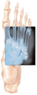
Acute Jones’ fractures are repaired with screw fixation of the broken bone using fluoroscopy. Patients may return to full activity when radiographs show that the bones were healing at the site of the fracture.
Recommendations from others
We were unable to locate any consensus statements or clinical guidelines regarding the treatment of Jones’ fractures.
DeLee and Drez’s Orthopaedic Sports Medicine recommends immobilization in a cast or below-the-knee boot with strict non-weightbearing for at least 6 weeks for acute Jones’ fractures.9 It recommends surgical treatment, followed by 6 weeks of cast immobilization, then progression to weight bearing based on radiographic findings, for nonoperative treatment failures or with desire to return high-performance athletes to activity.
In Fracture Management for Primary Care, the authors recommend posterior splinting and non-weightbearing with crutches for acute Jones’ fractures, followed by non-weightbearing short leg casting application at 3 to 5 days from injury.10 After a minimum of 6 to 8 weeks of casting, they recommend options of 4 additional weeks of casting or internal fixation for clinical or radiographic nonunion.
For tuberosity avulsion fractures, the authors recommend use of a firm-soled shoe for 4 to 8 weeks. For patients with discomfort at an initial 4- to 7-day follow-up, they give an option of using a walking short leg casting for 2 weeks, with follow-up every 2 to 4 weeks until clinical healing.
1. Mologne TS, Lundeen JM, Clapper MF, O’Brien TJ. Early screw fixation versus casting in the treatment of acute Jones fractures. Am J Sports Med 2005;33:970-975.
2. Clapper MF, O’Brien TJ, Lyons PM. Fractures of the fifth metatarsal: Analysis of a fracture registry. Clin Orthop Relat Res 1995;315:238-241.
3. Dameron TB. Fractures and anatomical variations of the proximal portion of the fifth metatarsal. J Bone Joint Surg Am 1975;57:788-792.
4. Torg JS, Balduini FC, Zelko RR, Pavlov H, Peff TC, Das M. Fractures of the base of the fifth metatarsal distal to the tuberosity. J Bone Joint Surg Am 1984;66:209-214.
5. Seitz WH, Grantham SA. The Jones’ fracture in the non-athlete. Foot Ankle 1985;6:97-100.
6. Josefsson PO, Karlsson M, Redlund-Johnell I, Wendeberg B. Closed treatment of Jones fracture: Good results in 40 cases after 11-26 years. Acta Orthop Scand 1994;65:545-547.
7. Josefsson PO, Karlsson M, Redlund-Johnell I, Wendeberg B. Jones fracture: Surgical versus nonsurgical treatment. Clin Orthop Related Res 1994;299:252-255.
8. Wiener BD, Linder JF, Giattini JF. Treatment of fractures of the fifth metatarsal: a prospective study. Foot Ankle Int 1997;18:267-269.
9. Brodsky JW, Krause JO. Stress fractures of the foot and ankle. In: Delee JC, Drez, D, Miller MD, eds. DeLee and Drez’s Orthopaedic Sports Medicine. Philadelphia, Pa: Saunders; 2003:2403-2406.
10. Metatarsal fractures. In: Eiff MP, Hatch RL, Calmbach WL. Fracture Management for Primary Care. Philadelphia, Pa: Saunders; 2003:345-349.
For acute Jones’ fractures in recreationally active patients, early intramedullary screw fixation results in lower failure rates and shorter times to both clinical union and return to sports than non-weightbearing short leg casting (strength of recommendation [SOR]: A, based on 2 randomized controlled trials (RCT)]. Non-weightbearing short leg casting achieves union in 56% to 100% of patients but can require prolonged casting (SOR: B, based on 2 prospective cohorts and multiple retrospective, follow-up studies). Stress fractures were not included in this review.
For avulsion fractures of the fifth metatarsal tuberosity, a soft Jones–dressing allows earlier return to pre-injury levels of activity than rigid short leg casting (SOR: B, based on a lower-quality RCT).
For athletes, surgical correction of all Jones-type fractures usually preferred
Douglas F. Aukerman, MD
Family and Community Medicine, The Milton S. Hershey, Medical Center, Penn State University
Fifth metatarsal fractures are frequently seen in clinical practice. When faced with a fifth metatarsal fracture, determine its exact location, which influences treatment. Acute fractures to the proximal end of the bone within the cancellous bone area, if nondisplaced, do very well with closed treatment.
Fractures between the insertion of the peroneus brevis and tertius tendons, which marks a transition from mostly cancellous to relatively avascular cortical bone, can be problematic. This injury, often called a Jones fracture, needs to be identified as a chronic stress injury, which uniformly does not heal well, an acute or chronic stress injury, or a pure acute injury. For athletes, both young and old, I prefer surgical correction of all Jones-type fractures to ensure a more definitive return to athletics. For the non-athlete, I allow the patient to make an informed decision for immediate surgical correction or for an attempt at closed treatment if it is not a chronic stress failure of the bone. I find that patients who choose closed treatment and understand the possible prolonged treatment course are not upset if they need surgical treatment for nonunion and are pleased with the option and attempt of not having surgery.
Evidence summary
Fractures within 1.5 cm of the fifth metatarsal tuberosity, without extension distal to the fourth-fifth intermetatarsal articulation, occurring with less than 2-week symptom prodrome and without a history of previous fracture, are defined as “acute Jones’ fractures” (FIGURE). In a recent RCT by Mologne et al,1 37 active-duty military personnel with acute Jones’ fractures were randomized to either 8 weeks of no weight-bearing in a short leg casting, followed by a walking cast or hard-soled shoe until clinical union; or to early outpatient intramedullary screw fixation followed by no weight-bearing for 2 weeks, then weight-bearing as tolerated in a hard-soled shoe until clinical union. Screw fixation significantly reduced both time to clinical union and time to return to sports—by nearly 50% when compared with non-weightbearing short leg casting. Furthermore, at 26 weeks the casting group saw a significant 44% failure rate compared with only 5% in the surgical group (number needed to treat [NNT]=2.6). Six patients in the surgical group had mild discomfort from the screw head, and 3 needed the screw to be removed. Generalization of the results was limited by the mostly male military population.
The rates and times of union with short leg casting vary over a wide range in the research literature. The casting group in the RCT above had union rates of 56% and median time to union of 14.5 weeks (lower and upper quartile range, 10.5–18.5).1 A prospective registry of 68 consecutive acute Jones’ fractures in primarily young military service members showed a 72% union rate with non-weightbearing short leg casting with average time to union of 21.2 weeks.2 A heterogeneous group of 5 retrospective follow-up studies of short leg casting reported wide ranges in union rates of 72% to 100%, and in time to healing of 7 weeks to 21 months.3-7 These studies varied in average age, sex, and athletic ability of their samples as well as type of immobilization and weight-bearing status during treatment.
Tuberosity avulsion fractures are proximal fifth metatarsal styloid fractures resulting from a forceful pull of the lateral band of the long plantar ligament or the peroneus brevis tendon during ankle inversion. A 12-week RCT in 89 consecutive patients presenting to an emergency department with fifth metatarsal tuberosity avulsion fractures compared a nonrigid, soft Jones’ dressing consisting of alternating layers of cast padding and elastic bandages with a rigid short leg casting.8 The Jones’ dressing had a significant 28% reduction in time to return to pre-injury levels of activity. Other outcomes—time in treatment modality, time to radiographic healing, and functional foot score—were not different between intervention groups. Validity was limited by the 32% lost to follow-up rate.
FIGURE
Acute fracture of the fifth metatarsal
Acute Jones’ fractures are repaired with screw fixation of the broken bone using fluoroscopy. Patients may return to full activity when radiographs show that the bones were healing at the site of the fracture.
Recommendations from others
We were unable to locate any consensus statements or clinical guidelines regarding the treatment of Jones’ fractures.
DeLee and Drez’s Orthopaedic Sports Medicine recommends immobilization in a cast or below-the-knee boot with strict non-weightbearing for at least 6 weeks for acute Jones’ fractures.9 It recommends surgical treatment, followed by 6 weeks of cast immobilization, then progression to weight bearing based on radiographic findings, for nonoperative treatment failures or with desire to return high-performance athletes to activity.
In Fracture Management for Primary Care, the authors recommend posterior splinting and non-weightbearing with crutches for acute Jones’ fractures, followed by non-weightbearing short leg casting application at 3 to 5 days from injury.10 After a minimum of 6 to 8 weeks of casting, they recommend options of 4 additional weeks of casting or internal fixation for clinical or radiographic nonunion.
For tuberosity avulsion fractures, the authors recommend use of a firm-soled shoe for 4 to 8 weeks. For patients with discomfort at an initial 4- to 7-day follow-up, they give an option of using a walking short leg casting for 2 weeks, with follow-up every 2 to 4 weeks until clinical healing.
1. Mologne TS, Lundeen JM, Clapper MF, O’Brien TJ. Early screw fixation versus casting in the treatment of acute Jones fractures. Am J Sports Med 2005;33:970-975.
2. Clapper MF, O’Brien TJ, Lyons PM. Fractures of the fifth metatarsal: Analysis of a fracture registry. Clin Orthop Relat Res 1995;315:238-241.
3. Dameron TB. Fractures and anatomical variations of the proximal portion of the fifth metatarsal. J Bone Joint Surg Am 1975;57:788-792.
4. Torg JS, Balduini FC, Zelko RR, Pavlov H, Peff TC, Das M. Fractures of the base of the fifth metatarsal distal to the tuberosity. J Bone Joint Surg Am 1984;66:209-214.
5. Seitz WH, Grantham SA. The Jones’ fracture in the non-athlete. Foot Ankle 1985;6:97-100.
6. Josefsson PO, Karlsson M, Redlund-Johnell I, Wendeberg B. Closed treatment of Jones fracture: Good results in 40 cases after 11-26 years. Acta Orthop Scand 1994;65:545-547.
7. Josefsson PO, Karlsson M, Redlund-Johnell I, Wendeberg B. Jones fracture: Surgical versus nonsurgical treatment. Clin Orthop Related Res 1994;299:252-255.
8. Wiener BD, Linder JF, Giattini JF. Treatment of fractures of the fifth metatarsal: a prospective study. Foot Ankle Int 1997;18:267-269.
9. Brodsky JW, Krause JO. Stress fractures of the foot and ankle. In: Delee JC, Drez, D, Miller MD, eds. DeLee and Drez’s Orthopaedic Sports Medicine. Philadelphia, Pa: Saunders; 2003:2403-2406.
10. Metatarsal fractures. In: Eiff MP, Hatch RL, Calmbach WL. Fracture Management for Primary Care. Philadelphia, Pa: Saunders; 2003:345-349.
Evidence-based answers from the Family Physicians Inquiries Network
Is DEET safe for children?
Reported evidence suggests that DEET use is safe for children older than 2 months, with only very rare incidence of major adverse effects (strength of recommendation [SOR]: C). Typically, a topical concentration between 10% and 30% should be used (SOR: C). Increasing DEET concentration does not improve protection, but does increase the duration of action (SOR: A).
Evidence summary
The increasing prevalence of mosquito-borne diseases, including West Nile virus, has raised concerns about safe and effective forms of prevention. For decades, parents have used the insect repellent DEET (N,N-diethyl-metatoluamide), but questions remain regarding adverse effects, including seizures, particularly when used in children.
Two large case series suggested that the risk of DEET is low. The first collected poison control center reports during the 1980s. The report concluded that DEET exposure rarely led to adverse effects and that the route of administration (ie, ingestion) was more closely linked to toxicity than age or gender.1 There were 5 major adverse reactions reported from 9086 exposures to DEET (0.05%); these included hypotension, hypotonic reaction, and syncope, and 1 death (a suicide ingestion).
The second series, also collected from poison control centers, included roughly 21,000 reports of DEET exposures during the 1990s. The authors concluded that the risk of toxicity was low and that there was no clear dose-dependent relationship between exposure and extent of severity of neurologic manifestations.2 This report found a rate of major adverse reactions (0.1%) from DEET that was similar to the first case series. The major reactions reported included hypotension, seizures, respiratory distress, and 2 deaths (0.01%). When limiting the data to infants and children only, there were 10 major events among 17,252 reported exposures (0.06%), and no deaths. Although infants and children accounted for 83.1% of all reported exposures, the majority of the serious outcomes (including the deaths) occurred in adults. About half of all those exposed reportedly had no ill effects, the other half had minor effects (transient effects that resolved without treatment). Only 4% experienced moderate effects (non–life threatening problem, but one that would likely require treatment). There were no data presented on the overall size of the exposed population, eg, users of DEET in the US.
Two recent narrative reviews also concluded that DEET toxicity is rare in children. The first review found that DEET posed essentially no risk in children.3 The second review was sponsored by SC Johnson and Company, the makers of OFF! brand insect repellent. It assessed animal studies, epidemiologic data, and case reports, and supported the safety of DEET in children.4
A theoretical risk is that DEET toxicity could be enhanced by coapplication with other agents. Some studies have uncovered dangerous interactions with military and industrial chemicals, but such exposures are unlikely in most children. The most practical concern regards sun-screen. One study reported that use of sun-screen increased the penetration of DEET.5 However, since the poison control center studies indicated that toxicity did not occur in a dose-dependent manner; the clinical significance of increased penetration is not clear.1,2
Increasing the concentration of DEET does not improve protection but does provide longer duration. Concentrations of 6.65% protect for about 2 hours while 23.8% DEET can last about 5 hours.6 By understanding this relationship, parents can apply lowest concentration necessary to provide the protection needed.
Recommendations from others
The American Academy of Pediatrics recommends avoiding DEET in children under 2 months of age. For all other children, it advises using DEET with a concentration between 10% and 30%.7
Counsel parents to take 3 steps to prevent bites—avoid, cover up, and repel
Paul Crawford, MD
USAF-Eglin Family Practice Residency, Eglin Air Force Base, Fla
The emergence of West Nile virus has heightened awareness of mosquitoes, and I often field questions about how to protect children from bites. I counsel parents to take 3 steps to prevent bites—avoid, cover up, and repel. Mosquitoes are active at dawn and dusk, so staying indoors during these times is protective. Covering up with long sleeves, pants, and socks protects from most bites. Lastly, DEET repellent protects exposed areas from mosquitoes. Lotions make it easier to apply DEET to children. Commonly, parents express fear of DEET due to media reports. This review will help me ease their fears.
1. Veltri JC, Osimitz TG, Bradford DC, Page BC. Retrospective analysis of calls to poison control centers resulting from exposure to the insect repellent N,N-diethyl-m-toluamide (DEET) from 1985–1989. J Toxicol Clin Toxicol 1994;32:1-16.
2. Bell JW, Veltri JC, Page BC. Human exposures to N,N-diethyl-m-toluamide insect repellents reported to the American Association of Poison Control Centers 1993–1997. Int J Toxicol 2002;21:341-352.
3. Koren G, Matsui D, Bailey B. DEET-based insect repellents: safety implications for children and pregnant and lactating women. CMAJ 2003;169:209-212.Erratum in: CMAJ 2003;169:283.
4. Osimitz TG, Murphy JV. Neurological effects associated with use of the insect repellent N,N-diethyl-m-toluamide (DEET). J Toxicol Clin Toxicol 1997;35:443-445.
5. Ross EA, Savage KA, Utley LJ, Tebbett IR. Insect repellent interactions: sunscreens enhance DEET (N,N-diethyl-m-toluamide) absorption. Drug Metab Dispos 2004;32:783-785.
6. Fradin MS, Day JF. Comparative efficacy of insect repellents against mosquito bites. N Engl J Med 2002;347:13-18.
7. American Academy of Pediatrics. West Nile virus information. Available at: www.aap.org/family/wnv-jun03.htm. Accessed on April 8, 2005.
Reported evidence suggests that DEET use is safe for children older than 2 months, with only very rare incidence of major adverse effects (strength of recommendation [SOR]: C). Typically, a topical concentration between 10% and 30% should be used (SOR: C). Increasing DEET concentration does not improve protection, but does increase the duration of action (SOR: A).
Evidence summary
The increasing prevalence of mosquito-borne diseases, including West Nile virus, has raised concerns about safe and effective forms of prevention. For decades, parents have used the insect repellent DEET (N,N-diethyl-metatoluamide), but questions remain regarding adverse effects, including seizures, particularly when used in children.
Two large case series suggested that the risk of DEET is low. The first collected poison control center reports during the 1980s. The report concluded that DEET exposure rarely led to adverse effects and that the route of administration (ie, ingestion) was more closely linked to toxicity than age or gender.1 There were 5 major adverse reactions reported from 9086 exposures to DEET (0.05%); these included hypotension, hypotonic reaction, and syncope, and 1 death (a suicide ingestion).
The second series, also collected from poison control centers, included roughly 21,000 reports of DEET exposures during the 1990s. The authors concluded that the risk of toxicity was low and that there was no clear dose-dependent relationship between exposure and extent of severity of neurologic manifestations.2 This report found a rate of major adverse reactions (0.1%) from DEET that was similar to the first case series. The major reactions reported included hypotension, seizures, respiratory distress, and 2 deaths (0.01%). When limiting the data to infants and children only, there were 10 major events among 17,252 reported exposures (0.06%), and no deaths. Although infants and children accounted for 83.1% of all reported exposures, the majority of the serious outcomes (including the deaths) occurred in adults. About half of all those exposed reportedly had no ill effects, the other half had minor effects (transient effects that resolved without treatment). Only 4% experienced moderate effects (non–life threatening problem, but one that would likely require treatment). There were no data presented on the overall size of the exposed population, eg, users of DEET in the US.
Two recent narrative reviews also concluded that DEET toxicity is rare in children. The first review found that DEET posed essentially no risk in children.3 The second review was sponsored by SC Johnson and Company, the makers of OFF! brand insect repellent. It assessed animal studies, epidemiologic data, and case reports, and supported the safety of DEET in children.4
A theoretical risk is that DEET toxicity could be enhanced by coapplication with other agents. Some studies have uncovered dangerous interactions with military and industrial chemicals, but such exposures are unlikely in most children. The most practical concern regards sun-screen. One study reported that use of sun-screen increased the penetration of DEET.5 However, since the poison control center studies indicated that toxicity did not occur in a dose-dependent manner; the clinical significance of increased penetration is not clear.1,2
Increasing the concentration of DEET does not improve protection but does provide longer duration. Concentrations of 6.65% protect for about 2 hours while 23.8% DEET can last about 5 hours.6 By understanding this relationship, parents can apply lowest concentration necessary to provide the protection needed.
Recommendations from others
The American Academy of Pediatrics recommends avoiding DEET in children under 2 months of age. For all other children, it advises using DEET with a concentration between 10% and 30%.7
Counsel parents to take 3 steps to prevent bites—avoid, cover up, and repel
Paul Crawford, MD
USAF-Eglin Family Practice Residency, Eglin Air Force Base, Fla
The emergence of West Nile virus has heightened awareness of mosquitoes, and I often field questions about how to protect children from bites. I counsel parents to take 3 steps to prevent bites—avoid, cover up, and repel. Mosquitoes are active at dawn and dusk, so staying indoors during these times is protective. Covering up with long sleeves, pants, and socks protects from most bites. Lastly, DEET repellent protects exposed areas from mosquitoes. Lotions make it easier to apply DEET to children. Commonly, parents express fear of DEET due to media reports. This review will help me ease their fears.
1. Veltri JC, Osimitz TG, Bradford DC, Page BC. Retrospective analysis of calls to poison control centers resulting from exposure to the insect repellent N,N-diethyl-m-toluamide (DEET) from 1985–1989. J Toxicol Clin Toxicol 1994;32:1-16.
2. Bell JW, Veltri JC, Page BC. Human exposures to N,N-diethyl-m-toluamide insect repellents reported to the American Association of Poison Control Centers 1993–1997. Int J Toxicol 2002;21:341-352.
3. Koren G, Matsui D, Bailey B. DEET-based insect repellents: safety implications for children and pregnant and lactating women. CMAJ 2003;169:209-212.Erratum in: CMAJ 2003;169:283.
4. Osimitz TG, Murphy JV. Neurological effects associated with use of the insect repellent N,N-diethyl-m-toluamide (DEET). J Toxicol Clin Toxicol 1997;35:443-445.
5. Ross EA, Savage KA, Utley LJ, Tebbett IR. Insect repellent interactions: sunscreens enhance DEET (N,N-diethyl-m-toluamide) absorption. Drug Metab Dispos 2004;32:783-785.
6. Fradin MS, Day JF. Comparative efficacy of insect repellents against mosquito bites. N Engl J Med 2002;347:13-18.
7. American Academy of Pediatrics. West Nile virus information. Available at: www.aap.org/family/wnv-jun03.htm. Accessed on April 8, 2005.
Evidence-based answers from the Family Physicians Inquiries Network