User login
Endosonography can help eliminate false negatives
BOSTON – Patients at high risk for non–small cell lung cancer (NSCLC) metastases were found to have a significant rate of unsuspected lymph node metastases upon endosonographic assessment, even in the presence of radiologically normal mediastinal lymph nodes, according to a study reported by Pravachan Hegde, MD, of the University of Montreal. Dr. Hegde presented the results at the 2017 AATS Centennial meeting.
Positron-emission tomography (PET) with computed tomography (CT) is routinely utilized to investigate lymph node (LN) metastases in non-small cell lung cancer, according to Dr. Hegde. However, this method has been found to be less sensitive in normal-sized LNs.
Dr. Hegde and his colleagues retrospectively reviewed a single-institution prospectively maintained database. Patients were identified from a cohort between January 2009 and December 2014. Consecutive patients with NSCLC were identified in whom both the pre-endosonography CT and PET-CT were negative for mediastinal LN metastases.
Patients were staged if they had central tumor, tumor size greater than 3 cm, N1 lymph node involvement on PET-CT/CT, or if there was low SUV in the primary tumor. Combined endosonography (EBUS+EUS-FNA) was performed in all patients.
A total of 22 out of 161 patients with radiologically normal mediastinum were found to be positive on combined EBUS/EUS staging. Out of 21 patients upstaged, 71% had tumor size greater than 3 cm; 28% had N1 disease; 61% had N2 disease; and 9% had adrenal involvement. None of the patients that were upstaged had N1 LN involvement on PET-CT or CT scan, according to Dr. Hegde.
A total of 416 lymph nodes were biopsied in the 161 patients by combined endosonography, 147 by EBUS and 269 by EUS. Of the 22 patients upstaged with endosonography, 12 were upstaged with EBUS and 10 were upstaged with EUS.
“Given the significant rate of unsuspected lymph node metastases, combined endosonographic lymph node staging should be routinely performed in staging of NSCLC in high risk patients even in the presence of radiologically normal mediastinal lymph nodes,” Dr. Hegde concluded.
In an interview, Moishe Liberman, MD, a coauthor of the study stated: “Pre-operative lymph node staging of lung cancer has dramatically changed over the last decade due to the availability and improvements in technology in PET, CT, EBUS, and EUS. While imaging (CT and PET) definitely help in staging, these tests are imperfect with significant false-negative rates as seen in our study.
“Minimally invasive, endoscopic techniques can help to significantly decrease these false- negative rates by providing biopsy of the target lymph nodes. Surprise intra-operative findings not accurately picked up by PET and CT should be almost nonexistant in 2017 with aggressive endosonographic pre-operative staging. This ensures that patients who actually benefit from surgery undergo resection and those with higher stage disease get appropriate treatment,” Dr. Liberman added. ■
Study suggests surgery is relatively safe for pediatric AAOCA
It’s relatively safe to surgically treat anomalous aortic origin of a coronary artery in children and adolescents, with essentially all patients cleared for full activity after 3 months, according to a study presented by Carlos M. Mery, MD.
Anomalous aortic origin of a coronary artery (AAOCA) is the second leading cause of sudden cardiac death (SCD) in young people, noted Dr. Mery of Texas Children’s Hospital at the plenary session of the 2017 AATS Centennial meeting. 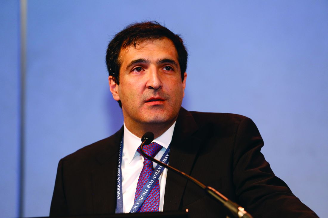
Surgical indications included an anomalous left coronary artery, ischemic symptoms, a positive nuclear perfusion test (NPT), or high-risk anatomy such as long intramural segment or ostial stenosis identified via CT angiography. Median patient age was 14 years.
Nine of the patients (20%) underwent surgery for an anomalous left coronary artery, with 32 patients (80%) receiving surgical intervention for an anomalous right coronary artery.
A total of 34 surgical procedures (77%) were unroofing of intramural segments, 7 were coronary translocations (16%), and 2 were ostioplasties (4%).
There were no operative deaths. One patient required coronary artery bypass grafting after developing ischemia following a coronary translocation. Minor complications were seen in eight other patients (18%).
One patient presented with a second episode of aborted sudden cardiac death 1 year after unroofing of an anomalous left coronary artery with a short intramural segment, and underwent successful coronary translocation and unroofing of a previously identified myocardial bridge.
At follow-up (median 2 years), 40 patients were asymptomatic (91%), while 4 patients had nonspecific chest pain (9%). Forty-one patients (93%) had returned to full activity, while 3 patients were waiting for their 3-month clearance to return to full activity. ■
trudd@frontlinemedcom.com
It’s relatively safe to surgically treat anomalous aortic origin of a coronary artery in children and adolescents, with essentially all patients cleared for full activity after 3 months, according to a study presented by Carlos M. Mery, MD.
Anomalous aortic origin of a coronary artery (AAOCA) is the second leading cause of sudden cardiac death (SCD) in young people, noted Dr. Mery of Texas Children’s Hospital at the plenary session of the 2017 AATS Centennial meeting.
Surgical indications included an anomalous left coronary artery, ischemic symptoms, a positive nuclear perfusion test (NPT), or high-risk anatomy such as long intramural segment or ostial stenosis identified via CT angiography. Median patient age was 14 years.
Nine of the patients (20%) underwent surgery for an anomalous left coronary artery, with 32 patients (80%) receiving surgical intervention for an anomalous right coronary artery.
A total of 34 surgical procedures (77%) were unroofing of intramural segments, 7 were coronary translocations (16%), and 2 were ostioplasties (4%).
There were no operative deaths. One patient required coronary artery bypass grafting after developing ischemia following a coronary translocation. Minor complications were seen in eight other patients (18%).
One patient presented with a second episode of aborted sudden cardiac death 1 year after unroofing of an anomalous left coronary artery with a short intramural segment, and underwent successful coronary translocation and unroofing of a previously identified myocardial bridge.
At follow-up (median 2 years), 40 patients were asymptomatic (91%), while 4 patients had nonspecific chest pain (9%). Forty-one patients (93%) had returned to full activity, while 3 patients were waiting for their 3-month clearance to return to full activity. ■
trudd@frontlinemedcom.com
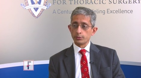
VIDEO: Wedge resection offers higher survival for NSCLC
BOSTON – High quality wedge resection results in higher survival for patients with early stage non–small cell lung cancer when compared with stereotactic body radiation therapy, according to new research.
The analysis, reported at the annual meeting of the American Association for Thoracic Surgery, evaluated the treatment of 7,337 patients in the National Cancer Database with clinical T1-T2, N0, M0 non–small cell lung cancer who were treated with either wedge resection or stereotactic body radiation therapy from 2005 to 2012.
In this video, Varun Puri, MD, of Washington University, St. Louis, discusses the study and the significance of the findings.
The video associated with this article is no longer available on this site. Please view all of our videos on the MDedge YouTube channel
agallegos@frontlinemedcom.com
On Twitter @legal_med
AT THE AATS ANNUAL MEETING
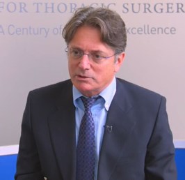
VIDEO: Lack of heart teams impact prevalence of PCI
BOSTON – Data show a marked bias in referral patterns for coronary revascularization in stand-alone interventional cardiology units lacking a heart team.
More patients underwent percutaneous coronary intervention (PCI) in hospitals without on-site cardiac surgery than patients at hospitals with on-site cardiac surgery, according to the analysis presented at the annual meeting of the American Association for Thoracic Surgery. The multivariate logistic regression analysis showed that the absence of on-site cardiac surgery and a heart team was an independent predictor for PCI.
In this video, Ehud Raanani, MD, of Tel Aviv University in Israel discusses referral patterns for coronary revascularization in stand-alone interventional cardiology units lacking a heart team and how this phenomenon could affect patients.
The video associated with this article is no longer available on this site. Please view all of our videos on the MDedge YouTube channel
agallegos@frontlinemedcom.com
On Twitter @legal_med
AT THE AATS ANNUAL MEETING
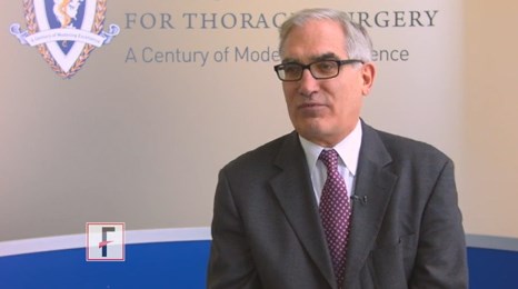
VIDEO: Surgeon case volume linked to mitral valve repair outcomes
BOSTON – Individual surgeon case volume is a significant factor in degenerative mitral valve repair rates and overall patient survival, according to a study presented at the annual meeting of the American Association for Thoracic Surgery.
The analysis, which reviewed the outcomes of 38,128 adult patients, found that higher surgeon volume of mitral cases is associated with better degenerative mitral repair rates and higher survival. In this video, Ralph Damiano Jr., MD, of Washington University, St. Louis, discusses the threshold of mitral cases that led to better repair rates and how further cardiac surgical subspecialization could improve outcomes in patients with degenerative mitral disease.
The video associated with this article is no longer available on this site. Please view all of our videos on the MDedge YouTube channel
allegros@frontlinemedcom.com
On Twitter @legal_med
AT THE AATS ANNUAL MEETING
VIDEO: Sutureless aortic valve shows promise in IDE trial
BOSTON – A new study highlights the success, safety, and effectiveness of a new sutureless aortic valve device in patients with severe symptomatic aortic valve stenosis (AS).
The findings, reported at the annual meeting of the American Association for Thoracic Surgery, were based on a prospective, single-arm clinical trial approved under a Food and Drug Administration Investigational Device Exemption (IDE) that aimed to assess the safety and efficacy of a new bovine pericardial sutureless aortic valve in patients with severe AS undergoing aortic valve replacement with or without concomitant procedures. In this video interview, Michael Borger, MD, explains how the study was conducted and what the findings mean for future use of the sutureless aortic valve device.
The video associated with this article is no longer available on this site. Please view all of our videos on the MDedge YouTube channel
agallegos@frontlinemedcom.com
On Twitter @legal_med
AT THE AATS ANNUAL MEETING
VIDEO: Incoming AATS president outlines goals
BOSTON – Strengthening member engagement is a top goal for incoming AATS President Duke E. Cameron, MD.
In this video, Dr. Cameron, of Massachusetts General Hospital, Boston, shared his objectives as the next AATS leader and the direction he envisions for the specialty over the next 100 years. Dr. Cameron also discussed his hope for new online educational efforts and the importance of physician collaboration with other health professionals.
On Twitter @legal_med
AT THE AATS ANNUAL MEETING
NSCLC Was a Key Focus of General Thoracic Session
The initial results of a phase III randomized trial comparing lobectomy to segmentectomy for small, peripheral non–small cell lung cancer (NSCLC) were presented by Kenji Suzuki, MD, of the Juntendo University Hospital, Japan.
Segmentectomy and lobectomy both proved feasible techniques for early-stage NSCLC. However, segmentectomy did not appear to be less invasive than lobectomy with regard to blood loss or the frequency of air leak, according to Dr. Suzuki.
A total of 1,106 patients (554 in lobectomy arm; 552 in segmentectomy arm) were enrolled between August 2009 and October 2014. There were 22 patients whose mode of surgery was converted from segmentectomy to lobectomy in the segmentectomy arm, resulting in 576 lobectomies and 530 segmentectomies. 
“The aim of the trial is to confirm the non-inferiority in overall survival (OS) of segmentectomy, compared with lobectomy,” said Dr. Suzuki.
Surgical complications were evaluated by the mode of surgery with an intention-to-treat analysis. As to a mode of surgery, segmentectomy was categorized into simple and complex in terms of technical difficulty; resection of the right or left S6, the left superior, and the lingular segment were defined as simple, because these procedures are easy and common.
Operative mortality was 0% in both groups. Postoperative complications, including pneumonia were not significantly different between the two groups. However, there was a significant difference in the rate of air leak detected: 3.8% in Group A and 6.5% in Group B (with no broncho-pleural fistulas being found).
Multivariate analysis showed that pack-year (PY) smoking greater than 20 vs. none was a significant predictor of postoperative complications. Significant predictors of pulmonary complications, including alveolar fistula and empyema, were typical segmentectomy (vs. lobectomy); and PY greater than 20 vs. none.
“The primary analysis of this study is planned for 2020,” said Dr. Suzuki. Those results should help to determine whether segmentectomy should be considered the standard of treatment, compared to lobectomy.
Previous research has shown that a wedge resection (WR) may be superior to stereotactic body radiation therapy (SBRT) for patients with early-stage non–small cell lung cancer (NSCLC). However, the role that the quality of the WR plays in improved outcomes is unknown, according to Seth Krantz, MD, of the NorthShore University Health System who presented the results of a database analysis of patients within the National Cancer Database (NCDB) with clinical T1-T2, N0, M0 NSCLC patients who were treated with either WR or SBRT from 2005-2012. These patients were analyzed for surgical quality markers, predictors of lymph node assessment and pathologic upstaging, and overall survival. Quality markers included the number of nodes examined and margins status.
Of more than 7,000 WR patients included (44%) had 0 LNs examined; 37% had 1-5 examined, and nearly 17% had more than 5 nodes examined. Significant predictors of having at least 5 nodes examined included younger age, fewer comorbidities, T2 tumors, and obtaining negative margins. Negative margins were obtained in the vast majority of WR patients.
“Our study confirms that nationwide, while most patients undergoing wedge resection for early stage disease receive a margin negative resection, fewer than 20% of patients had more than five lymph nodes assessed, and nearly half had no lymph nodes assessed. Pathologic assessment of lymph nodes was associated with improved long-term survival and greater utilization of adjuvant chemotherapy. Furthermore, the benefit of a wedge resection compared to SBRT, was significantly affected by the extent of lymph node assessment. 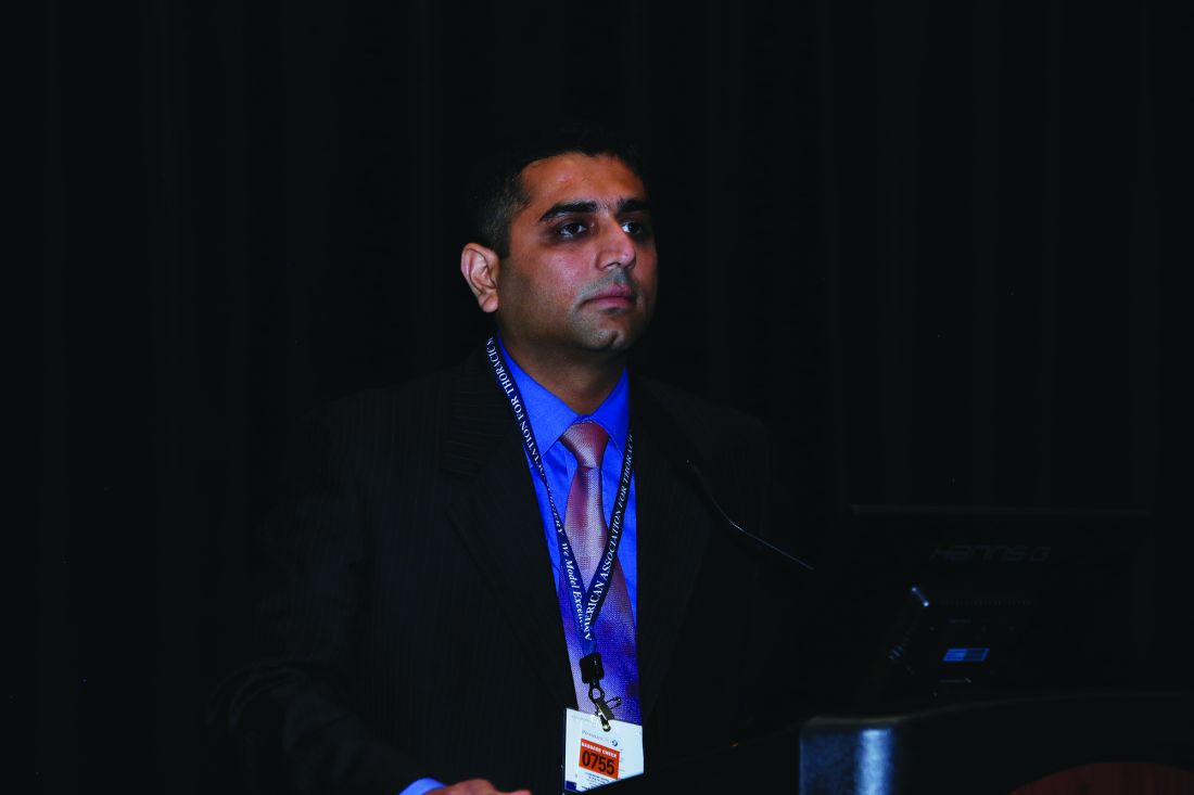
If patients are going to be offered a wedge resection for early stage non-small cell lung cancer, every effort should be made to perform a pathologic assessment of regional lymph nodes,” Dr. Krantz concluded.
Patients at high risk for non–small cell lung cancer (NSCLC) metastases were found to have a significant rate of unsuspected lymph node metastases upon endosonographic assessment, even in the presence of radiologically normal mediastinal lymph nodes, according to a study reported by Pravachan Hegde, MD, of the University of Montreal.
A total of 22 out of 161 patients with radiologically normal mediastinum were found to be positive on combined EBUS/EUS staging. “Given the significant rate of unsuspected lymph node metastases, combined endosonographic lymph node staging should be routinely performed in staging of NSCLC in high risk patients even in the presence of radiologically normal mediastinal lymph nodes,” Dr. Hedge concluded.
The initial results of a phase III randomized trial comparing lobectomy to segmentectomy for small, peripheral non–small cell lung cancer (NSCLC) were presented by Kenji Suzuki, MD, of the Juntendo University Hospital, Japan.
Segmentectomy and lobectomy both proved feasible techniques for early-stage NSCLC. However, segmentectomy did not appear to be less invasive than lobectomy with regard to blood loss or the frequency of air leak, according to Dr. Suzuki.
A total of 1,106 patients (554 in lobectomy arm; 552 in segmentectomy arm) were enrolled between August 2009 and October 2014. There were 22 patients whose mode of surgery was converted from segmentectomy to lobectomy in the segmentectomy arm, resulting in 576 lobectomies and 530 segmentectomies.
“The aim of the trial is to confirm the non-inferiority in overall survival (OS) of segmentectomy, compared with lobectomy,” said Dr. Suzuki.
Surgical complications were evaluated by the mode of surgery with an intention-to-treat analysis. As to a mode of surgery, segmentectomy was categorized into simple and complex in terms of technical difficulty; resection of the right or left S6, the left superior, and the lingular segment were defined as simple, because these procedures are easy and common.
Operative mortality was 0% in both groups. Postoperative complications, including pneumonia were not significantly different between the two groups. However, there was a significant difference in the rate of air leak detected: 3.8% in Group A and 6.5% in Group B (with no broncho-pleural fistulas being found).
Multivariate analysis showed that pack-year (PY) smoking greater than 20 vs. none was a significant predictor of postoperative complications. Significant predictors of pulmonary complications, including alveolar fistula and empyema, were typical segmentectomy (vs. lobectomy); and PY greater than 20 vs. none.
“The primary analysis of this study is planned for 2020,” said Dr. Suzuki. Those results should help to determine whether segmentectomy should be considered the standard of treatment, compared to lobectomy.
Previous research has shown that a wedge resection (WR) may be superior to stereotactic body radiation therapy (SBRT) for patients with early-stage non–small cell lung cancer (NSCLC). However, the role that the quality of the WR plays in improved outcomes is unknown, according to Seth Krantz, MD, of the NorthShore University Health System who presented the results of a database analysis of patients within the National Cancer Database (NCDB) with clinical T1-T2, N0, M0 NSCLC patients who were treated with either WR or SBRT from 2005-2012. These patients were analyzed for surgical quality markers, predictors of lymph node assessment and pathologic upstaging, and overall survival. Quality markers included the number of nodes examined and margins status.
Of more than 7,000 WR patients included (44%) had 0 LNs examined; 37% had 1-5 examined, and nearly 17% had more than 5 nodes examined. Significant predictors of having at least 5 nodes examined included younger age, fewer comorbidities, T2 tumors, and obtaining negative margins. Negative margins were obtained in the vast majority of WR patients.
“Our study confirms that nationwide, while most patients undergoing wedge resection for early stage disease receive a margin negative resection, fewer than 20% of patients had more than five lymph nodes assessed, and nearly half had no lymph nodes assessed. Pathologic assessment of lymph nodes was associated with improved long-term survival and greater utilization of adjuvant chemotherapy. Furthermore, the benefit of a wedge resection compared to SBRT, was significantly affected by the extent of lymph node assessment.
If patients are going to be offered a wedge resection for early stage non-small cell lung cancer, every effort should be made to perform a pathologic assessment of regional lymph nodes,” Dr. Krantz concluded.
Patients at high risk for non–small cell lung cancer (NSCLC) metastases were found to have a significant rate of unsuspected lymph node metastases upon endosonographic assessment, even in the presence of radiologically normal mediastinal lymph nodes, according to a study reported by Pravachan Hegde, MD, of the University of Montreal.
A total of 22 out of 161 patients with radiologically normal mediastinum were found to be positive on combined EBUS/EUS staging. “Given the significant rate of unsuspected lymph node metastases, combined endosonographic lymph node staging should be routinely performed in staging of NSCLC in high risk patients even in the presence of radiologically normal mediastinal lymph nodes,” Dr. Hedge concluded.
McChrystal Spoke on the Need for a New Kind of Team
Monday’s Honored Guest Lecturer Gen. Stanley A. McChrystal, a retired four-star general, and former commander of U.S. and International Security Assistance Forces in Afghanistan, and of the Joint Special Operations Command, spoke on the transforming nature of leadership and teams.
His address, “Team of Teams – Rules of Engagement for a Complex World,” discussed how old organizational frameworks with a centralized leadership and silos of responsibility, similar to the standard organization charts that everyone is familiar with, are no longer sufficiently functional and efficient in a changing world. He stated how the world is not just complicated, but complex, and the nature of complexity means that it cannot be predicted and a rapid, adaptive response is necessary.He highlighted his experiences in Iraq where a terrorist group, rather than using a hierarchical organization such as Al Queda, organically developed a highly adaptable and diffuse structure, which allowed them to have their leaders taken out without heavily impacting their ability to grow and rapidly respond. It wasn’t until the U.S. military under his leadership emulated such a diffuse response and geared up their efforts many-fold, that the anti-terrorism effort began to succeed.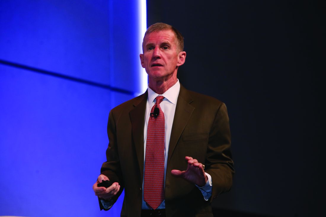
The cascade through the chain of command just wasn’t fast enough.“We had entered an environment where we had to resynchronize the organization every 24 hours. The entire organization.” So he established a daily conference call not just among the leadership but, instead, “connected everybody, all at the same time. We started by ordering 400 people to be on it and in a very short period of time it became 7,500, and went from 30 minutes to 90 minutes a day,” he said. And despite the fact that some people would call this ‘crazy,’ he said. “I’ll tell you it’s the most efficient thing I’ve ever been a part of in my life. In those 90 minutes, everyone in the organization got shared contextual understanding of what we were doing, what we were trying to do, and what we could do.” Leaders need to create an environment for a new kind of organization to grow.
Monday’s Lillehei Forum highlighted basic research
The Lillehei Forum is a series of presentations on basic research by residents in the field of cardiothoracic surgery.
A targeted near-infrared contrast agent specific for lung adenocarcinomas was assessed for effectiveness during intraoperative molecular imaging (IMI) in animal models. Use of the contrast agent proved significantly more effective at detecting positive margins than did surgery alone, according to a study presented by Jarrod D. Predina, MD.
Initial human experiences with IMI for NSCLC using a fluorescent contrast agent have been limited by technical hurdles including high background noise in inflammatory tissues and low signal output, according to Dr. Predina. As an alternative, he and his colleagues at the University of Pennsylvania hypothesized that a targeted near-infrared contrast agent specific for lung adenocarcinomas would improve sensitivity and specificity during surgery.
Using a mouse surgical model of non–small cell lung cancer (NSCLC) that recapitulates local and systemic post-operative recurrences, Dr. Predina and his colleagues at the University of Pennsylvania injected 140 mice intravenously prior to resection with a near-infrared imaging agent (OTL0038). This agent is specific for pulmonary adenocarcinomas due to its high affinity binding of the folate receptor alpha. Tumor-bearing mice were randomized to surgery with or without IMI. Suspicious residual disease was resected and analyzed by immunohistochemistry, flow cytometry, and immunofluorescence. Based on this data, OTL0038 was tested in a pilot study of five canines with spontaneously occurring lung cancer.
In a local recurrence model system, of 80 mice assessed, surgeons identified 10 positive margins in those randomized to imaging with IMI vs. three positive margins in mice undergoing surgery alone, a significant difference. In systemic recurrence models (60 mice), the mean number of pulmonary nodules located with IMI was 7.2 vs. 3.4 in controls, also a significant difference, according to Dr. Predina.
In five canines with a presumed diagnosis of NSCLC, no toxicity was observed. Four of five canines had fluorescent tumors; the non-fluorescing tumor was discovered to be a metastatic mammary tumor on final pathologic analysis. In one canine, an otherwise undetectable 8-mm pulmonary adenocarcinoma was discovered with IMI.
“Our data suggest that a targeted near-infrared contrast agent may improve IMI technology. Ultimately, this will enable accurate identification of residual disease that may otherwise be overlooked. These results are the basis of an ongoing phase I human trial,” concluded Dr. Predina.
The complexity of the lung has limited the prospects of bioengineering strategies utilizing stem cells and fully decellularized or bioartificial scaffolds, according to Brandon A. Guenthart, MD, of Columbia University.
Using standard protocols, Dr. Guenthart and his colleagues procured human lungs rejected for transplantation on the basis of standard clinical criteria. Lungs were placed on a custom-built EVLP system, ventilated and perfused. Utilizing video bronchoscopy, real-time transpleural imaging, and a custom designed micro-catheter delivery and occlusion system, the team delivered a decellularization solution into targeted lung regions that resulted in the selective removal of airway epithelium.
Human airway epithelial cells or human-derived alveolar progenitor cells were labeled (with a quantum dot or a near-infrared dye) and delivered into decellularized lung regions. Following delivery, EVLP was continued for 4-6 hours to allow for cell engraftment. Lung wedge samples were collected at each time point for histologic analysis.
Following decellularization, hematoxylin and eosin staining confirmed removal of pseudostratified and columnar epithelium in proximal airways and removal of type I and II pneumocytes in the distal lung. Delivered cells were retained in the lung after EVLP and fixation, and cellular morphology and distribution within the alveoli indicated early engraftment.
“Bioengineering human lungs utilizing advanced therapeutic interventions such as cell replacement may help combat the critical shortage of transplantable lungs,” said Dr. Guenthart. “Additionally, in the future, targeted intervention and patient-specific cell replacement strategies may have translational applications in vivo and eliminate the need for transplantation in select patients,” he concluded.
They used a previously validated porcine lung injury model of intravenous lipopolysaccharide (LPS) to induce a systemic inflammatory response and subsequent severe ARDS requiring ECMO support. A total of eight mature adult swine were administered LPS via the external jugular vein followed by sternotomy and central ECMO cannulation (right atrium to ascending aorta).
The left pulmonary artery (inflow) and left superior and inferior pulmonary veins (outflow) were dissected out and cannulated to isolate the left lung, which then underwent 4 hours normorthermic IVLP with Steen Solution followed by 4 hours of lung reperfusion after IVLP decannulation.
Dr. Mehaffey and his colleagues then compared the right (LPS control) and left lungs (LPS+IVLP) of the same animal.
All animals demonstrated a significant reduction in PaO2/FiO2 ratio and total lung compliance 2 hours after the start of LPS infusion. During IVLP, the left (treated) pulmonary vein oxygenation was superior to right (control) pulmonary vein oxygenation. After reperfusion and IVLP decannulation, six (75%) animals had improved lung function allowing for ECMO decannulation.
The left lung showed significant improvement of lung-specific oxygenation compared to the right control at 4 hours of reperfusion. Similarly, total lung compliance improved after targeted rehabilitation of the left lung, according to Dr. Mehaffey. In addition, the wet-to-dry ratio of lung tissue demonstrated significantly reduced edema in the rehabilitated left lungs compared to right controls.
Dr. Mehaffey and his colleagues concluded that IVLP may reduce the duration of ECMO and ventilator support resulting in major reduction in morbidity, mortality, and health care–related costs in patients with ARDS.