User login
KISSIMMEE, FLA. – Hypertrophy of port wine stains is more common than not in the majority of patients older than age 50 with the vascular anomaly, and is most often associated with red and purple lesions, according to findings from a retrospective patient-questionnaire study.
"By the age of 50, over 70% had hypertrophy of their port wine stain," said Dr. Anne Margreet van Drooge of the Netherlands Institute of Pigment Disorders at the University of Amsterdam.
Hypertrophy is known to be fairly common in port wine stains, but data are lacking on its pathogenesis and development. In this study, the incidence of hypertrophy in port wine stains was about 20% in all patients, and both color and hypertrophy were frequently reported as reasons for seeking treatment.
Only 4% of all hypertrophy cases were associated with pink lesions, Dr. van Drooge said at the annual meeting of the American Society for Laser Medicine and Surgery.
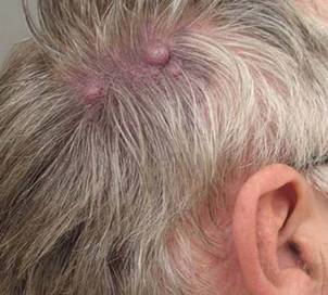
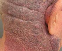
Of the 335 patients included in the review, 68 had hypertrophy, classified as thickening, nodular, or both. Cases were divided almost equally into age groups of under 18 years, 18-30 years, and greater than 30 years. The median age of those with hypertrophic port wine stains was 50 years, and the median age at the time of hypertrophic development was 31 years, although the ages of onset of nodular hypertrophy and diffuse thickening were 39 years and 12 years, respectively. Nodular hypertrophy was rarely seen in those younger than 18 years.
In all study patients, most of the port wine stains were on the face. Half were red in color, and 44% were purple. No differences were seen between those with and without hypertrophy with respect to gender, lesion size, or lesion location, she noted.
Patients included in the study had been referred to a single outpatient clinic between 2005 and 2009. Medical records and photographs were examined to identify and characterize hypertrophic port wine stains.
The findings of different ages at onset for different types of hypertrophy suggest that unique pathomechanics may exist for each type, Dr. van Drooge said. "There is definitely a correlation between the color of the port wine stain and the risk for the development of hypertrophy. In a multivariate analysis of all patient characteristics, we found that both darker color and older age were significantly associated with hypertrophy, independent from each other."
Improved understanding of hypertrophy in port wine stains is important given the adverse psychosocial effects these lesions can have on patients; in this study, 5% of patients said they avoided social interaction because of their port wine stain, she noted, adding that the findings have implications for treatment of port wine stains.
Although this study provided no direct support for the notion that treatment may prevent hypertrophy, some studies have suggested that it may, adding that treatment guidelines should be adapted to include more emphasis on hypertrophy and its prevention, keeping in mind the findings of this study with respect to age and lesion color.
"However, I don’t think it is possible to treat only darker port wine stains in the effort to prevent hypertrophy, since pink port wine stains – particularly in younger patients – eventually can darken, and with that, the risk for hypertrophy increases," she noted.
Dr. van Drooge had no disclosures to report.
KISSIMMEE, FLA. – Hypertrophy of port wine stains is more common than not in the majority of patients older than age 50 with the vascular anomaly, and is most often associated with red and purple lesions, according to findings from a retrospective patient-questionnaire study.
"By the age of 50, over 70% had hypertrophy of their port wine stain," said Dr. Anne Margreet van Drooge of the Netherlands Institute of Pigment Disorders at the University of Amsterdam.
Hypertrophy is known to be fairly common in port wine stains, but data are lacking on its pathogenesis and development. In this study, the incidence of hypertrophy in port wine stains was about 20% in all patients, and both color and hypertrophy were frequently reported as reasons for seeking treatment.
Only 4% of all hypertrophy cases were associated with pink lesions, Dr. van Drooge said at the annual meeting of the American Society for Laser Medicine and Surgery.
Of the 335 patients included in the review, 68 had hypertrophy, classified as thickening, nodular, or both. Cases were divided almost equally into age groups of under 18 years, 18-30 years, and greater than 30 years. The median age of those with hypertrophic port wine stains was 50 years, and the median age at the time of hypertrophic development was 31 years, although the ages of onset of nodular hypertrophy and diffuse thickening were 39 years and 12 years, respectively. Nodular hypertrophy was rarely seen in those younger than 18 years.
In all study patients, most of the port wine stains were on the face. Half were red in color, and 44% were purple. No differences were seen between those with and without hypertrophy with respect to gender, lesion size, or lesion location, she noted.
Patients included in the study had been referred to a single outpatient clinic between 2005 and 2009. Medical records and photographs were examined to identify and characterize hypertrophic port wine stains.
The findings of different ages at onset for different types of hypertrophy suggest that unique pathomechanics may exist for each type, Dr. van Drooge said. "There is definitely a correlation between the color of the port wine stain and the risk for the development of hypertrophy. In a multivariate analysis of all patient characteristics, we found that both darker color and older age were significantly associated with hypertrophy, independent from each other."
Improved understanding of hypertrophy in port wine stains is important given the adverse psychosocial effects these lesions can have on patients; in this study, 5% of patients said they avoided social interaction because of their port wine stain, she noted, adding that the findings have implications for treatment of port wine stains.
Although this study provided no direct support for the notion that treatment may prevent hypertrophy, some studies have suggested that it may, adding that treatment guidelines should be adapted to include more emphasis on hypertrophy and its prevention, keeping in mind the findings of this study with respect to age and lesion color.
"However, I don’t think it is possible to treat only darker port wine stains in the effort to prevent hypertrophy, since pink port wine stains – particularly in younger patients – eventually can darken, and with that, the risk for hypertrophy increases," she noted.
Dr. van Drooge had no disclosures to report.
FROM THE ANNUAL MEETING OF THE AMERICAN SOCIETY FOR LASER MEDICINE AND SURGERY