User login
Optimal timing for peripheral IV replacement?
Replace peripheral IV catheters as needed, rather than on a routine basis.1
STRENGTH OF RECOMMENDATION
A: Based on a randomized equivalence trial.
Rickard CM, Webster J, Wallis MC, et al. Routine versus clinically indicated replacement of peripheral intravenous catheters: a randomised controlled equivalence trial. Lancet. 2012;380:1066-1074.
ILLUSTRATIVE CASE
On Day 4 of her hospitalization for a wound infection requiring IV antibiotics, a 45-year-old patient is told by her nurse that her IV catheter must be replaced. It’s hospital policy, the RN says, to replace the catheter every 96 hours. The patient is afraid of needles and is not eager to have her catheter replaced every few days. Is it really necessary to replace the IV, she wants to know.
Each year, nearly 200 million peripheral IV catheters are placed in patients in hospitals throughout the United States.2 Many of the catheters need to be replaced due to phlebitis, infiltration, pain, or swelling at the IV site, but the rate of bloodstream infections associated with peripheral IVs is just 0.5 per 1000 catheter days.2
Timing of replacement is “unresolved”
The Centers for Disease Control and Prevention (CDC)’s 2011 guidelines state that it is not necessary to replace peripheral IV catheters in adults more than every 72 to 96 hours,3 but the CDC does not specify when the catheters should be replaced. For adult patients, the recommendation that a catheter be replaced only for clinical indications is an “unresolved issue,” according to the guidelines. For children, however, replacement only when clinically indicated is recommended by the CDC. Many hospitals have protocols that require replacement of IV catheters every 72 to 96 hours, regardless of clinical indication.
A 2008 study of 755 inpatients compared clinically indicated replacement of IV catheters with routine replacement and found no significant differences in phlebitis and infiltration rates between the 2 groups (38% vs 33%, respectively; relative risk [RR]=1.15; 95% confidence interval [CI], 0.95-1.40).4
A 2010 trial randomized 362 hospitalized patients to routine or clinically indicated replacement of peripheral IV lines, with median dwell times of 71 and 85 hours, respectively. There was no significant difference in rates of phlebitis between the routine replacement (7%) and clinically indicated (10%) groups (RR=1.44; 95% CI, 0.71-2.89; P=.34). No local infections or IV-related bloodstream infections occurred in either group.5
A 2010 Cochrane review included 5 randomized controlled trials (with a total of 3408 patients) that compared rates of suspected catheter-related phlebitis in patients whose catheters were routinely replaced with those in the clinically indicated group. The reviewers found no significant increase in phlebitis in the clinically indicated group (9%) vs the routine replacement group (7.2%) (odds ratio=1.24; 95% CI, 0.97-1.60; P=.09).6
Each of these studies had either a relatively small sample size or wide confidence intervals, raising the possibility of missing a real increase in infection due to inadequate statistical power. The study summarized here addressed these concerns.
STUDY SUMMARY: Forgoing routine replacement does not increase risk
Rickard et al1 conducted a multicenter, nonblinded randomized equivalence trial to determine whether routine or clinically indicated removal reduced rates of infection. In the routine group, catheters were replaced every 72 to 96 hours. In the clinically indicated group, catheters were replaced in instances of phlebitis, infiltration, occlusion, accidental removal, or suspected infection related to the catheter.
Participants (N=3283) were inpatients on medical and surgical units who had IV catheters in place and were expected to need treatment for at least 4 days. Individuals whose IV catheters had been placed in an emergency were excluded, as were those who had a known bloodstream infection or who were not expected to have the IV in place for at least 24 hours. Follow-up data were available for all participants.
The primary outcome was phlebitis, with a prespecified equivalence margin of 3%. In both groups, phlebitis occurred in 7% of patients (RR=1.06; 95% CI, 0.83-1.36; P=.64). The absolute risk difference was 0.41% (95% CI, -1.33 to 2.15), which was within the equivalence margin.
The mean IV catheter dwell time was 70 hours in the routine replacement group and 99 hours in the clinically indicated group. Nine patients in the routine replacement group developed bloodstream infections, vs 4 patients in the clinically indicated group (hazard ratio=0.46; 95% CI, 0.14-1.48; P=.19). One patient in the routine placement group had a catheter-related bloodstream infection; no one in the clinically indicated group did. The mortality rate for each group was <1%.
WHAT’S NEW: We can order clinically indicated IV replacement with confidence
The findings of this equivalence trial support prior studies and add greater statistical power. The results suggest that we can recommend clinically indicated replacement of peripheral IV catheters without increasing the rate of phlebitis. Implementing clinically indicated replacement of IVs could decrease hospital costs and improve patient satisfaction.
CAVEATS: Findings do not apply to patients with bacteremia
Patients with known bacteremia were excluded from this study, and the results are therefore not generalizable to this population.
The nonblinded nature of this trial raises the possibility of observer and reporting bias. However, measures were taken to minimize the potential for bias. A structured outcome assessment was used to standardize reporting of signs of phlebitis. Both patients’ pain scores and nurses’ assessments of the IV sites were used to determine whether an infection was present, and the investigators and research nurses were not involved in the removal of the IV catheters.
This study did not report on the daily maintenance protocols the investigators used for the peripheral IVs. The study was conducted in hospitals in Australia, and we don’t know whether the protocols used in that country are similar to standard protocols in US hospitals.
CHALLENGES TO IMPLEMENTATION: Changing hospital protocols won’t be easy
Implementing the findings of this study will require that physicians work with the nursing staff and administrators to create and implement new protocols for assessing peripheral IV catheters in hospitals with routine IV replacement policies already in place. It would be necessary to ensure that all clinicians who place peripheral IV catheters are taught the clinical signs of phlebitis and are using a standardized protocol. That said, we think that this is a worthwhile change to achieve the long-term benefits of fewer unnecessary IV catheter replacements.
Acknowledgement
The PURLs Surveillance System was supported in part by Grant Number UL1RR024999 from the National Center for Research Resources, a Clinical Translational Science Award to the University of Chicago. The content is solely the responsibility of the authors and does not necessarily represent the official views of the National Center for Research Resources or the National Institutes of Health.
1. Rickard CM, Webster J, Wallis MC, et al. Routine versus clinically indicated replacement of peripheral intravenous catheters: a randomised controlled equivalence trial. Lancet. 2012;380:1066-1074.
2. Maki DG, Kluger DM, Crnich CJ. The risk of bloodstream infection in adults with different intravascular devices: a systematic review of 200 published prospective studies. Mayo Clin Proc. 2006;81:1159-1171.
3. Centers for Disease Control and Prevention. 2011 guidelines for the prevention of intravascular catheter-related infections. Available at: http://www.cdc.gov/hicpac/BSI/-BSI-guidelines-2011.html. Accessed March 13, 2013.
4. Webster J, Clarke S, Paterson D, et al. Routine care of peripheral intravenous catheters versus clinically indicated replacement: randomised controlled trial. BMJ. 2008;337:a339.-
5. Rickard CM, McCann D, Munnings J, et al. Routine resite of peripheral intravenous devices every 3 days did not reduce complications compared with clinically indicated resite: a randomised controlled trial. BMC Med. 2010;8:53.-
6. Webster J, Osborne S, Rickard C, et al. Clinically-indicated replacement versus routine replacement of peripheral venous catheters. Cochrane Database Syst Rev. 2010;(3):CD007798.-
Replace peripheral IV catheters as needed, rather than on a routine basis.1
STRENGTH OF RECOMMENDATION
A: Based on a randomized equivalence trial.
Rickard CM, Webster J, Wallis MC, et al. Routine versus clinically indicated replacement of peripheral intravenous catheters: a randomised controlled equivalence trial. Lancet. 2012;380:1066-1074.
ILLUSTRATIVE CASE
On Day 4 of her hospitalization for a wound infection requiring IV antibiotics, a 45-year-old patient is told by her nurse that her IV catheter must be replaced. It’s hospital policy, the RN says, to replace the catheter every 96 hours. The patient is afraid of needles and is not eager to have her catheter replaced every few days. Is it really necessary to replace the IV, she wants to know.
Each year, nearly 200 million peripheral IV catheters are placed in patients in hospitals throughout the United States.2 Many of the catheters need to be replaced due to phlebitis, infiltration, pain, or swelling at the IV site, but the rate of bloodstream infections associated with peripheral IVs is just 0.5 per 1000 catheter days.2
Timing of replacement is “unresolved”
The Centers for Disease Control and Prevention (CDC)’s 2011 guidelines state that it is not necessary to replace peripheral IV catheters in adults more than every 72 to 96 hours,3 but the CDC does not specify when the catheters should be replaced. For adult patients, the recommendation that a catheter be replaced only for clinical indications is an “unresolved issue,” according to the guidelines. For children, however, replacement only when clinically indicated is recommended by the CDC. Many hospitals have protocols that require replacement of IV catheters every 72 to 96 hours, regardless of clinical indication.
A 2008 study of 755 inpatients compared clinically indicated replacement of IV catheters with routine replacement and found no significant differences in phlebitis and infiltration rates between the 2 groups (38% vs 33%, respectively; relative risk [RR]=1.15; 95% confidence interval [CI], 0.95-1.40).4
A 2010 trial randomized 362 hospitalized patients to routine or clinically indicated replacement of peripheral IV lines, with median dwell times of 71 and 85 hours, respectively. There was no significant difference in rates of phlebitis between the routine replacement (7%) and clinically indicated (10%) groups (RR=1.44; 95% CI, 0.71-2.89; P=.34). No local infections or IV-related bloodstream infections occurred in either group.5
A 2010 Cochrane review included 5 randomized controlled trials (with a total of 3408 patients) that compared rates of suspected catheter-related phlebitis in patients whose catheters were routinely replaced with those in the clinically indicated group. The reviewers found no significant increase in phlebitis in the clinically indicated group (9%) vs the routine replacement group (7.2%) (odds ratio=1.24; 95% CI, 0.97-1.60; P=.09).6
Each of these studies had either a relatively small sample size or wide confidence intervals, raising the possibility of missing a real increase in infection due to inadequate statistical power. The study summarized here addressed these concerns.
STUDY SUMMARY: Forgoing routine replacement does not increase risk
Rickard et al1 conducted a multicenter, nonblinded randomized equivalence trial to determine whether routine or clinically indicated removal reduced rates of infection. In the routine group, catheters were replaced every 72 to 96 hours. In the clinically indicated group, catheters were replaced in instances of phlebitis, infiltration, occlusion, accidental removal, or suspected infection related to the catheter.
Participants (N=3283) were inpatients on medical and surgical units who had IV catheters in place and were expected to need treatment for at least 4 days. Individuals whose IV catheters had been placed in an emergency were excluded, as were those who had a known bloodstream infection or who were not expected to have the IV in place for at least 24 hours. Follow-up data were available for all participants.
The primary outcome was phlebitis, with a prespecified equivalence margin of 3%. In both groups, phlebitis occurred in 7% of patients (RR=1.06; 95% CI, 0.83-1.36; P=.64). The absolute risk difference was 0.41% (95% CI, -1.33 to 2.15), which was within the equivalence margin.
The mean IV catheter dwell time was 70 hours in the routine replacement group and 99 hours in the clinically indicated group. Nine patients in the routine replacement group developed bloodstream infections, vs 4 patients in the clinically indicated group (hazard ratio=0.46; 95% CI, 0.14-1.48; P=.19). One patient in the routine placement group had a catheter-related bloodstream infection; no one in the clinically indicated group did. The mortality rate for each group was <1%.
WHAT’S NEW: We can order clinically indicated IV replacement with confidence
The findings of this equivalence trial support prior studies and add greater statistical power. The results suggest that we can recommend clinically indicated replacement of peripheral IV catheters without increasing the rate of phlebitis. Implementing clinically indicated replacement of IVs could decrease hospital costs and improve patient satisfaction.
CAVEATS: Findings do not apply to patients with bacteremia
Patients with known bacteremia were excluded from this study, and the results are therefore not generalizable to this population.
The nonblinded nature of this trial raises the possibility of observer and reporting bias. However, measures were taken to minimize the potential for bias. A structured outcome assessment was used to standardize reporting of signs of phlebitis. Both patients’ pain scores and nurses’ assessments of the IV sites were used to determine whether an infection was present, and the investigators and research nurses were not involved in the removal of the IV catheters.
This study did not report on the daily maintenance protocols the investigators used for the peripheral IVs. The study was conducted in hospitals in Australia, and we don’t know whether the protocols used in that country are similar to standard protocols in US hospitals.
CHALLENGES TO IMPLEMENTATION: Changing hospital protocols won’t be easy
Implementing the findings of this study will require that physicians work with the nursing staff and administrators to create and implement new protocols for assessing peripheral IV catheters in hospitals with routine IV replacement policies already in place. It would be necessary to ensure that all clinicians who place peripheral IV catheters are taught the clinical signs of phlebitis and are using a standardized protocol. That said, we think that this is a worthwhile change to achieve the long-term benefits of fewer unnecessary IV catheter replacements.
Acknowledgement
The PURLs Surveillance System was supported in part by Grant Number UL1RR024999 from the National Center for Research Resources, a Clinical Translational Science Award to the University of Chicago. The content is solely the responsibility of the authors and does not necessarily represent the official views of the National Center for Research Resources or the National Institutes of Health.
1. Rickard CM, Webster J, Wallis MC, et al. Routine versus clinically indicated replacement of peripheral intravenous catheters: a randomised controlled equivalence trial. Lancet. 2012;380:1066-1074.
2. Maki DG, Kluger DM, Crnich CJ. The risk of bloodstream infection in adults with different intravascular devices: a systematic review of 200 published prospective studies. Mayo Clin Proc. 2006;81:1159-1171.
3. Centers for Disease Control and Prevention. 2011 guidelines for the prevention of intravascular catheter-related infections. Available at: http://www.cdc.gov/hicpac/BSI/-BSI-guidelines-2011.html. Accessed March 13, 2013.
4. Webster J, Clarke S, Paterson D, et al. Routine care of peripheral intravenous catheters versus clinically indicated replacement: randomised controlled trial. BMJ. 2008;337:a339.-
5. Rickard CM, McCann D, Munnings J, et al. Routine resite of peripheral intravenous devices every 3 days did not reduce complications compared with clinically indicated resite: a randomised controlled trial. BMC Med. 2010;8:53.-
6. Webster J, Osborne S, Rickard C, et al. Clinically-indicated replacement versus routine replacement of peripheral venous catheters. Cochrane Database Syst Rev. 2010;(3):CD007798.-
Copyright © 2013 The Family Physicians Inquiries Network. All rights reserved.
Consider adding this drug to fight COPD that’s severe
Consider prescribing daily azithromycin for patients with chronic obstructive pulmonary disease (COPD) and a history of exacerbations—but do a careful risk-benefit analysis first.1
STRENGTH OF RECOMMENDATION
B: Based on one well-designed double-blind, randomized controlled trial (RCT).
Albert RK, Connett J, Bailey WC, et al. Azithromycin for prevention of exacerbations of COPD. N Engl J Med. 2011;365:689-698.
ILLUSTRATIVE CASE
A 65-year-old man with a history of moderate to severe COPD schedules an appointment soon after discharge from the hospital—his second hospitalization for COPD exacerbations in 4 months. The patient uses inhaled glucocorticoids, a long-acting beta-agonist (LABA), and a long-acting anticholinergic. Should you add a macrolide to his medication regimen?
Acute exacerbations of COPD—the third highest cause of death in the United States2—have a major effect on quality of life, often resulting in repeat trips to the emergency department (ED) and numerous hospitalizations, office visits, and days lost from work. According to a new study that used 2006 data, there were 1.25 million hospitalizations for COPD exacerbations that year, with health care costs of $11.9 billion.3 Preventing exacerbations and the associated morbidity and mortality is a major challenge that primary care physicians face.
Can a macrolide help?
Corticosteroids, long-acting beta-agonists (LABAs), and the anticholinergic tiotropium are known to reduce COPD exacerbations,4,5 but what about antibiotics? A Cochrane meta-analysis of 9 RCTs that assessed antibiotic use for COPD found that it did not decrease the number of exacerbations. Notably, however, macrolides were not used in any of the studies.6
Macrolides are known to have anti-inflammatory, antibacterial, and immunomodulatory properties that reduce pulmonary exacerbations in other chronic lung diseases. A recent meta-analysis found that patients with cystic fibrosis have fewer pulmonary exacerbations when they take azithromycin 3 times a week.7
Small studies of the effect of macrolides on the frequency of COPD exacerbations have had conflicting results.8,9 The larger study detailed here evaluated the ability of daily azithromycin therapy to reduce COPD exacerbations.
STUDY SUMMARY: Daily dose led to fewer exacerbations
This double-blind RCT included close to 1150 participants from 12 US academic health centers, randomly assigned to receive azithromycin 250 mg daily or placebo, in addition to their usual care. (About 10% of patients in both groups died, withdrew, or were lost to follow-up.)
To be included, patients had to be ≥40 years old and have a clinical diagnosis of COPD, defined as a smoking history of 10 pack-years or more, a decreased forced expiratory volume in one second/forced vital capacity (FEV1/FVC) ratio, and a decreased FEV1 after bronchodilation. In addition, participants had to be on long-term oxygen or have used systemic steroids within the previous year or have had an ED visit or hospital admission for COPD during that time frame. Exclusion criteria included a history of asthma, a resting heart rate >100 beats per minute, a prolonged corrected QT interval (QTc) on electrocardiogram or the use of a medication that might prolong QTc, and a documented hearing impairment.
At baseline, participants were similar in basic demographics, COPD severity, smoking history, and medication use: 49% of those in the azithromycin group and 46% of the placebo group were taking a combination of inhaled corticosteroids, LABAs, and a long-acting anticholinergic medication.
The primary outcome was the time to the first COPD exacerbation. This was defined as ≥3 days with 2 or more COPD symptoms—new onset or worsening cough, dyspnea, sputum production, chest tightness, or wheezing—for which antibiotics or steroids were required. Secondary outcomes were quality-of-life measurements on the St. George’s Respiratory Questionnaire (SGRQ) and the Medical Outcomes 36-item Short Form Health Survey (SF-36). Nasopharyngeal swabs were done every 3 months to check for colonization and resistance. Hearing was assessed with audiometry at the time of enrollment, and again at 3 and 12 months. All patients were followed for a year, with monthly telephone calls or clinic visits, to determine if an exacerbation had occurred in the previous month.
The median time to the first exacerbation in the azithromycin group was 266 days (95% confidence interval [CI], 227-313) vs 174 days (95% CI, 143-215) in the placebo group; P<.001. Frequency of acute exacerbations was 1.48 per patient-year for the azithromycin group compared with 1.83 for the placebo group (relative risk=0.83; 95% CI, 0.72–0.95; P=.01). The number needed to treat to prevent one acute exacerbation in a one-year period was 2.86.
There was no significant difference in the SGRQ and SF-36 scores for the azithromycin vs the placebo group. There was a small reduction in unscheduled office visits (0.11 per patient-year; P=.048) in the azithromycin group, and a decrease in hospitalization that was not statistically significant.
Azithromycin group had higher rates of adverse effects
Nasopharyngeal cultures from participants who became colonized during the course of the study found macrolide resistance in 81% of those in the azithromycin group vs 41% of the placebo group (P<.001). Twenty-five percent of patients in the azithromycin group developed measurable hearing loss, compared with 20% of those on placebo (P=0.04; number needed to harm=20).
WHAT’S NEW?: A better understanding of benefits and risks
This study shows that the addition of azithromycin (250 mg/d) to standard COPD treatment decreases the number of exacerbations, but does little to reduce hospital admissions. It also highlights the adverse effect profile of azithromycin and the importance of using the antibiotic only for carefully selected patients.
CAVEATS: Macrolide resistance is a key concern
Twenty-five percent of the azithromycin group had documented hearing loss—an additional one in 20 compared with patients in the placebo group. More importantly, there was an increase in the prevalence of macrolide-resistant respiratory pathogens in patients on daily azithromycin. The long-term impact of daily azithromycin on antibiotic resistance is unknown, both for patients themselves and the community at large.
Physicians will have to assess the benefit of a decrease in COPD exacerbations (approximately one every 3 years) vs the risk of an increase in hearing problems and macrolide resistance. A sensible approach would be to reserve daily use of azithromycin for patients with a history of multiple exacerbations, who potentially have more to gain.
CHALLENGES TO IMPLEMENTATION: There are none
There are no major challenges to implementation aside from the cost, which would be approximately $1200 per year (azithromycin 250 mg [30 tablets] at $98.99 per month).10
Acknowledgement
The PURLs Surveillance System is supported in part by Grant Number UL1RR024999 from the National Center for Research Resources, a Clinical Translational Science Award to the University of Chicago. The content is solely the responsibility of the authors and does not necessarily represent the official views of the National Center For Research Resources or the National Institutes of Health.
1. Albert RK, Connett J, Bailey WC, et al. Azithromycin for prevention of exacerbations of COPD. N Engl J Med. 2011;365:689-698.
2. Centers for Disease Control and Prevention. Injury prevention & control: data & statistics. Available at: http://www.cdc.gov/injury/wisqars/LeadingCauses.html. Accessed April 16, 2012.
3. Perera PN, Armstrong EP, Sherrill DL, et al. Acute exacerbations of COPD in the United States: inpatient burden and predictors of cost and mortality. COPD. 2012;9:131-141.
4. Jenkins CR, Jones PW, Calverley PM, et al. Efficacy of salmeterol/fluticasone propionate by GOLD stage of chronic obstructive pulmonary disease: analysis from the randomised, placebo-controlled TORCH study. Respir Res. 2009;10:59.-
5. Decramer M, Celli B, Kesten S, et al. Effect of tiotropium on outcomes in patients with moderate chronic obstructive pulmonary disease (UPLIFT); a prespecified subgroup analysis of a randomized controlled trial. Lancet. 2009;374:1171-1178.
6. Black PN, Staykova T, Chacko EE, et al. Prophylactic antibiotic therapy for chronic bronchitis. Cochrane Database Syst Rev. 2003;(1):CD004105.-
7. Southern KW, Barker PM, Solis-Moya A, et al. Macrolide antibiotics for cystic fibrosis. Cochrane Database Syst Rev. 2011;(12):CD002203.-
8. Seemungal TA, Wilkinson TM, Hurst JR, et al. Long-term erythromycin therapy is associated with decreased chronic obstructive pulmonary exacerbations. Am J Respir Crit Care Med. 2008;178:1139-1147.
9. Yamaya A, Azuma A, Tanaka H, et al. Inhibitory effects of macrolides on exacerbations and hospitalization in chronic obstructive pulmonary disease in Japan: a retrospective multicenter analysis. J Am Geriatri Soc. 2008;56:1358-1360.
10. www.Drugstore.com. Accessed March 28, 2012.
Consider prescribing daily azithromycin for patients with chronic obstructive pulmonary disease (COPD) and a history of exacerbations—but do a careful risk-benefit analysis first.1
STRENGTH OF RECOMMENDATION
B: Based on one well-designed double-blind, randomized controlled trial (RCT).
Albert RK, Connett J, Bailey WC, et al. Azithromycin for prevention of exacerbations of COPD. N Engl J Med. 2011;365:689-698.
ILLUSTRATIVE CASE
A 65-year-old man with a history of moderate to severe COPD schedules an appointment soon after discharge from the hospital—his second hospitalization for COPD exacerbations in 4 months. The patient uses inhaled glucocorticoids, a long-acting beta-agonist (LABA), and a long-acting anticholinergic. Should you add a macrolide to his medication regimen?
Acute exacerbations of COPD—the third highest cause of death in the United States2—have a major effect on quality of life, often resulting in repeat trips to the emergency department (ED) and numerous hospitalizations, office visits, and days lost from work. According to a new study that used 2006 data, there were 1.25 million hospitalizations for COPD exacerbations that year, with health care costs of $11.9 billion.3 Preventing exacerbations and the associated morbidity and mortality is a major challenge that primary care physicians face.
Can a macrolide help?
Corticosteroids, long-acting beta-agonists (LABAs), and the anticholinergic tiotropium are known to reduce COPD exacerbations,4,5 but what about antibiotics? A Cochrane meta-analysis of 9 RCTs that assessed antibiotic use for COPD found that it did not decrease the number of exacerbations. Notably, however, macrolides were not used in any of the studies.6
Macrolides are known to have anti-inflammatory, antibacterial, and immunomodulatory properties that reduce pulmonary exacerbations in other chronic lung diseases. A recent meta-analysis found that patients with cystic fibrosis have fewer pulmonary exacerbations when they take azithromycin 3 times a week.7
Small studies of the effect of macrolides on the frequency of COPD exacerbations have had conflicting results.8,9 The larger study detailed here evaluated the ability of daily azithromycin therapy to reduce COPD exacerbations.
STUDY SUMMARY: Daily dose led to fewer exacerbations
This double-blind RCT included close to 1150 participants from 12 US academic health centers, randomly assigned to receive azithromycin 250 mg daily or placebo, in addition to their usual care. (About 10% of patients in both groups died, withdrew, or were lost to follow-up.)
To be included, patients had to be ≥40 years old and have a clinical diagnosis of COPD, defined as a smoking history of 10 pack-years or more, a decreased forced expiratory volume in one second/forced vital capacity (FEV1/FVC) ratio, and a decreased FEV1 after bronchodilation. In addition, participants had to be on long-term oxygen or have used systemic steroids within the previous year or have had an ED visit or hospital admission for COPD during that time frame. Exclusion criteria included a history of asthma, a resting heart rate >100 beats per minute, a prolonged corrected QT interval (QTc) on electrocardiogram or the use of a medication that might prolong QTc, and a documented hearing impairment.
At baseline, participants were similar in basic demographics, COPD severity, smoking history, and medication use: 49% of those in the azithromycin group and 46% of the placebo group were taking a combination of inhaled corticosteroids, LABAs, and a long-acting anticholinergic medication.
The primary outcome was the time to the first COPD exacerbation. This was defined as ≥3 days with 2 or more COPD symptoms—new onset or worsening cough, dyspnea, sputum production, chest tightness, or wheezing—for which antibiotics or steroids were required. Secondary outcomes were quality-of-life measurements on the St. George’s Respiratory Questionnaire (SGRQ) and the Medical Outcomes 36-item Short Form Health Survey (SF-36). Nasopharyngeal swabs were done every 3 months to check for colonization and resistance. Hearing was assessed with audiometry at the time of enrollment, and again at 3 and 12 months. All patients were followed for a year, with monthly telephone calls or clinic visits, to determine if an exacerbation had occurred in the previous month.
The median time to the first exacerbation in the azithromycin group was 266 days (95% confidence interval [CI], 227-313) vs 174 days (95% CI, 143-215) in the placebo group; P<.001. Frequency of acute exacerbations was 1.48 per patient-year for the azithromycin group compared with 1.83 for the placebo group (relative risk=0.83; 95% CI, 0.72–0.95; P=.01). The number needed to treat to prevent one acute exacerbation in a one-year period was 2.86.
There was no significant difference in the SGRQ and SF-36 scores for the azithromycin vs the placebo group. There was a small reduction in unscheduled office visits (0.11 per patient-year; P=.048) in the azithromycin group, and a decrease in hospitalization that was not statistically significant.
Azithromycin group had higher rates of adverse effects
Nasopharyngeal cultures from participants who became colonized during the course of the study found macrolide resistance in 81% of those in the azithromycin group vs 41% of the placebo group (P<.001). Twenty-five percent of patients in the azithromycin group developed measurable hearing loss, compared with 20% of those on placebo (P=0.04; number needed to harm=20).
WHAT’S NEW?: A better understanding of benefits and risks
This study shows that the addition of azithromycin (250 mg/d) to standard COPD treatment decreases the number of exacerbations, but does little to reduce hospital admissions. It also highlights the adverse effect profile of azithromycin and the importance of using the antibiotic only for carefully selected patients.
CAVEATS: Macrolide resistance is a key concern
Twenty-five percent of the azithromycin group had documented hearing loss—an additional one in 20 compared with patients in the placebo group. More importantly, there was an increase in the prevalence of macrolide-resistant respiratory pathogens in patients on daily azithromycin. The long-term impact of daily azithromycin on antibiotic resistance is unknown, both for patients themselves and the community at large.
Physicians will have to assess the benefit of a decrease in COPD exacerbations (approximately one every 3 years) vs the risk of an increase in hearing problems and macrolide resistance. A sensible approach would be to reserve daily use of azithromycin for patients with a history of multiple exacerbations, who potentially have more to gain.
CHALLENGES TO IMPLEMENTATION: There are none
There are no major challenges to implementation aside from the cost, which would be approximately $1200 per year (azithromycin 250 mg [30 tablets] at $98.99 per month).10
Acknowledgement
The PURLs Surveillance System is supported in part by Grant Number UL1RR024999 from the National Center for Research Resources, a Clinical Translational Science Award to the University of Chicago. The content is solely the responsibility of the authors and does not necessarily represent the official views of the National Center For Research Resources or the National Institutes of Health.
1. Albert RK, Connett J, Bailey WC, et al. Azithromycin for prevention of exacerbations of COPD. N Engl J Med. 2011;365:689-698.
2. Centers for Disease Control and Prevention. Injury prevention & control: data & statistics. Available at: http://www.cdc.gov/injury/wisqars/LeadingCauses.html. Accessed April 16, 2012.
3. Perera PN, Armstrong EP, Sherrill DL, et al. Acute exacerbations of COPD in the United States: inpatient burden and predictors of cost and mortality. COPD. 2012;9:131-141.
4. Jenkins CR, Jones PW, Calverley PM, et al. Efficacy of salmeterol/fluticasone propionate by GOLD stage of chronic obstructive pulmonary disease: analysis from the randomised, placebo-controlled TORCH study. Respir Res. 2009;10:59.-
5. Decramer M, Celli B, Kesten S, et al. Effect of tiotropium on outcomes in patients with moderate chronic obstructive pulmonary disease (UPLIFT); a prespecified subgroup analysis of a randomized controlled trial. Lancet. 2009;374:1171-1178.
6. Black PN, Staykova T, Chacko EE, et al. Prophylactic antibiotic therapy for chronic bronchitis. Cochrane Database Syst Rev. 2003;(1):CD004105.-
7. Southern KW, Barker PM, Solis-Moya A, et al. Macrolide antibiotics for cystic fibrosis. Cochrane Database Syst Rev. 2011;(12):CD002203.-
8. Seemungal TA, Wilkinson TM, Hurst JR, et al. Long-term erythromycin therapy is associated with decreased chronic obstructive pulmonary exacerbations. Am J Respir Crit Care Med. 2008;178:1139-1147.
9. Yamaya A, Azuma A, Tanaka H, et al. Inhibitory effects of macrolides on exacerbations and hospitalization in chronic obstructive pulmonary disease in Japan: a retrospective multicenter analysis. J Am Geriatri Soc. 2008;56:1358-1360.
10. www.Drugstore.com. Accessed March 28, 2012.
Copyright © 2012 The Family Physicians Inquiries Network. All rights reserved.
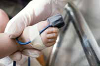
Pulse oximetry for newborns: Should it be routine?
Ensure that all newborns undergo pulse oximetry screening before discharge—and that abnormal results are immediately followed up with echocardiography.1
STRENGTH OF RECOMMENDATION
B: Based on a single cohort study consistent with multiple studies in other populations.
Ewer AK, Middleton LJ, Furmston AT, et al. Pulse oximetry screening for congenital heart defects in newborn infants (PulseOx): a test accuracy study. Lancet. 2011;378:785-794.
ILLUSTRATIVE CASE
A healthy, full-term baby is admitted to the newborn nursery. Antenatal surveillance, including routine ultrasound, was normal, as are physical examinations, both on admittance to the nursery and on the following day. Should the infant undergo pulse oximetry screening prior to discharge?
Congenital heart defects (CHD) are a leading cause of infant deaths in the developed world, occurring in approximately 9 of every 1000 live births.2 Roughly a quarter of those affected will have CHD serious enough to require either surgery or catheterization within the first year of life. These newborns are susceptible to sudden cardiovascular collapse due to changes in pulmonary vascular resistance and closure of the ductus arteriosus2—changes that often occur after the babies have gone home.
Delayed diagnosis is linked to worsening disease
A study evaluating 286 neonates admitted for cardiac surgery found that delayed diagnosis of CHD was associated with a worse preoperative condition. Cardiovascular compromise and end-organ dysfunction were most common in infants who presented with symptoms after they had gone home, the researchers found.3
In the past, screening for CHD hinged on mid-trimester ultrasound and postnatal physical examination. However, these methods do not reliably detect the condition in a timely fashion.4,5 More recently, pulse oximetry has been used for screening.
International studies prompt US recommendation
In a prospective multicenter trial in Saxony, Germany, more than 41,000 infants born between 2006 and 2008 underwent pulse oximetry screening at 24 to 72 hours of life.5 If the oxygen saturation was ≤95% and confirmed an hour later, echocardiography was performed. Pulse oximetry screening yielded true-positive results in 14 cases, false-positive results in 40, and false-negative results in 4. Sensitivity and specificity were 77.8% and 99.9%, respectively.
In another German study, 3364 term neonates underwent pulse oximetry screening between 6 and 36 hours of life.6 Eighteen neonates (0.5%) had abnormal results, 9 (50%) of whom were found to have heart defects. In this study, pulse oximetry had a sensitivity of 82% and a specificity of 99.9%.
A cohort study of 39,821 newborns in a single region of Sweden found that combining physical examination with pulse oximetry screening had a sensitivity of 82.8% and a specificity of 98%.7 No infants who underwent screening died from undiagnosed ductus arteriosus-dependent lung circulation, compared with 5 such deaths in regions where pulse oximetry screening was not done.
Encouraged by these findings, in late 2011 the US Secretary of Health and Human Services, with strong backing from the American Academy of Pediatrics, recommended universal pulse oximetry screening to detect critical CHD.8 The study detailed below took another look at its efficacy.
STUDY SUMMARY: Detection rate is higher for critical heart defects
The cohort study by Ewer et al enrolled 20,055 neonates born at >34 weeks’ gestation.1 All were screened with pulse oximetry on the right hand and on either foot and had a physical exam within their first 24 hours (The TABLE describes the screening protocol). Infants with a normal pulse oximetry and normal clinical exam were followed for a year to identify late-presenting heart defects.
One hundred ninety-five of the neonates who were screened had abnormal pulse oximetry test results; of these, 26 (13%) were found to have either critical (requiring intervention <28 days) or major (requiring intervention <12 months of age) CHD. Of the 169 infants who had positive pulse oximetry results but did not have critical or major heart defects, 6 were found to have less serious heart defects and 40 had infective or respiratory disorders that also required medical intervention.
Among the 19,860 infants with normal pulse oximetry, 27 (0.1%) were found to have either critical or major heart defects.
Pulse oximetry had a sensitivity of 75% (95% confidence interval [CI], 53.3-90.2) and a specificity of 99.1% (95% CI, 98.98-99.24) for detecting critical CHD. Sensitivity of pulse oximetry for all major CHD was 49% (95% CI, 35.0-63.2) and the specificity was 99.2% (95% CI, 99.02-99.28). The specificity may have been better if screening had been done after 24 hours of life; as seen in other studies,5,6 screening within the first 24 hours leads to more false-positive results.
The detection rate for critical CHD was higher than for major defects. However, most of the defects missed by screening were noncritical lesions, eg, ventricular septal defects.
The authors estimated that in a population of 100,000 newborns, about 120 would have critical CHD, and 90 of those 120 cases would be detected by pulse oximetry. There would be 843 false positives (although 229 of the infants with false-positive results would have other noncardiac conditions). It would be necessary to perform 10.4 echocardiograms to detect one patient with critical CHD.
WHAT’S NEW: A stronger case for newborn pulse oximetry screening
Pulse oximetry prior to discharge from the newborn nursery is not performed routinely in all institutions. And even when screening is done, there may not be a protocol addressing abnormal results. Pulse oximetry is a safe, noninvasive, inexpensive, and reasonably sensitive test that will detect many cases of critical CHD, some of which will not be diagnosed antenatally. Earlier diagnosis of CHD may lead to earlier interventions and improved patient outcomes.
CAVEATS: Timing of screening may alter results
This trial was a cohort study, not a randomized controlled trial (RCT)—the gold standard method of validating a screening test. It is unlikely, however, that an RCT will ever be done.
Screening occurred within the first 24 hours; other investigators have screened >24 hours (up to 38 hours), which may have better results. The critical lesions most likely to be missed by pulse oximetry screening were those causing obstruction to the aortic arch,1 a finding that was also seen in other studies.5,7
TABLE
Screening newborns for congenital heart defects: The protocol1
| Pulse oximetry outcome* | Physical exam outcome | Next step |
|---|---|---|
| Normal | Normal | No further action |
| Abnormal | Normal | Repeat pulse oximetry in 2 h:
|
| Abnormal | Abnormal | echocardiogram |
| *Test is normal if pulse oximetry >95% on right hand and difference between the right hand and either foot is <2%. | ||
CHALLENGES TO IMPLEMENTATION: Early discharge, lack of equipment may interfere
Determining the timing of pulse oximetry screening is important. That’s particularly true because early discharge is a common practice, and early screening may increase the number of false-positive results. As a screening tool, pulse oximetry is inexpensive, and follow-up echocardiography—which is needed to exclude serious cases of CHD in patients with positive pulse oximetry—is noninvasive and relatively inexpensive. Echocardiography is not readily available in all communities, however, and transportation to a facility that offers this test would likely increase the cost of screening.
Acknowledgement
The PURLs Surveillance System is supported in part by Grant Number UL1RR024999 from the National Center for Research Resources, a Clinical Translational Science Award to the University of Chicago. The content is solely the responsibility of the authors and does not necessarily represent the official views of the National Center for Research Resources or the National Institutes of Health.
1. Ewer AK, Middleton LJ, Furmston AT, et al. Pulse oximetry screening for congenital heart defects in newborn infants (PulseOx): a test accuracy study. Lancet. 201;378:785-794.
2. Mahle WT, Newburger JW, Matherne GP, et al. Role of pulse oximetry in examining newborns for congenital heart disease: a scientific statement from the AHA and AAP. Pediatrics. 2009;124:823-836.
3. Brown KL, Ridout DA, Hoskote A, et al. Delayed diagnosis of congenital heart disease worsens preoperative condition and outcome of surgery in neonates. Heart. 2006;92:1298-1302.
4. Acharya G, Sitras V, Maltau JM, et al. Major congenital heart disease in Northern Norway: shortcomings of pre- and postnatal diagnosis. Acta Obstet Gynecol Scand. 2004;83:1124-1129.
5. Riede FT, Worner C, Dahnert I, et al. Effectiveness of neonatal pulse oximetry screening for detection of critical congenital heart disease in daily clinical routine—results from a prospective multicenter study. Eur J Pediatr. 2010;169:975-981.
6. Tautz J, Merkel C, Loersch F, et al. Implication of pulse oxymetry screening for detection of congenital heart defects. Klin Padiatr. 2010;222:291-295.
7. de-Wahl GA, Wennergren M, Sandberg K, et al. Impact of pulse oximetry screening on the detection of duct dependent congenital heart disease: a Swedish prospective screening study in 39,821 newborns. BMJ. 2009;338:a3037.-
8. Mahle WT, Martin GR, Beekman RH III, et al. Endorsement of Health and Human Services recommendation for pulse oximetry screening for critical congenital heart disease. Pediatrics. 2012;129:190-192.
Ensure that all newborns undergo pulse oximetry screening before discharge—and that abnormal results are immediately followed up with echocardiography.1
STRENGTH OF RECOMMENDATION
B: Based on a single cohort study consistent with multiple studies in other populations.
Ewer AK, Middleton LJ, Furmston AT, et al. Pulse oximetry screening for congenital heart defects in newborn infants (PulseOx): a test accuracy study. Lancet. 2011;378:785-794.
ILLUSTRATIVE CASE
A healthy, full-term baby is admitted to the newborn nursery. Antenatal surveillance, including routine ultrasound, was normal, as are physical examinations, both on admittance to the nursery and on the following day. Should the infant undergo pulse oximetry screening prior to discharge?
Congenital heart defects (CHD) are a leading cause of infant deaths in the developed world, occurring in approximately 9 of every 1000 live births.2 Roughly a quarter of those affected will have CHD serious enough to require either surgery or catheterization within the first year of life. These newborns are susceptible to sudden cardiovascular collapse due to changes in pulmonary vascular resistance and closure of the ductus arteriosus2—changes that often occur after the babies have gone home.
Delayed diagnosis is linked to worsening disease
A study evaluating 286 neonates admitted for cardiac surgery found that delayed diagnosis of CHD was associated with a worse preoperative condition. Cardiovascular compromise and end-organ dysfunction were most common in infants who presented with symptoms after they had gone home, the researchers found.3
In the past, screening for CHD hinged on mid-trimester ultrasound and postnatal physical examination. However, these methods do not reliably detect the condition in a timely fashion.4,5 More recently, pulse oximetry has been used for screening.
International studies prompt US recommendation
In a prospective multicenter trial in Saxony, Germany, more than 41,000 infants born between 2006 and 2008 underwent pulse oximetry screening at 24 to 72 hours of life.5 If the oxygen saturation was ≤95% and confirmed an hour later, echocardiography was performed. Pulse oximetry screening yielded true-positive results in 14 cases, false-positive results in 40, and false-negative results in 4. Sensitivity and specificity were 77.8% and 99.9%, respectively.
In another German study, 3364 term neonates underwent pulse oximetry screening between 6 and 36 hours of life.6 Eighteen neonates (0.5%) had abnormal results, 9 (50%) of whom were found to have heart defects. In this study, pulse oximetry had a sensitivity of 82% and a specificity of 99.9%.
A cohort study of 39,821 newborns in a single region of Sweden found that combining physical examination with pulse oximetry screening had a sensitivity of 82.8% and a specificity of 98%.7 No infants who underwent screening died from undiagnosed ductus arteriosus-dependent lung circulation, compared with 5 such deaths in regions where pulse oximetry screening was not done.
Encouraged by these findings, in late 2011 the US Secretary of Health and Human Services, with strong backing from the American Academy of Pediatrics, recommended universal pulse oximetry screening to detect critical CHD.8 The study detailed below took another look at its efficacy.
STUDY SUMMARY: Detection rate is higher for critical heart defects
The cohort study by Ewer et al enrolled 20,055 neonates born at >34 weeks’ gestation.1 All were screened with pulse oximetry on the right hand and on either foot and had a physical exam within their first 24 hours (The TABLE describes the screening protocol). Infants with a normal pulse oximetry and normal clinical exam were followed for a year to identify late-presenting heart defects.
One hundred ninety-five of the neonates who were screened had abnormal pulse oximetry test results; of these, 26 (13%) were found to have either critical (requiring intervention <28 days) or major (requiring intervention <12 months of age) CHD. Of the 169 infants who had positive pulse oximetry results but did not have critical or major heart defects, 6 were found to have less serious heart defects and 40 had infective or respiratory disorders that also required medical intervention.
Among the 19,860 infants with normal pulse oximetry, 27 (0.1%) were found to have either critical or major heart defects.
Pulse oximetry had a sensitivity of 75% (95% confidence interval [CI], 53.3-90.2) and a specificity of 99.1% (95% CI, 98.98-99.24) for detecting critical CHD. Sensitivity of pulse oximetry for all major CHD was 49% (95% CI, 35.0-63.2) and the specificity was 99.2% (95% CI, 99.02-99.28). The specificity may have been better if screening had been done after 24 hours of life; as seen in other studies,5,6 screening within the first 24 hours leads to more false-positive results.
The detection rate for critical CHD was higher than for major defects. However, most of the defects missed by screening were noncritical lesions, eg, ventricular septal defects.
The authors estimated that in a population of 100,000 newborns, about 120 would have critical CHD, and 90 of those 120 cases would be detected by pulse oximetry. There would be 843 false positives (although 229 of the infants with false-positive results would have other noncardiac conditions). It would be necessary to perform 10.4 echocardiograms to detect one patient with critical CHD.
WHAT’S NEW: A stronger case for newborn pulse oximetry screening
Pulse oximetry prior to discharge from the newborn nursery is not performed routinely in all institutions. And even when screening is done, there may not be a protocol addressing abnormal results. Pulse oximetry is a safe, noninvasive, inexpensive, and reasonably sensitive test that will detect many cases of critical CHD, some of which will not be diagnosed antenatally. Earlier diagnosis of CHD may lead to earlier interventions and improved patient outcomes.
CAVEATS: Timing of screening may alter results
This trial was a cohort study, not a randomized controlled trial (RCT)—the gold standard method of validating a screening test. It is unlikely, however, that an RCT will ever be done.
Screening occurred within the first 24 hours; other investigators have screened >24 hours (up to 38 hours), which may have better results. The critical lesions most likely to be missed by pulse oximetry screening were those causing obstruction to the aortic arch,1 a finding that was also seen in other studies.5,7
TABLE
Screening newborns for congenital heart defects: The protocol1
| Pulse oximetry outcome* | Physical exam outcome | Next step |
|---|---|---|
| Normal | Normal | No further action |
| Abnormal | Normal | Repeat pulse oximetry in 2 h:
|
| Abnormal | Abnormal | echocardiogram |
| *Test is normal if pulse oximetry >95% on right hand and difference between the right hand and either foot is <2%. | ||
CHALLENGES TO IMPLEMENTATION: Early discharge, lack of equipment may interfere
Determining the timing of pulse oximetry screening is important. That’s particularly true because early discharge is a common practice, and early screening may increase the number of false-positive results. As a screening tool, pulse oximetry is inexpensive, and follow-up echocardiography—which is needed to exclude serious cases of CHD in patients with positive pulse oximetry—is noninvasive and relatively inexpensive. Echocardiography is not readily available in all communities, however, and transportation to a facility that offers this test would likely increase the cost of screening.
Acknowledgement
The PURLs Surveillance System is supported in part by Grant Number UL1RR024999 from the National Center for Research Resources, a Clinical Translational Science Award to the University of Chicago. The content is solely the responsibility of the authors and does not necessarily represent the official views of the National Center for Research Resources or the National Institutes of Health.
1. Ewer AK, Middleton LJ, Furmston AT, et al. Pulse oximetry screening for congenital heart defects in newborn infants (PulseOx): a test accuracy study. Lancet. 201;378:785-794.
2. Mahle WT, Newburger JW, Matherne GP, et al. Role of pulse oximetry in examining newborns for congenital heart disease: a scientific statement from the AHA and AAP. Pediatrics. 2009;124:823-836.
3. Brown KL, Ridout DA, Hoskote A, et al. Delayed diagnosis of congenital heart disease worsens preoperative condition and outcome of surgery in neonates. Heart. 2006;92:1298-1302.
4. Acharya G, Sitras V, Maltau JM, et al. Major congenital heart disease in Northern Norway: shortcomings of pre- and postnatal diagnosis. Acta Obstet Gynecol Scand. 2004;83:1124-1129.
5. Riede FT, Worner C, Dahnert I, et al. Effectiveness of neonatal pulse oximetry screening for detection of critical congenital heart disease in daily clinical routine—results from a prospective multicenter study. Eur J Pediatr. 2010;169:975-981.
6. Tautz J, Merkel C, Loersch F, et al. Implication of pulse oxymetry screening for detection of congenital heart defects. Klin Padiatr. 2010;222:291-295.
7. de-Wahl GA, Wennergren M, Sandberg K, et al. Impact of pulse oximetry screening on the detection of duct dependent congenital heart disease: a Swedish prospective screening study in 39,821 newborns. BMJ. 2009;338:a3037.-
8. Mahle WT, Martin GR, Beekman RH III, et al. Endorsement of Health and Human Services recommendation for pulse oximetry screening for critical congenital heart disease. Pediatrics. 2012;129:190-192.
Copyright © 2012 The Family Physicians Inquiries Network. All rights reserved.
Combatting lice in a single treatment
Consider prescribing spinosad—a recently approved topical pediculicide that is more effective than permethrin and eliminates the need for nit combing—for children with head lice.1
Stough D, Shellabarger S, Quiring J, et al. Efficacy and safety of spinosad and permethrin crème rinses for pediculosis capitis (head lice). Pediatrics. 2009;124:e389-e395.
STRENGTH OF RECOMMENDATION
B: Based on a single randomized controlled trial (RCT).
ILLUSTRATIVE CASE
A 6-year-old girl is brought in by her mother, who has just been notified of an outbreak of head lice in the child’s school. You examine her scalp and observe viable eggs (nits) at the hair shaft, and tell mom that her daughter has head lice. What’s the most effective treatment?
Head lice are common and easily spread among schoolchildren—and a source of frustration for patients, parents, and family physicians alike. Permethrin cream 1% (Nix), one of the most widely used and well-studied pediculicides, is available over the counter.2
Prescription pediculicides, including malathion (Ovide) and lindane (Kwell), are used less frequently due to their side effect profiles: Malathion is highly flammable due to a high alcohol content and lindane can cause neurotoxicity.2 Both are typically reserved for cases resistant to permethrin 1% cream. Local resistance patterns influence physician prescribing, of course, and greater resistance has increased the need for multiple treatments.3
Dual treatment—and combing— are often required
Permethrin is pediculicidal (ie, it kills only live lice); it is not ovicidal (ie, it does not kill nits at every stage).4,5 Thus, a second application is usually needed 7 to 10 days after the first to ensure complete eradication. And, because permethrin is not ovicidal, nit combing—a tedious, time-consuming, and often painful process—is required to remove lice and unhatched eggs from the hair shafts. Another downside: Children in districts that do not allow students to return to school until they are completely nit-free often miss school as a result of this lengthy process.
The US Food and Drug Administration (FDA) recently approved spinosad (Natroba topical suspension 0.9% cream rinse) for the treatment of lice in patients ≥4 years old.6 Spinosad, which is available only by prescription, eliminates the need for nit combing. But how well does it work?
STUDY SUMMARY: For most patients, a single application is sufficient
The study by Stough et al included 2 identical multicenter RCTs comparing 0.9% spinosad without nit combing vs 1% permethrin with combing in patients ≥6 months old who had active head lice. There were 446 patients in the primary treatment (spinosad) group and 470 in the control (permethrin) group. All participants within the same household were assigned to the same group.
Participants were evaluated on Days 1, 7, and 14. Those with active lice on Day 7 received another treatment (a second application with the same medication). The primary outcome was the percentage of participants who were lice free on Day 14.
The findings: After 14 days, 85% of the children in the spinosad group vs 44% of those in the permethrin group were lice free. In addition, 75% of those treated with spinosad required only one application for a cure, compared with 37% in the permethrin group.
There were no serious adverse events in either group. Participants in the permethrin group were more likely to experience scalp erythema than those in the spinosad group (6.8% vs 3.1%; P<.007). Complete metabolic panels and hematology studies were performed, and no significant differences pre- or posttreatment were found. The dropout rate was <10%, for similar reasons in both groups.

Spinosad is pediculicidal and ovicidal— killing lice and their eggs in all stages
WHAT’S NEW: Extensive nit combing no longer needed
Spinosad is pediculicidal and ovicidal— killing lice and their eggs in all stages—thereby eliminating the need for extensive combing in most cases. It has less resistance than current pediculicides, so a second course of treatment is needed only about one-quarter of the time. Spinosad received FDA approval for use in patients ≥4 years in 2011.
CAVEATS: Funding of study raises question of bias
The study by Stough et al was funded by ParaPRO, LLC, the manufacturer of Natroba. The company had access to the data and had a role in the interpretation of the findings and the decision to publish the study. However, the assessors were blinded to treatment group.
CHALLENGES TO iMPLEMENTATION: Cost is high and may not be covered
Cost will be a major barrier to treatment for many families. The average cost of an application of permethrin 1% is about $20; an application of spinosad costs $270 without insurance. Individual health plans may not cover it or may require prior authorization.
Acknowledgement
The PURLs Surveillance System is supported in part by Grant Number UL1RR024999 from the National Center for Research Resources, a Clinical Translational Science Award to the University of Chicago. The content is solely the responsibility of the authors and does not necessarily represent the official views of the National Center for Research Resources or the National Institutes of Health.
Click here to view PURL METHODOLOGY
1. Stough D, Shellabarger S, Quiring J, et al. Efficacy and Safety of spinosad and permethrin creme rinses for pediculosis capitis (head lice). Pediatrics. 2009;124:e389-e395.
2. Frankowski BL, Weiner LB. American Academy of Pediatrics, Committee on School Health Committee on Infectious Diseases. Head lice. Pediatrics. 2010;126:392-403.
3. Jones KN, English JC. 3rd. Review of common therapeutic options in the US for the treatment of pediculosis capitis. Clinl infect Dis. 2003;36:1355-1361.
4. Flinders D, Schweinitz P. Pediculosis and scabies. Am Fam Physician. 2004;69:341-350.
5. Centers for Disease Control and Prevention Parasites–lice. Available at http://www.cdc.gov/parasites/lice/. Accessed December 14, 2011.
6. US Food and Drug Administration Drugs@FDA. Available at: http://www.accessdata.fda.gov/scripts/cder/drugsatfda/index.cfm?fuseaction=Search. Drug Details. Accessed December 14, 2011.
Consider prescribing spinosad—a recently approved topical pediculicide that is more effective than permethrin and eliminates the need for nit combing—for children with head lice.1
Stough D, Shellabarger S, Quiring J, et al. Efficacy and safety of spinosad and permethrin crème rinses for pediculosis capitis (head lice). Pediatrics. 2009;124:e389-e395.
STRENGTH OF RECOMMENDATION
B: Based on a single randomized controlled trial (RCT).
ILLUSTRATIVE CASE
A 6-year-old girl is brought in by her mother, who has just been notified of an outbreak of head lice in the child’s school. You examine her scalp and observe viable eggs (nits) at the hair shaft, and tell mom that her daughter has head lice. What’s the most effective treatment?
Head lice are common and easily spread among schoolchildren—and a source of frustration for patients, parents, and family physicians alike. Permethrin cream 1% (Nix), one of the most widely used and well-studied pediculicides, is available over the counter.2
Prescription pediculicides, including malathion (Ovide) and lindane (Kwell), are used less frequently due to their side effect profiles: Malathion is highly flammable due to a high alcohol content and lindane can cause neurotoxicity.2 Both are typically reserved for cases resistant to permethrin 1% cream. Local resistance patterns influence physician prescribing, of course, and greater resistance has increased the need for multiple treatments.3
Dual treatment—and combing— are often required
Permethrin is pediculicidal (ie, it kills only live lice); it is not ovicidal (ie, it does not kill nits at every stage).4,5 Thus, a second application is usually needed 7 to 10 days after the first to ensure complete eradication. And, because permethrin is not ovicidal, nit combing—a tedious, time-consuming, and often painful process—is required to remove lice and unhatched eggs from the hair shafts. Another downside: Children in districts that do not allow students to return to school until they are completely nit-free often miss school as a result of this lengthy process.
The US Food and Drug Administration (FDA) recently approved spinosad (Natroba topical suspension 0.9% cream rinse) for the treatment of lice in patients ≥4 years old.6 Spinosad, which is available only by prescription, eliminates the need for nit combing. But how well does it work?
STUDY SUMMARY: For most patients, a single application is sufficient
The study by Stough et al included 2 identical multicenter RCTs comparing 0.9% spinosad without nit combing vs 1% permethrin with combing in patients ≥6 months old who had active head lice. There were 446 patients in the primary treatment (spinosad) group and 470 in the control (permethrin) group. All participants within the same household were assigned to the same group.
Participants were evaluated on Days 1, 7, and 14. Those with active lice on Day 7 received another treatment (a second application with the same medication). The primary outcome was the percentage of participants who were lice free on Day 14.
The findings: After 14 days, 85% of the children in the spinosad group vs 44% of those in the permethrin group were lice free. In addition, 75% of those treated with spinosad required only one application for a cure, compared with 37% in the permethrin group.
There were no serious adverse events in either group. Participants in the permethrin group were more likely to experience scalp erythema than those in the spinosad group (6.8% vs 3.1%; P<.007). Complete metabolic panels and hematology studies were performed, and no significant differences pre- or posttreatment were found. The dropout rate was <10%, for similar reasons in both groups.
Spinosad is pediculicidal and ovicidal— killing lice and their eggs in all stages
WHAT’S NEW: Extensive nit combing no longer needed
Spinosad is pediculicidal and ovicidal— killing lice and their eggs in all stages—thereby eliminating the need for extensive combing in most cases. It has less resistance than current pediculicides, so a second course of treatment is needed only about one-quarter of the time. Spinosad received FDA approval for use in patients ≥4 years in 2011.
CAVEATS: Funding of study raises question of bias
The study by Stough et al was funded by ParaPRO, LLC, the manufacturer of Natroba. The company had access to the data and had a role in the interpretation of the findings and the decision to publish the study. However, the assessors were blinded to treatment group.
CHALLENGES TO iMPLEMENTATION: Cost is high and may not be covered
Cost will be a major barrier to treatment for many families. The average cost of an application of permethrin 1% is about $20; an application of spinosad costs $270 without insurance. Individual health plans may not cover it or may require prior authorization.
Acknowledgement
The PURLs Surveillance System is supported in part by Grant Number UL1RR024999 from the National Center for Research Resources, a Clinical Translational Science Award to the University of Chicago. The content is solely the responsibility of the authors and does not necessarily represent the official views of the National Center for Research Resources or the National Institutes of Health.
Click here to view PURL METHODOLOGY
1. Stough D, Shellabarger S, Quiring J, et al. Efficacy and Safety of spinosad and permethrin creme rinses for pediculosis capitis (head lice). Pediatrics. 2009;124:e389-e395.
2. Frankowski BL, Weiner LB. American Academy of Pediatrics, Committee on School Health Committee on Infectious Diseases. Head lice. Pediatrics. 2010;126:392-403.
3. Jones KN, English JC. 3rd. Review of common therapeutic options in the US for the treatment of pediculosis capitis. Clinl infect Dis. 2003;36:1355-1361.
4. Flinders D, Schweinitz P. Pediculosis and scabies. Am Fam Physician. 2004;69:341-350.
5. Centers for Disease Control and Prevention Parasites–lice. Available at http://www.cdc.gov/parasites/lice/. Accessed December 14, 2011.
6. US Food and Drug Administration Drugs@FDA. Available at: http://www.accessdata.fda.gov/scripts/cder/drugsatfda/index.cfm?fuseaction=Search. Drug Details. Accessed December 14, 2011.
Copyright © 2012 The Family Physicians Inquiries Network.
All rights reserved.