User login
Painful red hands and feet
Q: What is the diagnosis?
- Fabry disease
- Peripheral neuropathy
- Polycythemia
- Primary idiopathic erythromelalgia
- Erythrodysesthesia syndrome
A: The correct diagnosis is primary idiopathic erythromelalgia.
Erythromelalgia is a relatively rare clinical condition of uncertain etiology, characterized by the triad of episodic redness, warmth, and burning pain in the extremities.1,2
The condition has primary (ie, no underlying cause is found) and secondary forms.1,3 The primary form may be inherited in an autosomal dominant manner (in which case the symptoms begin in childhood), or it may be idiopathic.4 On the other hand, erythromelalgia can also be secondary to polycythemia vera and other myeloproliferative disorders, connective tissue disorders, neuropathies, spinal cord diseases, carcinoma of the colon and thyroid, and astrocytomas.1–6
The common pathologic mechanism of erythromelalgia is thought to be microvascular arteriovenous shunting.5 A mutation in the voltage-gated sodium channel alpha subunit NaV1.7 may result in primary erythromelalgia. Small-fiber neuropathy2 can manifest as erythromelalgia and may respond to steroids.
The symptoms can be intermittent or, in rare cases, constant.3 The lower limbs are more commonly involved than the upper ones.3 Involvement is often bilateral and symmetric.3 The symptoms may worsen at night, after alcohol consumption, with higher environmental temperature, and with moderate exercise.3 In rare cases, ulceration and gangrene may occur. Patients may get relief by cooling the affected areas.2
DIAGNOSIS BY EXCLUSION OR BASED ON THE PRESENTATION
The diagnosis of erythromelalgia is based on a detailed history and physical examination during a painful episode.2,3 Because the condition is intermittent, only about two-thirds of patients have abnormal findings on physical examination at the time of presentation; in such cases, the diagnosis is based on the history alone.2
Testing is needed to exclude other diagnoses and to determine the cause of secondary erythromelalgia. Histopathologic study of lesions is not helpful, as the features are nonspecific and hence nondiagnostic.2
In our patient, the typical clinical features and the lack of an obvious cause on diagnostic testing confirmed the diagnosis.
THE DIFFERENTIAL DIAGNOSIS
Other conditions can be ruled out by clinical features and laboratory testing.
Fabry disease causes paresthesia and burning pain in the extremities but not erythema. Characteristic dark red keratotic papules are seen all over the body (angiokeratoma corporis diffusum). It is often associated with progressive renal insufficiency.
Peripheral neuropathy of varying causes may also cause pain in the extremities but not erythema. Neurologic examination and a nerve conduction velocity study can resolve the diagnostic problem. Clinical features and laboratory testing often help to pinpoint the cause of neuropathy.
Polycythemia may cause erythema in the hands, feet, and face and mucosal engorgement. Telangiectasia, petechiae, and cyanosis may also occur. Our patient’s normal complete blood cell count excluded this condition.
Erythrodysesthesia syndrome, typically caused by chemotherapeutic drugs, was not included in the differential diagnosis since our patient had taken no medications.
Other causes of palmar erythema to rule out, depending on the patient’s presentation, may include thyrotoxicosis, chronic febrile illness, leukemia, hepatic insufficiency, chronic alcoholism, and rheumatoid arthritis.
TREATMENT AND PROGNOSIS
There is no definitive therapy for erythromelalgia.4 Treatment is often difficult and needs a multidisciplinary approach. Simple measures such as cooling2 (eg, applying cold towels, immersion in cool water, walking on cold floors) or elevating the affected extremity often relieve symptoms. Patients should avoid precipitating factors such as warmth, dependency of extremities, exercise, tight footwear, and alcohol intake.
If there is an underlying disease, treating the disease may also alleviate the symptoms.7 Aspirin2,7 is the therapy of choice for erythromelalgia in patients with an underlying myeloproliferative disorder, and some authors have advocated it for all patients with erythromelalgia unless there is a contraindication.7
Other possible first-line treatments include the synthetic prostaglandin E1 analogue misoprostol (Cytotec) and prostacyclins. Gabapentin (Neurontin), serotonin reuptake inhibitors such as sertaline (Zoloft) and venlafaxine (Effexor), and intravenous nitroprusside (Nitro-press) are considered second-line drugs.7
Surgical sympathectomy8 has also been tried, with variable results.
Outcomes in patients with erythromelalgia
In a case series from Mayo Clinic,2 approximately equal numbers of patients with erythromelalgia became worse, stayed the same, or got better, and the disease resolved in 10% over a mean of 8.7 years.
THE OUTCOME IN OUR PATIENT
We advised our patient to avoid strenuous activity in a warm environment and to work in cooler areas as much as possible. We told him to wrap his affected extremities with cold towels during attacks, and we prescribed aspirin (650 mg/day) for 3 months. The treatment did not cure his condition, but his symptoms lessened within 2 months. We later referred him to a pain clinic.
- Kalgaard OM, Seem E, Kvernebo K. Erythromelalgia: a clinical study of 87 cases. Intern Med 1997; 242:191–197.
- Davis MD, O’Fallon WM, Rogers RS, Rooke TW. Natural history of erythromelalgia: presentation and outcome in 168 patients. Arch Dermatol 2000; 136:330–336.
- Galimberti D, Pontón A, Rubio L, et al. A case of primary erythromelalgia. J Eur Acad Dermatol Venereol 2009; 23:1338–1339.
- Ljubojevic S, Lipozencic J, Pustisek N. Erythromelalgia. Acta Dermatovenerol Croat 2004; 12:99–105.
- Mørk C, Kvernebo K, Asker CL, Salerud EG. Reduced skin capillary density during attacks of erythromelalgia implies arteriovenous shunting as pathogenetic mechanism. J Invest Dermatol 2002; 119:949–953.
- James WD, Berger TG, Elston DM. Andrews’ Diseases of the Skin: Clinical Dermatology. 10th ed. Philadelphia, PA: Saunders Elsevier; 2006.
- Mørk C, Kvernebo K. Erythromelalgia. In:Lebwohl MG, Heymann WR, Berth-Jones J, Koulson Y. editors. Treatment of Skin Disease: Comprehensive Therapeutic Strategies. 3rd ed. Philadelphia, PA: Saunders Elsevier; 2010:236–238.
- Seishima M, Kanoh H, Izumi T, et al. A refractory case of secondary erythermalgia successfully treated with lumbar sympathetic ganglion block. Br J Dermatol 2000; 143:868–872.
Q: What is the diagnosis?
- Fabry disease
- Peripheral neuropathy
- Polycythemia
- Primary idiopathic erythromelalgia
- Erythrodysesthesia syndrome
A: The correct diagnosis is primary idiopathic erythromelalgia.
Erythromelalgia is a relatively rare clinical condition of uncertain etiology, characterized by the triad of episodic redness, warmth, and burning pain in the extremities.1,2
The condition has primary (ie, no underlying cause is found) and secondary forms.1,3 The primary form may be inherited in an autosomal dominant manner (in which case the symptoms begin in childhood), or it may be idiopathic.4 On the other hand, erythromelalgia can also be secondary to polycythemia vera and other myeloproliferative disorders, connective tissue disorders, neuropathies, spinal cord diseases, carcinoma of the colon and thyroid, and astrocytomas.1–6
The common pathologic mechanism of erythromelalgia is thought to be microvascular arteriovenous shunting.5 A mutation in the voltage-gated sodium channel alpha subunit NaV1.7 may result in primary erythromelalgia. Small-fiber neuropathy2 can manifest as erythromelalgia and may respond to steroids.
The symptoms can be intermittent or, in rare cases, constant.3 The lower limbs are more commonly involved than the upper ones.3 Involvement is often bilateral and symmetric.3 The symptoms may worsen at night, after alcohol consumption, with higher environmental temperature, and with moderate exercise.3 In rare cases, ulceration and gangrene may occur. Patients may get relief by cooling the affected areas.2
DIAGNOSIS BY EXCLUSION OR BASED ON THE PRESENTATION
The diagnosis of erythromelalgia is based on a detailed history and physical examination during a painful episode.2,3 Because the condition is intermittent, only about two-thirds of patients have abnormal findings on physical examination at the time of presentation; in such cases, the diagnosis is based on the history alone.2
Testing is needed to exclude other diagnoses and to determine the cause of secondary erythromelalgia. Histopathologic study of lesions is not helpful, as the features are nonspecific and hence nondiagnostic.2
In our patient, the typical clinical features and the lack of an obvious cause on diagnostic testing confirmed the diagnosis.
THE DIFFERENTIAL DIAGNOSIS
Other conditions can be ruled out by clinical features and laboratory testing.
Fabry disease causes paresthesia and burning pain in the extremities but not erythema. Characteristic dark red keratotic papules are seen all over the body (angiokeratoma corporis diffusum). It is often associated with progressive renal insufficiency.
Peripheral neuropathy of varying causes may also cause pain in the extremities but not erythema. Neurologic examination and a nerve conduction velocity study can resolve the diagnostic problem. Clinical features and laboratory testing often help to pinpoint the cause of neuropathy.
Polycythemia may cause erythema in the hands, feet, and face and mucosal engorgement. Telangiectasia, petechiae, and cyanosis may also occur. Our patient’s normal complete blood cell count excluded this condition.
Erythrodysesthesia syndrome, typically caused by chemotherapeutic drugs, was not included in the differential diagnosis since our patient had taken no medications.
Other causes of palmar erythema to rule out, depending on the patient’s presentation, may include thyrotoxicosis, chronic febrile illness, leukemia, hepatic insufficiency, chronic alcoholism, and rheumatoid arthritis.
TREATMENT AND PROGNOSIS
There is no definitive therapy for erythromelalgia.4 Treatment is often difficult and needs a multidisciplinary approach. Simple measures such as cooling2 (eg, applying cold towels, immersion in cool water, walking on cold floors) or elevating the affected extremity often relieve symptoms. Patients should avoid precipitating factors such as warmth, dependency of extremities, exercise, tight footwear, and alcohol intake.
If there is an underlying disease, treating the disease may also alleviate the symptoms.7 Aspirin2,7 is the therapy of choice for erythromelalgia in patients with an underlying myeloproliferative disorder, and some authors have advocated it for all patients with erythromelalgia unless there is a contraindication.7
Other possible first-line treatments include the synthetic prostaglandin E1 analogue misoprostol (Cytotec) and prostacyclins. Gabapentin (Neurontin), serotonin reuptake inhibitors such as sertaline (Zoloft) and venlafaxine (Effexor), and intravenous nitroprusside (Nitro-press) are considered second-line drugs.7
Surgical sympathectomy8 has also been tried, with variable results.
Outcomes in patients with erythromelalgia
In a case series from Mayo Clinic,2 approximately equal numbers of patients with erythromelalgia became worse, stayed the same, or got better, and the disease resolved in 10% over a mean of 8.7 years.
THE OUTCOME IN OUR PATIENT
We advised our patient to avoid strenuous activity in a warm environment and to work in cooler areas as much as possible. We told him to wrap his affected extremities with cold towels during attacks, and we prescribed aspirin (650 mg/day) for 3 months. The treatment did not cure his condition, but his symptoms lessened within 2 months. We later referred him to a pain clinic.
- Kalgaard OM, Seem E, Kvernebo K. Erythromelalgia: a clinical study of 87 cases. Intern Med 1997; 242:191–197.
- Davis MD, O’Fallon WM, Rogers RS, Rooke TW. Natural history of erythromelalgia: presentation and outcome in 168 patients. Arch Dermatol 2000; 136:330–336.
- Galimberti D, Pontón A, Rubio L, et al. A case of primary erythromelalgia. J Eur Acad Dermatol Venereol 2009; 23:1338–1339.
- Ljubojevic S, Lipozencic J, Pustisek N. Erythromelalgia. Acta Dermatovenerol Croat 2004; 12:99–105.
- Mørk C, Kvernebo K, Asker CL, Salerud EG. Reduced skin capillary density during attacks of erythromelalgia implies arteriovenous shunting as pathogenetic mechanism. J Invest Dermatol 2002; 119:949–953.
- James WD, Berger TG, Elston DM. Andrews’ Diseases of the Skin: Clinical Dermatology. 10th ed. Philadelphia, PA: Saunders Elsevier; 2006.
- Mørk C, Kvernebo K. Erythromelalgia. In:Lebwohl MG, Heymann WR, Berth-Jones J, Koulson Y. editors. Treatment of Skin Disease: Comprehensive Therapeutic Strategies. 3rd ed. Philadelphia, PA: Saunders Elsevier; 2010:236–238.
- Seishima M, Kanoh H, Izumi T, et al. A refractory case of secondary erythermalgia successfully treated with lumbar sympathetic ganglion block. Br J Dermatol 2000; 143:868–872.

Multiple facial bumps with weight loss
A Good-quality patient-oriented evidence
B Inconsistent or limited-quality patient-oriented evidence
C Consensus, usual practice, opinion, disease-oriented evidence, case series
A 12-Year-old girl came into our hospital for treatment of multiple bumps that had developed around her eyes and other areas of her face 2 months earlier. She had difficulty opening her eyes and complained of gradual weight loss.
On examination, we noted numerous skin-colored, shiny, dome-shaped, coalescing papules and nodules with central umbilications that were distributed mostly on her periocular and perinasal areas (FIGURE). When we expressed the papules with forceps, they exuded a cheesy material. We also noticed crusting and signs of inflammation on her eyelids.
The systemic examination was unremarkable.
FIGURE
Opening her eyes was difficult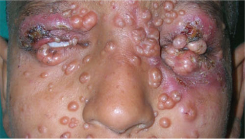
This 12-year-old patient had multiple dome-shaped, coalescing papules and nodules with central umbilications in the periocular and perinasal areas.
WHAT IS YOUR DIAGNOSIS?
HOW WOULD YOU TREAT THIS PATIENT?
Diagnosis: Giant molluscum contagiosum
Molluscum contagiosum (MC) is a relatively common, benign, viral cutaneous infection that primarily affects children, sexually active adults, and immunodeficient individuals. MC accounts for approximately 1% of all diagnosed skin disorders in the United States; internationally, however, the incidence is higher.1 The causative organism of MC is a member of the Poxviridae family2 and is thought to be transmitted by close personal contact, autoinoculation, and fomites.3
MC is clinically characterized by the presence of pearly white, dome-shaped papules or nodules with central dells. The lesions are typically located on the trunk, body folds, extremities, and genitalia (particularly when the infection is sexually acquired).2,3 Pruritus and an eczematous reaction can develop around the lesions.
MC is a recognized ocular complication of acquired immune deficiency syndrome (AIDS). Periocular MC can also occur after eyebrow shaping in beauty salons.4 In human immunodeficiency virus (HIV)-positive patients, lesions are usually widespread, tend to be large, and usually occur during the advanced stage of HIV infection.2,5
The differential includes carcinoma
When considering a diagnosis of MC, you’ll need to rule out the following causes of similar-looking papules and nodules:
Nodular basal cell carcinoma presents as a slow-growing, firm, shiny, pearly nodule with fine telangiectasia. It may also present as a cystic lesion that can be mistaken for inclusion cysts of the eyelid. If left untreated, the tumor may ulcerate.
Juvenile xanthogranulomas are rubbery, tan-orange papules or nodules. Patients may have one or several papules or nodules in the head and neck region; these lesions may appear elsewhere, as well.
Cryptococcosis may present as painless papules or pustules, which then become nodules that may ulcerate. The lesions may show central umbilications.
Keratoacanthoma begins as a firm, roundish, skin-colored or reddish papule that rapidly progresses to a dome-shaped nodule, with a smooth, shiny surface and a central crateriform ulceration or keratin plug. Patients typically have a solitary lesion that may appear on the face, neck, or dorsum of the upper extremities.
Penicillosis often presents with MC-like skin lesions, in addition to fever, anemia, hepatomegaly, lymphadenopathy, and productive cough.
History and lab work clinch the Dx
Diagnosis is made by the distinctive clinical appearance, but can be confirmed by skin biopsy demonstrating eosinophilic molluscum bodies packed into the cells of the spinous layer of the epidermis.3 Giemsa stain of the material obtained from a crushed papule will reveal the presence of pathognomonic “molluscum bodies” in the cells of the epidermis.2,3
Our patient’s Giemsa stain revealed molluscum bodies. And since it is always wise to rule out concomitant HIV infection in patients who have giant MC, we tested our patient. Her results were positive; she had a CD4+ count of 93 cells/mm3.
Many treatment options from which to choose
MC is usually self-limiting, although it can take several months—or even a few years—to resolve on its own6 (strength of recommendation [SOR]: B). However, most patients with MC should receive treatment to obtain relief from symptoms, prevent autoinoculation or transmission to close contacts, decrease occurrence of scarring, reduce secondary bacterial infections, and improve cosmesis.
Several treatment options are available, and most rely on destruction of the lesions. Manual extrusion is a simple but effective therapy6 (SOR: B). Cryotherapy and curettage are also effective treatment options5 (SOR: C). Pretreatment topical anesthesia is often helpful if these therapies are used in children.
Topical imiquimod2 (1%-5%) cream applied 3 to 7 times a week can be used to treat generalized MC infection or MC localized to the anogenital area6 (SOR: A). Some patients may improve with topical tretinoin therapy6 (SOR: C).
Chemical cauterization with 10% povidone iodine with 50% salicylic acid7 (SOR: B), 10% potassium hydroxide8 (SOR: B), cantharidin2 (SOR: C), or 25% to 50% trichloroacetic acid6 (SOR: C) is also effective. Treatment with flashlamp pulsed dye laser is a safe and efficient treatment modality9 (SOR: C). Cidofovir10 (1%-3%) cream or ointment, electron beam therapy, and photodynamic therapy have also been used with variable success rates6 (SOR: C).
MC is particularly difficult to treat in patients with poorly managed HIV and AIDS. Pairing proper antiretroviral therapy with lesion-destroying therapies is usually helpful for these patients.3
If you are caring for a patient with giant MC, you’ll need to stress the benign—but potentially contagious—nature of the disease. Tell the patient to wash his or her hands frequently, to avoid scratching the lesions, and to keep infected areas covered with clothing (when possible). In suspected sexually transmitted cases, the patient should adopt safe sexual practices or abstinence, if necessary. It is unclear whether condoms or other barrier methods provide adequate protection.1
Our patient transfers to the HIV clinic
We sequentially expressed the large lesions on our patient’s face and put her on a course of cefadroxil to control the secondary infection of MC. Her facial lesions gradually improved over 2 months.
We also referred the patient to our institution’s HIV clinic, where she was put on highly active antiretroviral therapy (HAART). We advised her mother to get tested for HIV, and she turned out to be HIV positive, as well.
CORRESPONDENCE Sudip Kumar Ghosh, MD, DNB, Department of Dermatology, Venereology, and Leprosy, R.G. Kar Medical College, 1 Khudiram Bose Sarani, Kolkata-700004, West Bengal, India; dr_skghosh@yahoo.co.in
1. Taillac PP. Molluscum contagiosum: eMedicine, Emergency Medicine. Available at: . Accessed October 29, 2010.
2. Tom W, Friedlander SF. Poxvirus infections. In: Wolff K, Goldsmith LA, Katz SI, et al, eds. Fitzpatrick’s Dermatology in General Medicine. 7th ed. New York, NY: McGraw-Hill; 2008: 1899-1913.
3. Turchin I, Barankin B. Dermacase. Molluscum contagiosum. Can Fam Physician. 2006;52:1395-1407.
4. Ghosh SK, Bandyopadhyay D. Molluscum contagiosum after eyebrow shaping: a beauty salon hazard. Clin Exp Dermatol. 2009;34:e339-e340.
5. Gur I. The epidemiology of molluscum contagiosum in HIVseropositive patients: a unique entity or insignificant finding? Int J STD AIDS. 2008;19:503-506.
6. Mckenna DB, Benton EC. Molluscum contagiosum. In: Lebwohl MG, Heymann WR, Berth-Jones J, et al, eds. Treatment of Skin Disease: Comprehensive Therapeutic Strategies. 2nd ed. London: Mosby; 2002: 399-401.
7. Ohkuma M. Molluscum contagiosum treated with iodine solution and salicylic acid plaster. Int J Dermatol. 1990;29:443-445.
8. Mahajan BB, Pall A, Gupta RR. Topical 20% KOH—an effective therapeutic modality for molluscum contagiosum in children. Indian J Dermatol Venereol Leprol. 2003;69:175-177.
9. Binder B, Weger W, Komericki P, et al. Treatment of molluscum contagiosum with a pulsed dye laser: pilot study with 19 children. J Dtsch Dermatol Ges. 2008;6:121-125.
10. Watanabe T, Tamaki K. Cidofovir diphosphate inhibits molluscum contagiosum virus DNA polymerase activity. J Invest Dermatol. 2008;128:1327-1329.
A Good-quality patient-oriented evidence
B Inconsistent or limited-quality patient-oriented evidence
C Consensus, usual practice, opinion, disease-oriented evidence, case series
A 12-Year-old girl came into our hospital for treatment of multiple bumps that had developed around her eyes and other areas of her face 2 months earlier. She had difficulty opening her eyes and complained of gradual weight loss.
On examination, we noted numerous skin-colored, shiny, dome-shaped, coalescing papules and nodules with central umbilications that were distributed mostly on her periocular and perinasal areas (FIGURE). When we expressed the papules with forceps, they exuded a cheesy material. We also noticed crusting and signs of inflammation on her eyelids.
The systemic examination was unremarkable.
FIGURE
Opening her eyes was difficult
This 12-year-old patient had multiple dome-shaped, coalescing papules and nodules with central umbilications in the periocular and perinasal areas.
WHAT IS YOUR DIAGNOSIS?
HOW WOULD YOU TREAT THIS PATIENT?
Diagnosis: Giant molluscum contagiosum
Molluscum contagiosum (MC) is a relatively common, benign, viral cutaneous infection that primarily affects children, sexually active adults, and immunodeficient individuals. MC accounts for approximately 1% of all diagnosed skin disorders in the United States; internationally, however, the incidence is higher.1 The causative organism of MC is a member of the Poxviridae family2 and is thought to be transmitted by close personal contact, autoinoculation, and fomites.3
MC is clinically characterized by the presence of pearly white, dome-shaped papules or nodules with central dells. The lesions are typically located on the trunk, body folds, extremities, and genitalia (particularly when the infection is sexually acquired).2,3 Pruritus and an eczematous reaction can develop around the lesions.
MC is a recognized ocular complication of acquired immune deficiency syndrome (AIDS). Periocular MC can also occur after eyebrow shaping in beauty salons.4 In human immunodeficiency virus (HIV)-positive patients, lesions are usually widespread, tend to be large, and usually occur during the advanced stage of HIV infection.2,5
The differential includes carcinoma
When considering a diagnosis of MC, you’ll need to rule out the following causes of similar-looking papules and nodules:
Nodular basal cell carcinoma presents as a slow-growing, firm, shiny, pearly nodule with fine telangiectasia. It may also present as a cystic lesion that can be mistaken for inclusion cysts of the eyelid. If left untreated, the tumor may ulcerate.
Juvenile xanthogranulomas are rubbery, tan-orange papules or nodules. Patients may have one or several papules or nodules in the head and neck region; these lesions may appear elsewhere, as well.
Cryptococcosis may present as painless papules or pustules, which then become nodules that may ulcerate. The lesions may show central umbilications.
Keratoacanthoma begins as a firm, roundish, skin-colored or reddish papule that rapidly progresses to a dome-shaped nodule, with a smooth, shiny surface and a central crateriform ulceration or keratin plug. Patients typically have a solitary lesion that may appear on the face, neck, or dorsum of the upper extremities.
Penicillosis often presents with MC-like skin lesions, in addition to fever, anemia, hepatomegaly, lymphadenopathy, and productive cough.
History and lab work clinch the Dx
Diagnosis is made by the distinctive clinical appearance, but can be confirmed by skin biopsy demonstrating eosinophilic molluscum bodies packed into the cells of the spinous layer of the epidermis.3 Giemsa stain of the material obtained from a crushed papule will reveal the presence of pathognomonic “molluscum bodies” in the cells of the epidermis.2,3
Our patient’s Giemsa stain revealed molluscum bodies. And since it is always wise to rule out concomitant HIV infection in patients who have giant MC, we tested our patient. Her results were positive; she had a CD4+ count of 93 cells/mm3.
Many treatment options from which to choose
MC is usually self-limiting, although it can take several months—or even a few years—to resolve on its own6 (strength of recommendation [SOR]: B). However, most patients with MC should receive treatment to obtain relief from symptoms, prevent autoinoculation or transmission to close contacts, decrease occurrence of scarring, reduce secondary bacterial infections, and improve cosmesis.
Several treatment options are available, and most rely on destruction of the lesions. Manual extrusion is a simple but effective therapy6 (SOR: B). Cryotherapy and curettage are also effective treatment options5 (SOR: C). Pretreatment topical anesthesia is often helpful if these therapies are used in children.
Topical imiquimod2 (1%-5%) cream applied 3 to 7 times a week can be used to treat generalized MC infection or MC localized to the anogenital area6 (SOR: A). Some patients may improve with topical tretinoin therapy6 (SOR: C).
Chemical cauterization with 10% povidone iodine with 50% salicylic acid7 (SOR: B), 10% potassium hydroxide8 (SOR: B), cantharidin2 (SOR: C), or 25% to 50% trichloroacetic acid6 (SOR: C) is also effective. Treatment with flashlamp pulsed dye laser is a safe and efficient treatment modality9 (SOR: C). Cidofovir10 (1%-3%) cream or ointment, electron beam therapy, and photodynamic therapy have also been used with variable success rates6 (SOR: C).
MC is particularly difficult to treat in patients with poorly managed HIV and AIDS. Pairing proper antiretroviral therapy with lesion-destroying therapies is usually helpful for these patients.3
If you are caring for a patient with giant MC, you’ll need to stress the benign—but potentially contagious—nature of the disease. Tell the patient to wash his or her hands frequently, to avoid scratching the lesions, and to keep infected areas covered with clothing (when possible). In suspected sexually transmitted cases, the patient should adopt safe sexual practices or abstinence, if necessary. It is unclear whether condoms or other barrier methods provide adequate protection.1
Our patient transfers to the HIV clinic
We sequentially expressed the large lesions on our patient’s face and put her on a course of cefadroxil to control the secondary infection of MC. Her facial lesions gradually improved over 2 months.
We also referred the patient to our institution’s HIV clinic, where she was put on highly active antiretroviral therapy (HAART). We advised her mother to get tested for HIV, and she turned out to be HIV positive, as well.
CORRESPONDENCE Sudip Kumar Ghosh, MD, DNB, Department of Dermatology, Venereology, and Leprosy, R.G. Kar Medical College, 1 Khudiram Bose Sarani, Kolkata-700004, West Bengal, India; dr_skghosh@yahoo.co.in
1. Taillac PP. Molluscum contagiosum: eMedicine, Emergency Medicine. Available at: . Accessed October 29, 2010.
2. Tom W, Friedlander SF. Poxvirus infections. In: Wolff K, Goldsmith LA, Katz SI, et al, eds. Fitzpatrick’s Dermatology in General Medicine. 7th ed. New York, NY: McGraw-Hill; 2008: 1899-1913.
3. Turchin I, Barankin B. Dermacase. Molluscum contagiosum. Can Fam Physician. 2006;52:1395-1407.
4. Ghosh SK, Bandyopadhyay D. Molluscum contagiosum after eyebrow shaping: a beauty salon hazard. Clin Exp Dermatol. 2009;34:e339-e340.
5. Gur I. The epidemiology of molluscum contagiosum in HIVseropositive patients: a unique entity or insignificant finding? Int J STD AIDS. 2008;19:503-506.
6. Mckenna DB, Benton EC. Molluscum contagiosum. In: Lebwohl MG, Heymann WR, Berth-Jones J, et al, eds. Treatment of Skin Disease: Comprehensive Therapeutic Strategies. 2nd ed. London: Mosby; 2002: 399-401.
7. Ohkuma M. Molluscum contagiosum treated with iodine solution and salicylic acid plaster. Int J Dermatol. 1990;29:443-445.
8. Mahajan BB, Pall A, Gupta RR. Topical 20% KOH—an effective therapeutic modality for molluscum contagiosum in children. Indian J Dermatol Venereol Leprol. 2003;69:175-177.
9. Binder B, Weger W, Komericki P, et al. Treatment of molluscum contagiosum with a pulsed dye laser: pilot study with 19 children. J Dtsch Dermatol Ges. 2008;6:121-125.
10. Watanabe T, Tamaki K. Cidofovir diphosphate inhibits molluscum contagiosum virus DNA polymerase activity. J Invest Dermatol. 2008;128:1327-1329.