User login
Restrictive transfusion will likely be standard, doc says
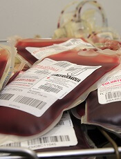
MUNICH—A restrictive transfusion strategy during cardiovascular surgery will likely become the standard of care, according to an investigator from the TRICS III trial.
The study showed that a restrictive approach to transfusion did not increase the risk of poor outcomes at 6 months after cardiac surgery.
David Mazer, MD, of St. Michael’s Hospital, University of Toronto in Ontario, Canada, presented these findings at the 2018 ESC Congress. The results were published simultaneously in NEJM.
Dr. Mazer and his colleagues previously reported results from the TRICS III trial showing that 28-day outcomes were similar whether patients undergoing cardiac surgery were treated with a restrictive or a liberal transfusion strategy.
However, the team wanted to look into 6-month results to rule out latent problems, such as sequelae from perioperative organ hypoxia.
“Our research question was, ‘At what point does the risk of anemia, or the risk of a lower hemoglobin, outweigh the risk of transfusion?’” Dr. Mazer said. “We wanted to know whether it is safe to let your hemoglobin go to a lower level before you transfuse. The answer is yes. It’ll save blood, make blood more available, reduce costs of transfusion, and result in similar or better outcomes.”
Patients
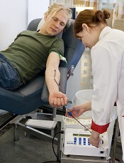
The trial included 2317 patients who were randomized to a restrictive transfusion strategy, which meant they received red cell transfusions if their hemoglobin concentrations fell below 7.5 g/dL intraoperatively or postoperatively.
Another 2347 patients were randomized to the liberal approach, which meant they received transfusions if their hemoglobin fell below 9.5 g/dL in the operating room and intensive care unit (ICU) and below 8.5 g/dL outside the ICU.
Baseline characteristics were well balanced between the arms. Patients were a mean of 72 years old, and 35% were female. The majority of patients in both arms underwent coronary artery bypass surgery, valve surgery, or both. Heart transplants were excluded.
Results
At 6 months, 17.4% of patients in the restrictive arm and 17.1% in the liberal arm met the primary composite outcome of death from any cause, myocardial infarction, stroke, or new-onset renal failure with dialysis (P=0.006 for noninferiority).
Unexpectedly, patients age 75 and older had a lower risk of the primary outcome with the restrictive strategy, while the liberal strategy was associated with lower risk in younger patients.
For all age groups, there were no significant differences between the treatment arms for the individual components of the primary composite outcome or for secondary outcomes.
The mortality rate was 6.2% in the restrictive arm and 6.4% in the liberal arm. The rate of myocardial infarction was 7.3% in both arms.
The rate of stroke was 4% in the restrictive arm and 3.3% in the liberal arm. The incidence of new-onset renal failure with dialysis was 3.9% and 4.2%, respectively.
The secondary composite outcome included the components of the primary outcome plus hospital readmissions, emergency department visits, and coronary revascularization. This outcome occurred in 43.8% of patients in the restrictive arm and 42.8% in the liberal arm.
The incidence of hospital readmissions/emergency visits was 35.5% in the restrictive arm and 33.6% in the liberal arm. The incidence of coronary revascularization was 0.7% and 0.9%, respectively.
The investigators also found the restrictive transfusion strategy effectively saved blood. Just over half of patients (52.3%) in the restrictive arm and almost three-quarters (72.6%) of those in the liberal arm were transfused after randomization.
“This research has already started to change transfusion practice around the world,” Dr. Mazer said. “With this data at six months, we’ve proven the longer-term safety of restrictive therapy. This approach has already been adopted into guidelines and will likely become the standard of care worldwide.”
This study was funded by the Canadian Institutes of Health Research, Canadian Blood Services, the National Health and Medical Research Council in Australia, and the Health Research Council of New Zealand. Dr. Mazer had no relevant disclosures.
MUNICH—A restrictive transfusion strategy during cardiovascular surgery will likely become the standard of care, according to an investigator from the TRICS III trial.
The study showed that a restrictive approach to transfusion did not increase the risk of poor outcomes at 6 months after cardiac surgery.
David Mazer, MD, of St. Michael’s Hospital, University of Toronto in Ontario, Canada, presented these findings at the 2018 ESC Congress. The results were published simultaneously in NEJM.
Dr. Mazer and his colleagues previously reported results from the TRICS III trial showing that 28-day outcomes were similar whether patients undergoing cardiac surgery were treated with a restrictive or a liberal transfusion strategy.
However, the team wanted to look into 6-month results to rule out latent problems, such as sequelae from perioperative organ hypoxia.
“Our research question was, ‘At what point does the risk of anemia, or the risk of a lower hemoglobin, outweigh the risk of transfusion?’” Dr. Mazer said. “We wanted to know whether it is safe to let your hemoglobin go to a lower level before you transfuse. The answer is yes. It’ll save blood, make blood more available, reduce costs of transfusion, and result in similar or better outcomes.”
Patients
The trial included 2317 patients who were randomized to a restrictive transfusion strategy, which meant they received red cell transfusions if their hemoglobin concentrations fell below 7.5 g/dL intraoperatively or postoperatively.
Another 2347 patients were randomized to the liberal approach, which meant they received transfusions if their hemoglobin fell below 9.5 g/dL in the operating room and intensive care unit (ICU) and below 8.5 g/dL outside the ICU.
Baseline characteristics were well balanced between the arms. Patients were a mean of 72 years old, and 35% were female. The majority of patients in both arms underwent coronary artery bypass surgery, valve surgery, or both. Heart transplants were excluded.
Results
At 6 months, 17.4% of patients in the restrictive arm and 17.1% in the liberal arm met the primary composite outcome of death from any cause, myocardial infarction, stroke, or new-onset renal failure with dialysis (P=0.006 for noninferiority).
Unexpectedly, patients age 75 and older had a lower risk of the primary outcome with the restrictive strategy, while the liberal strategy was associated with lower risk in younger patients.
For all age groups, there were no significant differences between the treatment arms for the individual components of the primary composite outcome or for secondary outcomes.
The mortality rate was 6.2% in the restrictive arm and 6.4% in the liberal arm. The rate of myocardial infarction was 7.3% in both arms.
The rate of stroke was 4% in the restrictive arm and 3.3% in the liberal arm. The incidence of new-onset renal failure with dialysis was 3.9% and 4.2%, respectively.
The secondary composite outcome included the components of the primary outcome plus hospital readmissions, emergency department visits, and coronary revascularization. This outcome occurred in 43.8% of patients in the restrictive arm and 42.8% in the liberal arm.
The incidence of hospital readmissions/emergency visits was 35.5% in the restrictive arm and 33.6% in the liberal arm. The incidence of coronary revascularization was 0.7% and 0.9%, respectively.
The investigators also found the restrictive transfusion strategy effectively saved blood. Just over half of patients (52.3%) in the restrictive arm and almost three-quarters (72.6%) of those in the liberal arm were transfused after randomization.
“This research has already started to change transfusion practice around the world,” Dr. Mazer said. “With this data at six months, we’ve proven the longer-term safety of restrictive therapy. This approach has already been adopted into guidelines and will likely become the standard of care worldwide.”
This study was funded by the Canadian Institutes of Health Research, Canadian Blood Services, the National Health and Medical Research Council in Australia, and the Health Research Council of New Zealand. Dr. Mazer had no relevant disclosures.
Lots of blood collection system recalled
The US Food and Drug Administration (FDA) has announced a recall of 10 lots of the Leukotrap RC System with RC2D Filter, a blood collection system for leukoreduced red blood cells.
Haemonetics Corporation issued the recall due to reports of higher than expected residual white blood cells in blood processed with certain lot numbers of Leukotrap RC Systems with RC2D Filter.
The FDA said the problem is the result of a manufacturing assembly issue, and use of the affected lots may result in a higher than expected level of leukocytes in transfused blood.
Therefore, these lots should not be used, unused product can be returned to Haemonetics, and the company will replace these recalled lots. Customers can contact their local customer service representative to coordinate returns and shipments of replacement product.
The FDA said blood processed using the affected lots should not be re-filtered. If blood products processed by the affected lots are shown to have levels of leukocytes above recognized standards, the products should be labeled as non-leukoreduced. However, if the blood products have levels of leukocytes within recognized standards, they can still be labeled as leukoreduced.
The affected lots, which were shipped between April 2018 and July 2018, are:
- 1856199, Product ID 129-62
- 1856113, Product ID 129-63
- 1856114, Product ID 129-63
- 1856131, Product ID 129-63
- 1856134, Product ID 129-63
- 1856135, Product ID 129-63
- 1856183, Product ID 129-63
- 1856185, Product ID 129-63
- 1856186, Product ID 129-63
- 1856201, Product ID 129-63.
Enzymes convert blood from type A to O more efficiently

BOSTON—New research suggests enzymes from the human gut can turn type A blood into type O more efficiently than previously studied enzymes.
Researchers found this new family of enzymes—whose name has not been made public—can remove the A antigens from red blood cells (RBCs), thereby converting type A blood to O.
Stephen Withers, PhD, of the University of British Columbia in Vancouver, Canada, presented details on the enzymes at the 256th National Meeting & Exposition of the American Chemical Society (abstract CARB105).
A metagenomics approach
Dr. Withers said researchers have been studying the use of enzymes to modify blood as far back as 1982. However, it has been difficult to identify efficient, selective enzymes that are also safe and economical.
To assess potential enzyme candidates quickly, Dr. Withers and his colleagues used metagenomics.
“With metagenomics, you take all of the organisms from an environment and extract the sum total DNA of those organisms all mixed up together,” Dr. Withers explained.
Casting such a wide net allowed the researchers to sample the genes of millions of microorganisms without the need for individual cultures. The researchers then used E. coli to select for genes that code for enzymes that can cleave sugar residues.
“This is a way of getting that genetic information out of the environment and into the laboratory setting and then screening for the activity we are interested in,” Dr. Withers said.
Focusing on the gut
Dr. Withers and his colleagues had considered sampling DNA from mosquitoes and leeches, the types of organisms that degrade blood, but the researchers ultimately found successful candidate enzymes in the human gut microbiome.
Glycosylated proteins called mucins line the gut wall, providing sugars that serve as attachment points for gut bacteria while also feeding them as they assist in digestion. Some of the mucin sugars are similar in structure to the antigens on RBCs in type A and B blood.
Dr. Withers and his colleagues studied the enzymes the bacteria use to pluck the sugars off mucin and identified a family of enzymes that could convert type A blood to type O.
The name of this enzyme family has not been released to the public, as the patent for the enzymes has not been submitted.
“By homing in on the bacteria feeding on those sugars, we isolated the enzymes the bacteria use to pluck off the sugar molecules,” Dr. Withers explained. “We then produced quantities of those enzymes through cloning and found that they were capable of performing a similar action on blood antigens.”
The enzymes could convert type A blood to type O by removing N-Acetyl galactosamine from the surface of RBCs.
In fact, the new enzymes were 30 times more effective for converting A to O than the alpha-N-acetylgalactosaminidase from Elizabethkingia meningospeticum that was described in a 2007 Nature Biotechnology paper.
Dr. Withers said he and his colleagues needed to use 30 times less of the new enzymes. This means the new enzymes are more economical, and it is easier to remove all traces of the added enzymes, which would need to be done before transfusion.
Looking ahead
Dr. Withers noted that the new enzymes work in whole blood. So if they prove safe for use in humans, the enzymes could potentially be introduced to whole blood donations and left to make the conversion from A to O. Then, the enzymes could be removed by washing RBCs before transfusion.
Dr. Withers and his colleagues are now working to validate these enzymes and test them on a larger scale for potential clinical testing. He also plans to carry out directed evolution, a protein engineering technique that simulates natural evolution, with the goal of creating the most efficient sugar-removing enzyme.
“I am optimistic that we have a very interesting candidate to adjust donated blood to a common type,” Dr. Withers said. “Of course, it will have to go through lots of clinical trials to make sure that it doesn’t have any adverse consequences, but it is looking very promising.”
The researchers acknowledged support and funding from the Canadian Institutes of Health Research.
FDA approves assay to screen blood for Zika
The US Food and Drug Administration (FDA) has approved the Procleix® Zika Virus Assay for blood screening.
The assay is approved to detect Zika virus in individual or pooled plasma specimens from human donors, including volunteer donors of whole blood and blood components for transfusion.
The assay is also approved for testing plasma or serum specimens to screen other living or cadaveric organ donors and human cells, tissues, and cellular and tissue-based products.
The Procleix Zika Virus Assay, which was developed by Hologic, Inc. and Grifols, is designed to run on the Procleix Panther System. The Procleix Panther System automates all aspects of nucleic acid technology-based blood screening on a single, integrated platform.
The Procleix Zika Virus Assay has been in use in the US since June 2016, when it was authorized under an investigational new drug protocol to screen donated blood collected in the US.
Also in 2016, the assay was CE-marked for use in European countries conforming to CE Mark regulations.
Plasma transfusion during air transport can reduce mortality
Receiving a plasma transfusion during emergency air transport can improve survival for trauma patients, according to a study published in NEJM.
Trauma patients with severe bleeding had a significant decrease in 30-day mortality when they received a plasma transfusion while being airlifted to a hospital.
Transfusion-related reactions and allergic reactions were more common among plasma recipients than patients who only received standard care.
However, this difference was not significant, and most of these reactions were considered minor.
“These results have the power to significantly alter trauma resuscitation, and their importance to the trauma community cannot be overstated,” said study author Jason Sperry, MD, of the University of Pittsburgh School of Medicine in Pennsylvania.
“This is the first trial in a quarter century to have the potential to alter prehospital care so considerably.”
Patients and intervention
This trial, known as PAMPer (Prehospital Air Medical Plasma), was a phase 3, randomized study enrolling 501 trauma patients at risk of hemorrhagic shock.
Most patients were male (72.7%), and most had suffered blunt trauma (82.4%). About half of patients (51.1%) had prehospital intubation, and more than a third (34.7%) received a prehospital transfusion of red blood cells.
Air medical bases participating in this study were randomized to administer plasma or standard care to eligible patients for 1-month intervals. When the air transport teams were in their plasma interval, they’d begin administering 2 units of thawed plasma to a patient as soon as trial eligibility was confirmed.
If the 2 units were completed during the flight, the team would revert to standard care. If the transfusions weren’t completed, the plasma would continue to be administered when the patient arrived at the trauma center.
The teams administered the assigned treatment 99% of the time (496/501).
In the plasma group, there were 205 patients (89.1%) who received 2 units of plasma, 21 (9.1%) who received 1 unit, and 4 patients (1.7%) who did not receive plasma due to logistical challenges.
In 84.4% of the patients, the plasma infusion was completed during air transport. The remaining patients completed their plasma transfusions at the trauma center.
There was 1 patient (0.4%) in the standard-care group who received plasma before transport began.
Primary outcome
The study’s primary outcome was 30-day mortality. Ninety-six percent of patients (n=481) had data for this outcome—220 patients in the plasma group and 261 in the standard-care group.
Thirty-day mortality was significantly lower in the plasma group than the standard-care group—23.2% and 33.0%, respectively (P=0.03).
In an adjusted analysis, the administration of prehospital plasma was associated with a 39% lower risk for 30-day mortality than standard care (adjusted odds ratio, 0.61; P=0.02).
Secondary outcomes
Initially, there were significant differences between the plasma (n=230) and standard-care groups (n=271) when it came to:
- Mortality at 24 hours—13.9% and 22.1%, respectively (P=0.02)
- In-hospital mortality—22.2% and 32.5%, respectively (P=0.01)
- Median volume of blood components transfused in the first 24 hours—3 and 4 units, respectively (P=0.02)
- Median volume of red cells transfused in the first 24 hours—3 and 4 units, respectively (P=0.03).
- Median prothrombin-time ratio at first blood sampling—1.2 and 1.3, respectively (P<0.001).
When the researchers adjusted P values for multiple comparisons, the between-group difference in prothrombin-time ratio remained significant (P<0.001).
However, the differences in 24-hour mortality (P=0.55), in-hospital mortality (P=0.33), blood components transfused (P=0.41), and red cells transfused (P=0.69) did not retain significance.
Likewise, there were no significant between-group differences (in adjusted or unadjusted analyses) when it came to multi-organ failure, acute lung injury/acute respiratory distress syndrome, nosocomial infections, or allergic/transfusion-related reactions.
There were 10 adverse events (AEs) considered related to the trial regimen. In the standard-care group, the 4 AEs were sepsis (a serious AE), adult respiratory distress syndrome (a serious AE), fever, and pain.
In the plasma group, the 6 AEs were 2 allergic reactions, 1 case of anaphylaxis, 1 case of hypotension, 1 case of urticaria, and 1 transfusion-related reaction (a serious AE).
Turbulence aids platelet production
Turbulence promotes large-scale production of functional platelets from human induced pluripotent stem cells (hiPSCs), according to research published in Cell.
Exposure to turbulent energy in a bioreactor stimulated hiPSC-derived megakaryocytes to produce 100 billion platelets.
Transfusion of these platelets in 2 animal models promoted blood clotting and prevented bleeding just as well as platelets from human donors.
“The discovery of turbulent energy provides a new physical mechanism and ex vivo production strategy for the generation of platelets that should impact clinical-scale cell therapies for regenerative medicine,” said study author Koji Eto, MD, PhD, of Kyoto University in Japan.
Previous attempts to generate platelets from hiPSC-derived megakaryocytes failed to achieve a scale suitable for clinical manufacturing.
While searching for a solution to this problem, Dr Eto and his colleagues noticed that hiPSC-derived megakaryocytes produced more platelets when being rotated in a flask than under static conditions in a petri dish.
This observation suggested that physical stress from horizontal shaking under liquid conditions enhances platelet generation.
Following up on this discovery, the researchers tested a rocking-bag-based bioreactor followed by a new microfluidic system with a flow chamber and multiple pillars, but these devices generated fewer than 20 platelets per hiPSC-derived megakaryocyte.
To examine the ideal physical conditions for generating platelets, Dr Eto and his team conducted live-imaging studies of mouse bone marrow.
These experiments revealed that megakaryocytes release platelets only when they are exposed to turbulent blood flow. In support of this idea, simulations confirmed that the bioreactor and microfluidic system the researchers previously tested lacked sufficient turbulent energy.
“The discovery of the crucial role of turbulence in platelet production significantly extends past research showing that shear stress from blood flow is also a key physical factor in this process,” Dr Eto said.
“Our findings also show that iPS cells are not the end-all be-all for producing platelets. Understanding fluid dynamics in addition to iPS cell technology was necessary for our discovery.”
After testing various devices, the researchers discovered that large-scale production of high-quality platelets was possible using a bioreactor called VerMES. This system consists of 2 oval-shaped, horizontally oriented mixing blades that generate relatively high levels of turbulence by moving up and down in a cylinder.
With the optimal level of turbulent energy and shear stress created by the blade motion, the hiPSC-derived megakaryocytes generated 100 billion platelets—enough to satisfy clinical requirements.
Transfusion experiments in 2 animal models of thrombocytopenia showed that these platelets perform similarly to platelets from human donors. Both types of platelets promoted blood clotting and reduced bleeding times to a comparable extent after ear vein incisions in rabbits and tail artery punctures in mice.
Dr Eto and his team are taking this work further by designing automated protocols, lowering manufacturing costs, and optimizing platelet yields. They are also developing universal platelets lacking human leukocyte antigens in order to reduce the risk of immune-mediated transfusion reactions.
“We expect clinical trials to begin within a year or two,” Dr Eto said. “We believe these findings will be a last scientific step to receiving permission for clinical trials using our platelets.”
Study links gut bacteria and TRALI
A new study has revealed a previously unknown link between gastrointestinal flora and transfusion-related acute lung injury (TRALI).
“We observed that the composition of the gastrointestinal flora drives the pathogenic immune response in the lungs during TRALI,” said Rick Kapur, PhD, of Lund University in Lund, Sweden.
Dr Kapur and his colleagues described this discovery in Blood Advances.
The researchers compared 2 groups of mice. One group was kept in a strictly sterile environment, allowing the gastrointestinal flora to be minimally affected by external factors. The other group was raised in a normal, less sterile environment.
“We saw that the mice kept in a more sterile environment were resistant to TRALI development, while the less sterile-raised mice developed severe TRALI,” said study author John W. Semple, PhD, of Lund University.
The composition of the gastrointestinal flora was significantly different between the 2 groups of mice, as determined by genetic sequencing of stool.
When the researchers wiped out the gastrointestinal flora with several types of antibiotics, they found that mice in the less sterile environment did not develop TRALI.
The researchers also transplanted stool from mice that developed TRALI into TRALI-resistant mice. After the stool transplant, the resistant mice were also able to develop TRALI, which confirmed the link between the composition of the gastrointestinal flora and the onset of TRALI.
The researchers still need to clarify which specific gut bacteria are directly involved, but the knowledge that intestinal bacteria may affect the lungs could facilitate diagnostics and the development of potential new drugs.
The ability to easily assess the risk for TRALI via analysis of gastrointestinal flora is equally important, according to the researchers.
“Knowing the composition of the gastrointestinal flora of people who will receive blood transfusions, an analysis which can be easily performed today, would allow you to assess who may be at increased risk for developing TRALI,” Dr Kapur said.
“The TRALI model in mice is very similar to the condition in humans, and the next step will be to validate these findings in humans,” Dr Semple said. “It’s not often that these types of findings in mice can lead directly to clinical studies in humans, but that will be our aim.”
FDA grants EUA for freeze-dried plasma product

The US Food and Drug Administration (FDA) has granted emergency use authorization (EUA) for a freeze-dried plasma product to be used by the military.
The product is Pathogen-Reduced Leukocyte-Depleted Freeze-Dried Plasma manufactured by the Centre de Transfusion Sanguine des Armées, referred to in the EUA as “French FDP.”
Under the EUA, French FDP is authorized for the treatment of hemorrhage or coagulopathy in US military personnel during an emergency involving agents of military combat (eg, firearms, projectiles, and explosive devices) when plasma is not available or when the use of plasma is not practical.
The use of plasma in combat settings is severely limited by logistical and operational challenges, such as the need for refrigeration and, in the case of frozen plasma, a long thawing period.
French FDP is a powdered, freeze-dried product that can be used following reconstitution in settings where refrigeration is not available, thus enabling the rapid availability of plasma for use at the point of injury.
The FDA issued the EUA for French FDP in response to a request from the US Department of Defense (DoD) and after receiving the required determination by DoD and a declaration by the Secretary of the Department of Health and Human Services.
This action is the result of a collaboration between the FDA and the DoD to prioritize development of medical products intended to help save the lives of American military personnel. The FDA outlined its approach to advancing the development of such products in a work plan developed with DoD.
“Earlier this year, we reaffirmed our commitment to the Department of Defense and to the dedicated men and women protecting our country by expediting the development and availability of safe and effective priority medical products that are essential to the health of our military service members,” said FDA Commissioner Scott Gottlieb, MD.
“Through our collaborative program with the DoD, they’ve made clear the importance of access to freeze-dried plasma in initial efforts to control hemorrhage from battlefield trauma. Granting this authorization will support access to this important product in the event it’s needed. The FDA remains deeply committed to implementing an enduring pathway to ensure that these potentially life-saving medical products are made available in the most expeditious, safe, and effective manner possible.”
FDA revises guidance on screening blood for Zika

The US Food and Drug Administration (FDA) has released a revised guidance on testing donated blood and blood components for Zika virus.
The revised guidance states that it is no longer necessary to screen every donation individually.
Pooled donations can be tested for Zika virus in most cases, although, in areas where there is an increased risk of mosquito-borne transmission of Zika, donations should be tested individually.
The FDA said the revised guidance is a result of careful consideration of all available scientific evidence, including consultation with other public health agencies, and following the recommendations of the December 2017 meeting of the Blood Products Advisory Committee.
“When Zika virus first emerged, the unknown course of the epidemic and the observed severe effects from the disease indicated that individual donor testing was needed to ensure the continued safety of the blood supply,” said Peter Marks, MD, PhD, director of the FDA’s Center for Biologics Evaluation and Research.
“Now, given the significant decrease in cases of Zika virus infection in the US and its territories, we are moving away from testing each individual donation to testing pooled donations. This is usually more cost-effective and less burdensome for blood establishments. However, the FDA will continue to monitor the situation closely, and as appropriate, reconsider what measures are needed to maintain the safety of the blood supply.”
The FDA’s revised guidance replaces the August 2016 guidance, which recommended universal nucleic acid testing of individual units of blood donated in US states and territories.
The revised guidance explains that, to comply with applicable testing regulations, blood establishments must continue to test all donated whole blood and blood components for Zika virus using a nucleic acid test.
However, in many cases, pooled testing of donations using an FDA-licensed screening test is a sufficient method for complying with these regulations. If there is an increased risk of local mosquito-borne transmission of Zika virus in a specific area, donations should be tested individually.
As an alternative to pooled or individual testing, blood establishments can use an FDA-approved pathogen-reduction device for plasma and certain platelet products.
The FDA said these recommendations will continue to ensure the safety of the US blood supply by reducing the risk of Zika virus transmission while also reducing the burden of testing for blood establishments.
Perioperative RBC transfusions linked to VTE
Patients who receive red blood cell (RBC) transfusions before, during, or immediately after surgery may have an increased risk of venous thromboembolism (VTE), a new study suggests.
In this retrospective study, transfusion recipients had twice the risk of VTE as patients who did not receive RBC transfusions during the perioperative period.
Investigators say these findings require validation, but they suggest a need for physicians to consider non-transfusion alternatives to treat anemia in patients undergoing surgery.
“These findings reinforce the importance of following rigorous perioperative patient blood management practices for transfusions,” said Ruchika Goel, MD, of Weill Cornell Medicine in New York, New York.
Dr Goel and her colleagues reported their findings in JAMA Surgery.
The investigators analyzed data on 750,937 patients who underwent surgery in 2014. In all, 6.3% (n=47,410) of patients received at least one RBC transfusion.
Within 30 days of surgery, 0.8% (n=6309) of all patients had a VTE. This included 0.6% (n=4336) with deep vein thrombosis (DVT), 0.3% (n=2514) with pulmonary embolism (PE), and 0.1% (n=541) with DVT and PE.
Analyses showed that patients who received RBC transfusions were twice as likely to develop VTE as those who did not receive transfusions (adjusted odds ratio [aOR]=2.1). This was true for both DVT (aOR=2.2) and PE (aOR=1.9).
“We also saw this risk across different surgical disciplines, ranging from neurosurgery to cardiac surgery,” said study author Aaron Tobian, MD, PhD, of Johns Hopkins University School of Medicine in Baltimore, Maryland.
Furthermore, VTE risk increased with the number of RBC transfusions a patient received. The aORs were 2.1 for 1 transfusion, 3.1 for 2 transfusions, and 4.5 for 3 or more transfusions (compared to patients who did not receive any transfusions).
“This retrospective study demonstrates that there may be additional risks to blood transfusion that are not generally recognized in the community,” Dr Tobian said. “While additional research is needed to confirm these results, the findings reinforce the need to limit blood transfusions to only when necessary.”