User login
Since the 1940s, scientists have been stymied by the cause of the “sliding stones” in California’s Death Valley. How, without human intervention, does a 700-lb boulder move from one place to another, leaving a distinctive, meandering trail? Finally, in August 2014, recorded pictures showed that the power of water, freezing and melting, actually moves these boulders.1 Those who treat kidney stones (nephrolithiasis) have always known the power of liquid to move “boulders.”
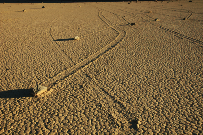
The Sliding Stones of Death Valley, California
The incidence of kidney stones in the United States has risen from 3.8% of the population in the 1970s to 8.8% of the population in the 2010s.2,3 Stones are three times more common in whites than in nonwhites and twice as common in men as in women.4 The cost of kidney stones in the US, including hospitalizations, surgery, and time lost from work, is calculated at $5 billion per year.5
Next page: History of treatment >>
Until the early 1980s, the treatment of choice for a kidney stone was “watchful waiting,” with hydration and pain management. A patient would be given a piece of cheesecloth or a basket, and instructed to urinate through it in order to “catch” the stone. When a stone finally passed, its chemical composition was analyzed. In patients with stones that were too large to pass or found in a location that made passage unlikely, surgical attempts were made to retrieve the stones. These surgeries could be open or “closed” (endoscopic), but they often caused permanent damage to the ureters and/or renal pelvis. Not surprisingly, the introduction of extracorporeal shock wave lithotripsy (ESWL) in the 1980s caused an immediate sensation.6
Stones can remain asymptomatic for some time—only to be found incidentally on radiologic exam for another condition.2 But when a patient presents with “classic” symptoms of kidney stones—colicky flank pain, hematuria, testicular pain (males only!), urinary frequency and urgency, nausea and vomiting—a helical CT is ordered to determine stone position; knowing this is vital to treatment. If the stone is non-obstructing and measures less than 10 mm, medical management is the first choice.7,8 This consists of IV or oral fluids, accompanied by narcotic and/or non-narcotic pain medications, as kidney stone pain can be excruciating. NSAIDs alone are rarely strong enough, and their use incurs a risk for intrinsic kidney damage.
If conservative care does not allow the stone to pass, alpha-adrenergic blockers and/or calcium channel blockers are added.9 In the case of cysteine stones, alkalization of the urine will help dissolve the stone.2 Only 20% of stones are found in the ureter; the vast majority (up to 70%) are lower urethral stones (LUS). Use of tamsulosin has been shown to move LUS stones at a faster rate, so long as they measure less than 10 mm.10
Before treating the stone patient with acute presentation, the urology practitioner may wait a couple of days to see whether the stone passes. The treatment choice then depends on the size of the stone and the position at presentation. If a stone measures less than 6 mm, medical management will be chosen.2 In fact, for smaller, nonobstructing stones, fluids, pain control, and alpha-blockers have been shown in the literature to produce a better outcome than other treatment options.9
For stones larger than 6 mm, or those causing an obstruction or a complication (pyelonephritis or urosepsis), removal is imperative.4 Modality choice depends on the position of the stone and the size of the patient. ESWL, the least invasive means, is the treatment of choice.2 However, as obesity becomes more prevalent (with its underlying metabolic abnormalities), the effectiveness of ESWL may be hindered by the obese patient’s body mass. That said, some manufacturers are increasing the reach of their lithotripsy machines for just this reason.11
Continue for more treatment options >>
Stenting, another option to allow stone fragments to pass, can be uncomfortable, and it requires anesthesia; however, its use is associated with minimal damage to the ureter.2 Percutaneous nephrolithotomy, with or without a basket or a nephrostomy tube, can also be used.12 This method is often needed in patients with a large “stone burden.”2 Open procedures to remove stones, though the gold standard in the early 1980s, are rarely required today.
Recurrence rates for stones can be as high as 50%.13 Depending on the type of stone, certain interventions are essential to reduce recurrence. The ROKS stone calculator can be used to identify patients at increased risk for stone recurrence.14
REFERENCES
1. Norris RD, Norris JM, Lorenz RD, et al. Sliding rocks on racetrack playa, Death Valley National Park: first observation of rocks in motion. PLoS One. 2014;9(8):e105948.
2. Curhan G. Nephrolithiasis. In: Gilbert SJ, Weiner DE, eds. National Kidney Foundation’s Primer on Kidney Diseases. 6th ed. Philadelphia, PA: Elsevier; 2013:405-411.
3. Trinchieri A. Epidemiology of urolithiasis: an update. Clin Cases Miner Bone Metab. 2008;5(2):101-106.
4. Worcester EM, Coe FL. Clinical practice: calcium kidney stones. N Engl J Med. 2010;363(10):954-963.
5. Saigal CS, Joyce G, Timilsina AR; Urologic Diseases in America Project. Direct and indirect costs of nephrolithiasis in an employed population: opportunity for disease management? Kidney Int. 2005;68(4):1808-1814.
6. Segura JW, Patterson DE, LeRoy AJ, et al. Percutaneous removal of kidney stones: review of 1,000 cases. J Urol. 1985;134(6):1077-1081.
7. Wells CG, Chandrashekar KB, Jyothirmayi GN, et al. Kidney stones: current diagnosis and management. Clinician Reviews. 2012;22(2):31-37.
8. Coll DM, Varanelli MJ, Smith RC. Relationship of spontaneous passage of ureteral calculi to stone size and location as revealed by unenhanced helical CT. AJR Am J Roentgenol. 2002;178(1):101-103.
9. Campschroer T, Zhu Y, Duijvesz D, et al. Alpha-blockers as medical expulsive therapy for ureteral stones. Cochrane Database Syst Rev. 2014;4:CD008509.
10. Erturhan S, Erbagci A, Yagci F, et al. Comparative evaluation of efficacy of use of tamsulosin and/or tolterodine for medical treatment of distal ureteral stones. Urology. 2007;69(4):633-636.
11. Mezentsev VA. Extracorporeal shock wave lithotripsy in the treatment of renal pelvicalyceal stones in morbidly obese patients. Int Braz J Urol. 2005;31(2):105-110.
12. Amer T, Ahmed K, Bultitude M, et al. Standard versus tubeless percutaneous nephrolithotomy: a systematic review. Urol Int. 2012;88(4):373-82.
13. Ljunghall S. Incidence of upper urinary tract stones. Miner Electrolyte Metab. 1987;13(4):220-227.
14. Rule AD, Lieske JC, Li X, et al. The ROKS Nomogram for Predicting a Second Symptomatic Stone Episode. J Am Soc Nephrol. 2014 Aug 7. [Epub ahead of print]
Since the 1940s, scientists have been stymied by the cause of the “sliding stones” in California’s Death Valley. How, without human intervention, does a 700-lb boulder move from one place to another, leaving a distinctive, meandering trail? Finally, in August 2014, recorded pictures showed that the power of water, freezing and melting, actually moves these boulders.1 Those who treat kidney stones (nephrolithiasis) have always known the power of liquid to move “boulders.”
The Sliding Stones of Death Valley, California
The incidence of kidney stones in the United States has risen from 3.8% of the population in the 1970s to 8.8% of the population in the 2010s.2,3 Stones are three times more common in whites than in nonwhites and twice as common in men as in women.4 The cost of kidney stones in the US, including hospitalizations, surgery, and time lost from work, is calculated at $5 billion per year.5
Next page: History of treatment >>
Until the early 1980s, the treatment of choice for a kidney stone was “watchful waiting,” with hydration and pain management. A patient would be given a piece of cheesecloth or a basket, and instructed to urinate through it in order to “catch” the stone. When a stone finally passed, its chemical composition was analyzed. In patients with stones that were too large to pass or found in a location that made passage unlikely, surgical attempts were made to retrieve the stones. These surgeries could be open or “closed” (endoscopic), but they often caused permanent damage to the ureters and/or renal pelvis. Not surprisingly, the introduction of extracorporeal shock wave lithotripsy (ESWL) in the 1980s caused an immediate sensation.6
Stones can remain asymptomatic for some time—only to be found incidentally on radiologic exam for another condition.2 But when a patient presents with “classic” symptoms of kidney stones—colicky flank pain, hematuria, testicular pain (males only!), urinary frequency and urgency, nausea and vomiting—a helical CT is ordered to determine stone position; knowing this is vital to treatment. If the stone is non-obstructing and measures less than 10 mm, medical management is the first choice.7,8 This consists of IV or oral fluids, accompanied by narcotic and/or non-narcotic pain medications, as kidney stone pain can be excruciating. NSAIDs alone are rarely strong enough, and their use incurs a risk for intrinsic kidney damage.
If conservative care does not allow the stone to pass, alpha-adrenergic blockers and/or calcium channel blockers are added.9 In the case of cysteine stones, alkalization of the urine will help dissolve the stone.2 Only 20% of stones are found in the ureter; the vast majority (up to 70%) are lower urethral stones (LUS). Use of tamsulosin has been shown to move LUS stones at a faster rate, so long as they measure less than 10 mm.10
Before treating the stone patient with acute presentation, the urology practitioner may wait a couple of days to see whether the stone passes. The treatment choice then depends on the size of the stone and the position at presentation. If a stone measures less than 6 mm, medical management will be chosen.2 In fact, for smaller, nonobstructing stones, fluids, pain control, and alpha-blockers have been shown in the literature to produce a better outcome than other treatment options.9
For stones larger than 6 mm, or those causing an obstruction or a complication (pyelonephritis or urosepsis), removal is imperative.4 Modality choice depends on the position of the stone and the size of the patient. ESWL, the least invasive means, is the treatment of choice.2 However, as obesity becomes more prevalent (with its underlying metabolic abnormalities), the effectiveness of ESWL may be hindered by the obese patient’s body mass. That said, some manufacturers are increasing the reach of their lithotripsy machines for just this reason.11
Continue for more treatment options >>
Stenting, another option to allow stone fragments to pass, can be uncomfortable, and it requires anesthesia; however, its use is associated with minimal damage to the ureter.2 Percutaneous nephrolithotomy, with or without a basket or a nephrostomy tube, can also be used.12 This method is often needed in patients with a large “stone burden.”2 Open procedures to remove stones, though the gold standard in the early 1980s, are rarely required today.
Recurrence rates for stones can be as high as 50%.13 Depending on the type of stone, certain interventions are essential to reduce recurrence. The ROKS stone calculator can be used to identify patients at increased risk for stone recurrence.14
REFERENCES
1. Norris RD, Norris JM, Lorenz RD, et al. Sliding rocks on racetrack playa, Death Valley National Park: first observation of rocks in motion. PLoS One. 2014;9(8):e105948.
2. Curhan G. Nephrolithiasis. In: Gilbert SJ, Weiner DE, eds. National Kidney Foundation’s Primer on Kidney Diseases. 6th ed. Philadelphia, PA: Elsevier; 2013:405-411.
3. Trinchieri A. Epidemiology of urolithiasis: an update. Clin Cases Miner Bone Metab. 2008;5(2):101-106.
4. Worcester EM, Coe FL. Clinical practice: calcium kidney stones. N Engl J Med. 2010;363(10):954-963.
5. Saigal CS, Joyce G, Timilsina AR; Urologic Diseases in America Project. Direct and indirect costs of nephrolithiasis in an employed population: opportunity for disease management? Kidney Int. 2005;68(4):1808-1814.
6. Segura JW, Patterson DE, LeRoy AJ, et al. Percutaneous removal of kidney stones: review of 1,000 cases. J Urol. 1985;134(6):1077-1081.
7. Wells CG, Chandrashekar KB, Jyothirmayi GN, et al. Kidney stones: current diagnosis and management. Clinician Reviews. 2012;22(2):31-37.
8. Coll DM, Varanelli MJ, Smith RC. Relationship of spontaneous passage of ureteral calculi to stone size and location as revealed by unenhanced helical CT. AJR Am J Roentgenol. 2002;178(1):101-103.
9. Campschroer T, Zhu Y, Duijvesz D, et al. Alpha-blockers as medical expulsive therapy for ureteral stones. Cochrane Database Syst Rev. 2014;4:CD008509.
10. Erturhan S, Erbagci A, Yagci F, et al. Comparative evaluation of efficacy of use of tamsulosin and/or tolterodine for medical treatment of distal ureteral stones. Urology. 2007;69(4):633-636.
11. Mezentsev VA. Extracorporeal shock wave lithotripsy in the treatment of renal pelvicalyceal stones in morbidly obese patients. Int Braz J Urol. 2005;31(2):105-110.
12. Amer T, Ahmed K, Bultitude M, et al. Standard versus tubeless percutaneous nephrolithotomy: a systematic review. Urol Int. 2012;88(4):373-82.
13. Ljunghall S. Incidence of upper urinary tract stones. Miner Electrolyte Metab. 1987;13(4):220-227.
14. Rule AD, Lieske JC, Li X, et al. The ROKS Nomogram for Predicting a Second Symptomatic Stone Episode. J Am Soc Nephrol. 2014 Aug 7. [Epub ahead of print]
