User login
The adage, “the sun should never set on an untapped pleural effusion,” was instilled in physicians for generations. However, anyone who practices medicine currently knows the sun often rises and sets several times before a pleural effusion is tapped. Why the change in mindset? Since the American Board of Internal Medicine removed the requirement for internal medicine residents to perform a minimum number of bedside procedures for certification, fewer graduating residents feel comfortable performing thoracentesis.
Additionally, the fear of litigation and institutional persecution from a postprocedure complication has caused many frontline clinicians to shy away from performing thoracentesis. Most important, we now appreciate that not all pleural effusions need to be tapped immediately, and the clinical decision making about the timing and technique to drain a pleural effusion is more complex than previously thought.
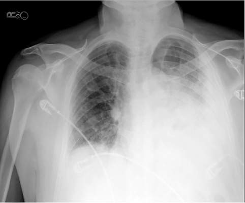
In recent years, the availability of portable ultrasound for bedside diagnostics and procedural guidance has revolutionized the practice of medicine, including the management of pleural effusions. When confronted with an obscured lower lobe on chest radiograph (Figure, left), clinicians were previously relegated to primitive bedside maneuvers, such as percussion or auscultation, to make critical decisions about the clinical management. Now, clinicians are able to look inside the body with point-of-care ultrasound and visually assess a pleural effusion before making any decisions. Point-of-care ultrasound has shifted the paradigm in the management of pleural effusions in several ways.
Ultrasound allows rapid detection and differentiation of pleural effusions from other pathologic findings.
Chest radiographs cannot accurately differentiate a pleural effusion from other common conditions, such as pneumonia, atelectasis, or an elevated hemidiaphragm. Ultrasound is the only bedside diagnostic modality that can rapidly differentiate these conditions within seconds and may reveal unsuspected findings, such as a mass or pericardial effusion.
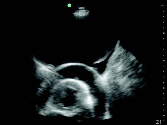
For example, the pleural ultrasound exam of a patient in the confirmed the presence of a large, left-sided pleural effusion (Figure, right) but also revealed an unsuspected large pericardial effusion (asterisk) that was causing hemodynamic compromise. The management of this patient shifted focus from the pleural effusion to the pericardial effusion, and urgent pericardiocentesis was performed. The sensitivity of ultrasound to detect a pleural effusion is proportional to the volume of fluid, reaching 100% with as little as 100 mL of fluid (Kalokairinou-Motogna et al. Med Ultra. 2010;12[1]:12). The diagnostic accuracy of ultrasound for detection of pleural effusions is comparable to CT scans of the chest and superior to portable chest radiographs (Lichtenstein et al. Anesthesiology. 2004;100[1]:9).
Ultrasound characterizes pleural effusions to determine the most appropriate management strategy.
Any clinician with basic ultrasonography skills can learn to evaluate pleural effusions and categorize them as simple or complex based on the sonographic appearance. Visualization of fibrinous stranding or loculations increases the probability of pleural fluid being exudative and often drives the decision to drain the fluid. The density and distribution of loculations can guide decisions about the most appropriate type of drainage procedure – thoracentesis versus tube thoracostomy versus surgical intervention. Use of color flow Doppler ultrasound allows clinicians to assess whether or not pleural fluid is free flowing and amenable to drainage, potentially saving the patient from an unnecessary attempt at drainage.
Ultrasound affords frontline clinicians the ability to streamline consultation with the most appropriate specialist based on the type of drainage procedure indicated and potentially prevent duplicate procedures on the same patient from different specialists.
Ultrasound reduces the risk of postprocedure complications from thoracentesis.
The risk of postthoracentesis pneumothorax was reported to be as high as 20%-39% before the routine use of point-of-care ultrasound (Grogan et al. Arch Int Med. 1990;150:873). Ultrasound guidance has been shown to increase procedural success rates and decrease the risk of postprocedure pneumothorax (2.7%), cost of hospitalization, and length of stay (Mercaldi et al. Chest. 2015;143[2]:532).
Regardless of the chest radiograph or CT scan findings, if the ultrasound exam reveals a scant volume of pleural fluid, or densely loculated pleural fluid, clinicians can avoid unnecessary attempts at bedside drainage, which likely partly accounts for the reduction in postprocedure pneumothorax. Use of ultrasound for needle site selection may prevent up to 10% of potential accidental organ punctures and increases accurate site selection by 26%, compared with chest radiograph and physical examination findings combined (Diacon et al. Chest. 2003;123:436).
Ultrasound facilitates patient-centered care.
Point-of-care ultrasound is the only new technology that has taken clinicians back to the bedside to spend more time with patients. Clinicians can simultaneously perform an ultrasound exam and converse with patients to gather a medical history. The ultrasound image serves as a tool to help patients understand their condition and facilitates shared decision making with clinicians at the bedside.
As more specialties have gained expertise in thoracic ultrasonography, the use of ultrasound guidance for thoracentesis has evolved to become the standard of care in many hospitals in the United States. Besides pulmonary specialists, several acute care specialists, including hospitalists, intensivists, and emergency medicine physicians, are routinely using point-of-care ultrasound to guide diagnostic decision making and procedures. Over the past 10 years, nearly a dozen procedure services led by internal medicine-trained hospitalists have been created at academic institutions that are routinely performing ultrasound-guided thoracenteses with low complication rates (Franco-Sadud et al. SGIM Forum. 2016;39[5]:13). More important, ultrasound is being used on the front lines to expeditiously evaluate pleural effusions and perform a diagnostic thoracentesis or consult with the appropriate subspecialist. Even though demonstration of competency in bedside procedures is no longer required for board certification in internal medicine, many internal medicine residency programs have incorporated diagnostic and procedural point-of-care ultrasound training into their education curriculum (Schnobrich et al. JGME. 2013;5[3]:498). Further, approximately 62% of medical schools report integrating ultrasound education in their medical student curriculum, and in coming years, most medical students will likely graduate with a basic skill set in point-of-care ultrasonography (Bahner et al. Academic Med. 2014;89[12]:1681). As point-of-care ultrasound education becomes integrated in training of physicians and other health-care providers, use of ultrasound to guide management of pleural effusions could become universally practiced and accepted as the new standard of care. Thus, it is plausible that a day will come in the near future when the sun will not set on an “un-ultrasound-ed” pleural effusion.
Dr. Franco-Sadud is with the section of hospital medicine/division of general internal medicine, Medical College of Wisconsin, Milwaukee, Wisconsin; Dr. Soni is with the section of hospital medicine and the section of pulmonary and critical care medicine, South Texas Veterans Health Care System and University of Texas Health Science Center, San Antonio.
The adage, “the sun should never set on an untapped pleural effusion,” was instilled in physicians for generations. However, anyone who practices medicine currently knows the sun often rises and sets several times before a pleural effusion is tapped. Why the change in mindset? Since the American Board of Internal Medicine removed the requirement for internal medicine residents to perform a minimum number of bedside procedures for certification, fewer graduating residents feel comfortable performing thoracentesis.
Additionally, the fear of litigation and institutional persecution from a postprocedure complication has caused many frontline clinicians to shy away from performing thoracentesis. Most important, we now appreciate that not all pleural effusions need to be tapped immediately, and the clinical decision making about the timing and technique to drain a pleural effusion is more complex than previously thought.
In recent years, the availability of portable ultrasound for bedside diagnostics and procedural guidance has revolutionized the practice of medicine, including the management of pleural effusions. When confronted with an obscured lower lobe on chest radiograph (Figure, left), clinicians were previously relegated to primitive bedside maneuvers, such as percussion or auscultation, to make critical decisions about the clinical management. Now, clinicians are able to look inside the body with point-of-care ultrasound and visually assess a pleural effusion before making any decisions. Point-of-care ultrasound has shifted the paradigm in the management of pleural effusions in several ways.
Ultrasound allows rapid detection and differentiation of pleural effusions from other pathologic findings.
Chest radiographs cannot accurately differentiate a pleural effusion from other common conditions, such as pneumonia, atelectasis, or an elevated hemidiaphragm. Ultrasound is the only bedside diagnostic modality that can rapidly differentiate these conditions within seconds and may reveal unsuspected findings, such as a mass or pericardial effusion.
For example, the pleural ultrasound exam of a patient in the confirmed the presence of a large, left-sided pleural effusion (Figure, right) but also revealed an unsuspected large pericardial effusion (asterisk) that was causing hemodynamic compromise. The management of this patient shifted focus from the pleural effusion to the pericardial effusion, and urgent pericardiocentesis was performed. The sensitivity of ultrasound to detect a pleural effusion is proportional to the volume of fluid, reaching 100% with as little as 100 mL of fluid (Kalokairinou-Motogna et al. Med Ultra. 2010;12[1]:12). The diagnostic accuracy of ultrasound for detection of pleural effusions is comparable to CT scans of the chest and superior to portable chest radiographs (Lichtenstein et al. Anesthesiology. 2004;100[1]:9).
Ultrasound characterizes pleural effusions to determine the most appropriate management strategy.
Any clinician with basic ultrasonography skills can learn to evaluate pleural effusions and categorize them as simple or complex based on the sonographic appearance. Visualization of fibrinous stranding or loculations increases the probability of pleural fluid being exudative and often drives the decision to drain the fluid. The density and distribution of loculations can guide decisions about the most appropriate type of drainage procedure – thoracentesis versus tube thoracostomy versus surgical intervention. Use of color flow Doppler ultrasound allows clinicians to assess whether or not pleural fluid is free flowing and amenable to drainage, potentially saving the patient from an unnecessary attempt at drainage.
Ultrasound affords frontline clinicians the ability to streamline consultation with the most appropriate specialist based on the type of drainage procedure indicated and potentially prevent duplicate procedures on the same patient from different specialists.
Ultrasound reduces the risk of postprocedure complications from thoracentesis.
The risk of postthoracentesis pneumothorax was reported to be as high as 20%-39% before the routine use of point-of-care ultrasound (Grogan et al. Arch Int Med. 1990;150:873). Ultrasound guidance has been shown to increase procedural success rates and decrease the risk of postprocedure pneumothorax (2.7%), cost of hospitalization, and length of stay (Mercaldi et al. Chest. 2015;143[2]:532).
Regardless of the chest radiograph or CT scan findings, if the ultrasound exam reveals a scant volume of pleural fluid, or densely loculated pleural fluid, clinicians can avoid unnecessary attempts at bedside drainage, which likely partly accounts for the reduction in postprocedure pneumothorax. Use of ultrasound for needle site selection may prevent up to 10% of potential accidental organ punctures and increases accurate site selection by 26%, compared with chest radiograph and physical examination findings combined (Diacon et al. Chest. 2003;123:436).
Ultrasound facilitates patient-centered care.
Point-of-care ultrasound is the only new technology that has taken clinicians back to the bedside to spend more time with patients. Clinicians can simultaneously perform an ultrasound exam and converse with patients to gather a medical history. The ultrasound image serves as a tool to help patients understand their condition and facilitates shared decision making with clinicians at the bedside.
As more specialties have gained expertise in thoracic ultrasonography, the use of ultrasound guidance for thoracentesis has evolved to become the standard of care in many hospitals in the United States. Besides pulmonary specialists, several acute care specialists, including hospitalists, intensivists, and emergency medicine physicians, are routinely using point-of-care ultrasound to guide diagnostic decision making and procedures. Over the past 10 years, nearly a dozen procedure services led by internal medicine-trained hospitalists have been created at academic institutions that are routinely performing ultrasound-guided thoracenteses with low complication rates (Franco-Sadud et al. SGIM Forum. 2016;39[5]:13). More important, ultrasound is being used on the front lines to expeditiously evaluate pleural effusions and perform a diagnostic thoracentesis or consult with the appropriate subspecialist. Even though demonstration of competency in bedside procedures is no longer required for board certification in internal medicine, many internal medicine residency programs have incorporated diagnostic and procedural point-of-care ultrasound training into their education curriculum (Schnobrich et al. JGME. 2013;5[3]:498). Further, approximately 62% of medical schools report integrating ultrasound education in their medical student curriculum, and in coming years, most medical students will likely graduate with a basic skill set in point-of-care ultrasonography (Bahner et al. Academic Med. 2014;89[12]:1681). As point-of-care ultrasound education becomes integrated in training of physicians and other health-care providers, use of ultrasound to guide management of pleural effusions could become universally practiced and accepted as the new standard of care. Thus, it is plausible that a day will come in the near future when the sun will not set on an “un-ultrasound-ed” pleural effusion.
Dr. Franco-Sadud is with the section of hospital medicine/division of general internal medicine, Medical College of Wisconsin, Milwaukee, Wisconsin; Dr. Soni is with the section of hospital medicine and the section of pulmonary and critical care medicine, South Texas Veterans Health Care System and University of Texas Health Science Center, San Antonio.
