Screening is the testing of an individual who is at risk for a disease, but who does not exhibit signs or symptoms of the disease. The goal of screening is to detect disease at a stage when cure or control is possible, and an effective screening program should reduce the number of disease-specific deaths in the screened population. Screening should focus on diseases that are associated with potentially serious consequences and that are detectable in the preclinical phase, yet it should avoid identifying “pseudodisease” (ie, positive test findings that would not be expected to affect the patient’s health) or causing morbidity due to the test procedure itself.1 Finally, screening is only worthwhile when treatment of the disease is more effective when administered early.
Since lung cancer screening began in the 1950s,2,3 many studies have attempted to define the medical benefits and economic impact of widespread screening. Many important unresolved issues remain, including the effectiveness of lung cancer screening for reducing disease-specific mortality, the potential harms of screening, its cost-effectiveness, and the potential impact of new research methods on the early identification of lung cancer.
DOES LUNG CANCER SCREENING REDUCE DISEASE-SPECIFIC MORTALITY?
Early studies examined the usefulness of large-scale chest radiograph programs, either with or without sputum cytology, for lung cancer screening. Although several studies reported that radiographic screening identified patients with early lung cancer and reported higher survival rates, reviews and meta-analyses of these reports concluded that screening did not significantly reduce disease-specific mortality.4,5
The utility of chest radiography for the detection of early lung cancer is limited by several factors, including poor sensitivity for the detection of small or subtle nodules and a relatively high false-positive rate.6–8 More recently, several cohort studies and randomized, controlled trials have shown that computed tomography (CT) screening is effective for the identification of early lung cancer in high-risk patients (eg, individuals with chronic, heavy tobacco use or asbestos exposure).9–11 A recent meta-analysis concluded that CT-based screening significantly increases the number of early lung cancers identified, but also increases the number of false-positive findings (nodules) and unnecessary thoracotomies for benign lesions.12
Lung cancer screening should increase the number of patients identified at early disease stages. Treatment of early-stage lung cancer should decrease the number of patients identified with late-stage cancer, resulting in a stage shift toward earlier disease for the population as a whole. Although lung cancer screening cohort studies and randomized, controlled trials have demonstrated that screening increases the number of early-stage lung cancer cases identified, these studies have generally not demonstrated decreased rates of late-stage lung cancers or stage shifting in the populations studied. In the 1970s, the National Cancer Institute began three large-scale screening trials at Mayo Clinic, Memorial Sloan-Kettering Cancer Institute, and The Johns Hopkins University, each enrolling approximately 10,000 patients. In the Mayo trial, the incidence of advanced-stage tumors was nearly identical for the screened versus unscreened patients, with 303 cancer cases detected in the screened group versus 304 cases in the control group.13 CT-based cohort studies have also reported increased rates of early recognition of lung cancer and accompanying large increases in the number of diagnostic procedures performed. However, early controlled trials of CT screening showed no differences between screened and unscreened groups in the numbers of patients with late-stage tumors or deaths due to lung cancer.14
Results such as these have led some researchers to argue that survival benefits of screening largely reflect observational biases. For example:
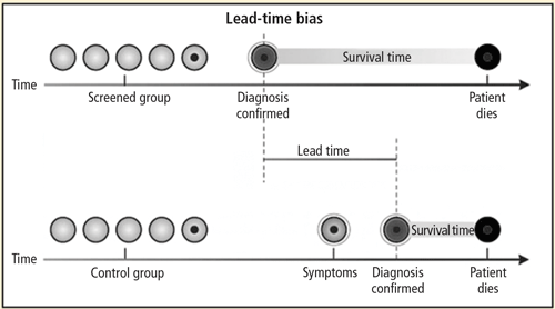
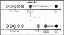
Figure 1. Lead-time bias. Patients identified by screening may live longer with disease than patients diagnosed clinically, although overall survival time is not improved.Lead-time bias occurs when screening results in earlier recognition of disease, but does not change the patient’s eventual lifespan, creating the appearance that the patient’s survival time with the disease is longer (Figure 1).15 Longer lead times should be observed in a successful screening program even if eventual mortality remains exactly the same, and lead time bias is therefore an expected outcome of screening.
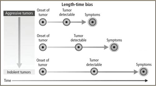
Figure 2. Length-time bias. Indolent tumors move more gradually from the detectable stage to the onset of symptoms. These tumors are therefore more likely to be identified by intermittent screening.Length-time bias arises from the observation that any screening test that is applied intermittently is more likely to detect indolent tumors than aggressive, fast-growing tumors that would result in clinical symptoms(Figure 2).15 Indolent tumors move more gradually from the detectable state to the onset of clinical symptoms, and are therefore especially likely to be identified by screening.
Overdiagnosis bias occurs when a screening test identifies disease that never would have affected the patient’s life in the absence of screening. This type of bias might occur if screening identifies a lesion that is so indolent that it would never cause clinical disease, or if the population is otherwise in such poor health that successfully screened patients would die from other causes.
There is no question that these biases affect reports of survival in lung cancer screening, although it is unclear whether they explain the reported benefit of screening observed in cohort studies. Screening advocates have argued that the failure to screen high-risk patients for lung cancer has the potential for significant harm. In contrast, opponents of screening have argued that there was a lack of data showing a reduction in the number of patients diagnosed with late-stage cancers or in cancer-related mortality.
IS LUNG CANCER OVERDIAGNOSED IN SCREENED POPULATIONS?
Although the apparent benefit of lung cancer screening is susceptible to different sources of bias, overdiagnosis has received the greatest attention on the basis of both theoretical concerns and observations from screening studies. Estimates of lung cancer growth suggest that a typical 10-cm tumor, which is usually large enough to be fatal, has progressed through approximately 40 volume doublings during the course of its existence. In contrast, a more survivable—and clinically detectable—1-cm tumor has progressed through approximately 30 volume doublings.16,17 A lung tumor therefore spends most of its existence relatively undetectable. It has been estimated that the median doubling time is approximately 181 days, and that 22% of lung cancers have doubling times more than 465 days.18 The appearance of tumors on CT may suggest the growth rate, with 1 study showing that solid malignant nodules had a mean doubling time of 149 days, compared with 457 days for partial ground-glass–opacity nodules, and 813 days for pure ground-glass nodules.19
These estimates suggest that if a 1-cm tumor with a history of 30 volume doublings continues to grow at a typical rate (ie, a 181-day doubling time), the patient will die of cancer within 5 years. If the tumor is among the 22% of those with a 465-day doubling time, the survival time would be 12.7 years. For malignant pure ground-glass nodules, the projected time to death is 22 years. Individuals with lung cancer are often elderly, long-term cigarette smokers with emphysema or other chronic health problems—many of whom would die of other causes before their lung cancers progressed enough to cause significant health problems.
Reprinted with permission from the American College of Chest Physicians (Raz DJ, et al. Natural history of stage I non-small cell lung cancer: implications for early detection. Chest 2007; 132:193–199).
Figure 3. Survival is worse in untreated than in treated non–small cell lung cancer patients, arguing against overdiagnosis bias. Blue line: patients receiving surgery; green line: untreated patients who refused surgery.
As an argument against the significance of overdiagnosis in lung cancer screening, it has been noted that outcomes are worse for patients identified with early-stage lung cancer in screening studies who do not receive treatment. For example, the results of a study of 1,432 patients with stage I non–small cell lung cancer (NSCLC) are illustrated in Figure 3. Survival was much better in screened patients who were treated than in those who were untreated, with almost all the untreated patients dying within 10 years of diagnosis.20 However, the subjects in this study were atypical of those in most screening studies. Thirty-three percent of the patients had squamous cell carcinoma and 61% had relatively large T2 lesions, compared with a typical screening study comprised of patients with more than 50% T1 lesions and a smaller percentage of squamous cell carcinoma.
Another argument against overdiagnosis comes from gene profiling studies that have compared genetic tumor markers for tumors identified by screening with tumors identified clinically. One study found that the expression profile of 3,231 genes was similar for patients with lung cancer identified by screening or by symptoms.21 However, these investigators also found that nine genes known to be important in tumor growth differed between screened and nonscreened populations.
The significance of overdiagnosis is supported by a long-term follow-up study from the Mayo Clinic chest radiography screening trial, which found that the number of lung cancer cases remained higher in the screening group than the control group (585 vs 500 cases) for up to 28 years after screening, suggesting an overdiagnosis of lung cancer by approximately 85 cases per 500 patients screened (approximately 17%).22 Several studies have also demonstrated that screening populations may have tumors with more favorable histology or clinical characteristics, including higher levels of bronchioloalveolar carcinoma or well-differentiated adenocarcinoma.23–25 Finally, autopsy series have found undiagnosed lung tumors in as many as 1% of patients who died from natural causes, with fewer advanced tumors found in the 1970s than in the 1950s.26,27
These arguments led most to believe randomized controlled trials of CT-based screening were needed. The largest of these, the National Lung Screening Trial (NLST), has recently reported results that will clarify the impact of lung cancer screening on cancer-related mortality.28 This study enrolled 53,456 subjects between the ages of 55 and 74 years with a history of at least 30 pack-years of smoking. Patients were randomized to baseline screening followed by annual screening for 2 years using either low-dose helical CT or chest radiography and outcome follow-up 5 years after randomization. Data analysis after 6 to 8 years of follow-up found 442 lung cancer deaths in the chest radiograph arm versus 354 in the CT arm, representing a 20.3% reduction with CT.29 Screening of 320 patients using low-dose helical CT would be required to avoid each lung cancer death. Thus, after years of debate, it has been demonstrated that it is possible to reduce lung cancer-specific mortality with CT-based screening.
ARE THERE SIGNIFICANT RISKS WITH CT-BASED SCREENING?
Reprinted with kind permission from Springer Science+Business Media (Fischbach F, et al. Detection of pulmonary nodules by multislice computed tomography: improved detection rate with reduced slice thickness. Eur Radiol 2003; 13:2378–2383).
Figure 4. Benign lung nodules visualized on computed tomography.Lung cancer screening using chest CT may be associated with certain risks. The detailed high-resolution images produced by contemporary CT reveal small benign lung nodules in as many as 74% of patients(Figure 4).24,30 Although these nodules rarely represent a significant health problem, they require follow-up procedures and contribute to patient anxiety.31 In one study, every 1,000 individuals screened with CT imaging resulted in the identification of nine cases of stage I NSCLC, 235 false-positive nodules measuring at least 5 mm, and four thoracotomies for benign lesions.12
Radiation from CT tests is a potential concern, although it is difficult to quantify the importance of this risk. One estimate of CT-related radiation exposure found that annual CT screening of 50% of the eligible population between 50 and 75 years of age in the United States would result in approximately 36,000 new cancers, or a 1.8% increase in the rate of cancer over the expected rate.32 Many patients and health care professionals are already concerned about the degree of radiation exposure from medical diagnostics. A recent study that examined cumulative radiation exposure due to medical imaging in 952,420 adults aged 18 to 64 years found that approximately 57.9% of men and 78.7% of women receive at least some annual health care-related radiation exposure.33 Radiation exposure was considered moderate (> 3–20 mSv/yr) for 18.1% of men and 20.3% of women, and was considered high (> 20–50 mSv/yr) or very high (>50 mSv/hr) for 2.3% of men and 2.1% of women.
IS SCREENING COST-EFFECTIVE?
It is difficult to calculate the cost-effectiveness of CT screening because the impact of screening on mortality and the economic implications of false-positive findings are not well understood. A cost-effectiveness analysis of helical CT screening assumed that screening would result in a 50% stage shift and a 13% reduction in mortality.34 Under these assumptions, the cost-effectiveness was greater among current smokers ($116,300 per quality-adjusted life year saved by screening) than among currently quitting smokers ($558,600) or former smokers ($2,322,700). These investigators concluded that lung cancer screening is unlikely to be cost-effective, especially among those with the lowest levels of current tobacco exposure (quitting or former smokers).
Larger stage shifts or reductions in mortality would be expected to translate into greater cost-effectiveness, although the real-world effects of screening on these parameters are uncertain. Data from a US nationwide survey suggested that only about one-half of all current smokers would opt for surgery following a positive screening result, which might significantly decrease the cost-effectiveness of treatment.35
It is unclear how well the methods used in screening studies such as the NLST would translate to actual clinical practice at a national level, or how the health care system would manage the many small lung nodules that would be identified using this approach.
HOW WILL FUTURE DEVELOPMENTS AFFECT LUNG CANCER SCREENING?
Ongoing studies will continue to refine our understanding of the impact of lung cancer screening. For example, the randomized Prostate, Lung, Colorectal, and Ovarian Screening Trial is examining chest radiograph screening versus control in both smokers and never-smokers between 55 and 74 years of age.36 It is anticipated that this study will provide important information about how well chest radiographs perform for the identification of lung cancer in high- and lower-risk populations. Large randomized trials in Europe are comparing CT with no imaging for lung cancer screening.37 Efforts to better characterize specific patient populations who are at the greatest risk of lung cancer may help to improve the efficiency and cost-effectiveness of screening. Advances in molecular testing may help to identify molecular and genetic tumor biomarkers that herald increased lung cancer risk and greater need for screening. More research is needed to better understand the optimal management of patients with small lung nodules on screening tests. Professional societies are poised to publish revised screening recommendations as data from the NLST become available. Finally, insurers will need to evaluate the evidence and develop reimbursement policies.
SUMMARY AND CONCLUSIONS
Lung cancer screening efforts conducted over the last several decades have shown that it is possible to identify early lung cancer in high-risk patient populations. However, demonstrating a clear improvement in cancer-related mortality has been more difficult. Biases inherent to noncontrolled trials of screening may explain some of the beneficial effects on survival observed in some studies. Recent results from the NLST have for the first time demonstrated a significant reduction in lung cancer mortality in high-risk patients screened for lung cancer with chest CT, although there are continuing concerns about the cost of screening, the risks from radiation exposure, and the additional testing resulting from the identification of small benign lung nodules. Ongoing research will help to maximize the benefit of lung cancer screening and minimize the related risks.
References
Holin SM, Dwork RE, Glaser S, Rikli AE, Stocklen JB. Solitary pulmonary nodules found in a community-wide chest roentgenographic survey: a five-year follow-up study. Am Rev Tuberc1959; 79:427–439.
Nash FA, Morgan JM, Tomkins JG. South London Lung Cancer Study. Br Med J1968; 2:715–721.
Obuchowski NA, Graham RJ, Baker ME, Powell KA. Ten criteria for effective screening: their application to multislice CT screening for pulmonary and colorectal cancers. AJR Am J Roentgenol2001; 176:1357–1362.
Eddy DM. Screening for lung cancer. Ann Intern Med1989; 111:232–237.
Manser RL, Irving LB, Byrnes G, Abramson MJ, Stone CA, Campbell DA. Screening for lung cancer: a systematic review and meta-analysis of controlled trials. Thorax2003; 58:784–789.
Krupinski EA, Berger WG, Dallas WJ, Roehrig H. Searching for nodules: what features attract attention and influence detection?Acad Radiol2003; 10:861–868.
Yoshida H. Local contralateral subtraction based on bilateral symmetry of lung for reduction of false positives in computerized detection of pulmonary nodules. IEEE Trans Biomed Eng2004; 51:778–789.
Shiraishi J, Abe H.Engelmann R, Doi K. Effect of high sensitivity in a computerized scheme for detecting extremely subtle solitary pulmonary nodules in chest radiographs: observer performance study. Acad Radiol2003; 10:1302–1311.
Veronesi G, Bellomi M, Scanagatta P, et al. Difficulties encountered managing nodules detected during a computed tomography lung cancer screening program. J Thorac Cardiovasc Surg2008; 136:611–617.
Wilson DO, Weissfeld JL, Fuhrman CR, et al. The Pittsburgh Lung Screening Study (PLuSS): outcomes within 3 years of a first computed tomography scan [published online ahead of print July 17, 2008]. Am J Respir Crit Care Med2008; 178:956–961. doi: 10.1164/rccm.200802-336OC
Fasola G, Belvedere O, Aita M, et al. Low-dose computed tomography screening for lung cancer and pleural mesothelioma in an asbestos-exposed population: baseline results of a prospective, nonrandomized feasibility trial—an Alpe-adria Thoracic Oncology Multidisciplinary Group Study (ATOM 002). Oncologist2007; 12:1215–1224.
Gopal M, Abdullah SE, Grady JJ, Goodwin JS. Screening for lung cancer with low-dose computed tomography: a systematic review and meta-analysis of the baseline findings of randomized controlled trials. J Thorac Oncol2010; 5:1233–1239.
Fontana RS, Sanderson DR, Woolner LB, et al. Screening for lung cancer: a critique of the Mayo Lung Project. Cancer1991; 67( suppl 4):1155–1164.
Bach PB, Jett JR, Pastorino U, Tockman MS, Swensen SJ, Begg CB. Computed tomography screening and lung cancer outcomes. JAMA2007; 297:953–961.
Patz EF, Goodman PC, Bepler G. Screening for lung cancer. N Engl J Med2000; 343:1627–1633.
Weiss W. Implications of tumor growth rate for the natural history of lung cancer. J Occup Med1984; 26:345–352.
Reich JM. A critical appraisal of overdiagnosis: estimates of its magnitude and implications for lung cancer screening. Thorax2008; 63:377–383.
Winer-Muram HT, Jennings SG, Tarver RD, et al. Volumetric growth rate of stage I lung cancer prior to treatment: serial CT scanning. Radiology2002; 223:798–805.
Hasegawa M, Sone S, Takashima S, et al. Growth rate of small lung cancers detected on mass CT screening. Br J Radiol2000; 73:1252–1259.
Raz DJ, Zell JA, Ou SH, Gandara DR, Anton-Culver H, Jablons DM. Natural history of stage I non-small cell lung cancer: implications for early detection [published online ahead of print May 15, 2007]. Chest2007; 132:193–199. doi: 10.1378/chest.06-3096
Bianchi F, Hu J, Pelosi G, et al. Lung cancers detected by screening with spiral computed tomography have a malignant phenotype when analyzed by cDNA microarray. Clin Cancer Res2004; 10( 18 Pt 1):6023–6028.
Marcus PM, Bergstralh EJ, Zweig MH, Harris A, Offord KP, Fontana RS. Extended lung cancer incidence follow-up in the Mayo Lung Project and overdiagnosis. J Natl Cancer Inst2006; 98:748–756.
Sone S, Li F, Yang ZG, et al. Results of three-year mass screening programme for lung cancer using mobile low-dose spiral computed tomography scanner. Br J Cancer2001; 84:25–32.
Swensen SJ, Jett JR, Hartman TE, et al. CT screening for lung cancer: five-year prospective experience [published online ahead of print February 4, 2005]. Radiology2005; 235:259–265. doi: 10.1148/radiol.2351041662
International Early Lung Cancer Action Program Investigators, Henschke CI, Yankelevitz DF, Libby DM, et al. Survival of patients with stage I lung cancer detected on CT screening. N Engl J Med2006; 355:1763–1771.
Manser RL, Dodd M, Byrnes G, Irving LB, Campbell DA. Incidental lung cancers identified at coronial autopsy: implications for overdiagnosis of lung cancer by screening. Respir Med2005; 99:501–507.
Chan CK, Wells CK, McFarlane MJ, Feinstein AR. More lung cancer but better survival: implications of secular trends in “necropsy surprise” rates. Chest1989; 96:291–296.
National Lung Screening Trial Research Team, Aberle DR, Adams AM, Berg CD, et al. Baseline characteristics of participants in the randomized national lung screening trial [published correction appears in J Natl Cancer Inst 2011; 103:1560]. J Natl Cancer Inst2010; 102:1771–1779.
Fischbach F, Knollmann F, Griesshaber V, Freund T, Akkol E, Felix R. Detection of pulmonary nodules by multislice computed tomography: improved detection rate with reduced slice thickness [published online ahead of print May 13, 2003]. Eur Radiol2003; 13:2378–2383. doi: 10.1007/s00330-003-1915-7
van den Bergh KA, Essink-Bot ML, Borsboom GJ, et al. Short-term health-related quality of life consequences in a lung cancer CT screening trial (NELSON) [published online ahead of pring November 24, 2009]. Br J Cancer2010; 102:27–34. doi: 10.1038/sj.bjc.6605459
Brenner DJ. Radiation risks potentially associated with low-dose CT screening of adult smokers for lung cancer. Radiology2004; 231:440–445.
Fazel R, Krumholz HM, Wang Y, et al. Exposure to low-dose ionizing radiation from medical imaging procedures. N Engl J Med2009; 361:849–857.
Mahadevia PJ, Fleisher LA, Frick KD, Eng J, Goodman SN, Powe NR. Lung cancer screening with helical computed tomography in older adult smokers: a decision and cost-effectiveness analysis. JAMA2003; 289:313–322.
Silvestri GA, Nietert PJ, Zoller J, Carter C, Bradford D. Attitudes towards screening for lung cancer among smokers and their nonsmoking counterparts. [published online ahead of print November 13, 2006]Thorax2007; 62:126–130. doi: 10.1136/thx.2005.056036
Tammemagi CM, Pinsky PF, Caporaso NE, et al. Lung cancer risk prediction: Prostate, Lung, Colorectal and Ovarian Cancer Screening Trial models and validation [published online ahead of print May 23, 2011]. J Natl Cancer Inst2011; 103:1058–1068. doi: 10.1093/jnci/djr173
van Klaveren RJ, Oudkerk M, Prokop M, et al. Management of lung nodules detected by volume CT scanning. N Engl J Med2009; 361:2221–2229.
Peter Mazzone, MD, MPH, FCCP Director of Education, Lung Cancer Program, and Pulmonary Rehabilitation Program; Respiratory Institute, Cleveland Clinic, Cleveland, OH
Correspondence: Peter Mazzone, MD, MPH, FCCP, Critical Care Medicine, Cleveland Clinic, 9500 Euclid Avenue, A90, Cleveland, OH 44195; mazzonp@ccf.org
Dr. Mazzone reported that he has been a member of advisory committees for Boehringer Ingelheim and Oncimmune. He has research supported by Metabolomx.
This article was developed from an audio transcript of Dr. Mazzone’s presentation at the “Advances in Lung Cancer Evaluation and Management” symposium held in Cleveland, Ohio, on April 30, 2011. The transcript was formatted and edited by Cleveland Clinic Journal of Medicine staff for clarity and conciseness and was then reviewed, revised, and approved by Dr. Mazzone.
Author and Disclosure Information
Peter Mazzone, MD, MPH, FCCP Director of Education, Lung Cancer Program, and Pulmonary Rehabilitation Program; Respiratory Institute, Cleveland Clinic, Cleveland, OH
Correspondence: Peter Mazzone, MD, MPH, FCCP, Critical Care Medicine, Cleveland Clinic, 9500 Euclid Avenue, A90, Cleveland, OH 44195; mazzonp@ccf.org
Dr. Mazzone reported that he has been a member of advisory committees for Boehringer Ingelheim and Oncimmune. He has research supported by Metabolomx.
This article was developed from an audio transcript of Dr. Mazzone’s presentation at the “Advances in Lung Cancer Evaluation and Management” symposium held in Cleveland, Ohio, on April 30, 2011. The transcript was formatted and edited by Cleveland Clinic Journal of Medicine staff for clarity and conciseness and was then reviewed, revised, and approved by Dr. Mazzone.
Screening is the testing of an individual who is at risk for a disease, but who does not exhibit signs or symptoms of the disease. The goal of screening is to detect disease at a stage when cure or control is possible, and an effective screening program should reduce the number of disease-specific deaths in the screened population. Screening should focus on diseases that are associated with potentially serious consequences and that are detectable in the preclinical phase, yet it should avoid identifying “pseudodisease” (ie, positive test findings that would not be expected to affect the patient’s health) or causing morbidity due to the test procedure itself.1 Finally, screening is only worthwhile when treatment of the disease is more effective when administered early.
Since lung cancer screening began in the 1950s,2,3 many studies have attempted to define the medical benefits and economic impact of widespread screening. Many important unresolved issues remain, including the effectiveness of lung cancer screening for reducing disease-specific mortality, the potential harms of screening, its cost-effectiveness, and the potential impact of new research methods on the early identification of lung cancer.
DOES LUNG CANCER SCREENING REDUCE DISEASE-SPECIFIC MORTALITY?
Early studies examined the usefulness of large-scale chest radiograph programs, either with or without sputum cytology, for lung cancer screening. Although several studies reported that radiographic screening identified patients with early lung cancer and reported higher survival rates, reviews and meta-analyses of these reports concluded that screening did not significantly reduce disease-specific mortality.4,5
The utility of chest radiography for the detection of early lung cancer is limited by several factors, including poor sensitivity for the detection of small or subtle nodules and a relatively high false-positive rate.6–8 More recently, several cohort studies and randomized, controlled trials have shown that computed tomography (CT) screening is effective for the identification of early lung cancer in high-risk patients (eg, individuals with chronic, heavy tobacco use or asbestos exposure).9–11 A recent meta-analysis concluded that CT-based screening significantly increases the number of early lung cancers identified, but also increases the number of false-positive findings (nodules) and unnecessary thoracotomies for benign lesions.12
Lung cancer screening should increase the number of patients identified at early disease stages. Treatment of early-stage lung cancer should decrease the number of patients identified with late-stage cancer, resulting in a stage shift toward earlier disease for the population as a whole. Although lung cancer screening cohort studies and randomized, controlled trials have demonstrated that screening increases the number of early-stage lung cancer cases identified, these studies have generally not demonstrated decreased rates of late-stage lung cancers or stage shifting in the populations studied. In the 1970s, the National Cancer Institute began three large-scale screening trials at Mayo Clinic, Memorial Sloan-Kettering Cancer Institute, and The Johns Hopkins University, each enrolling approximately 10,000 patients. In the Mayo trial, the incidence of advanced-stage tumors was nearly identical for the screened versus unscreened patients, with 303 cancer cases detected in the screened group versus 304 cases in the control group.13 CT-based cohort studies have also reported increased rates of early recognition of lung cancer and accompanying large increases in the number of diagnostic procedures performed. However, early controlled trials of CT screening showed no differences between screened and unscreened groups in the numbers of patients with late-stage tumors or deaths due to lung cancer.14
Results such as these have led some researchers to argue that survival benefits of screening largely reflect observational biases. For example:
Figure 1. Lead-time bias. Patients identified by screening may live longer with disease than patients diagnosed clinically, although overall survival time is not improved.Lead-time bias occurs when screening results in earlier recognition of disease, but does not change the patient’s eventual lifespan, creating the appearance that the patient’s survival time with the disease is longer (Figure 1).15 Longer lead times should be observed in a successful screening program even if eventual mortality remains exactly the same, and lead time bias is therefore an expected outcome of screening.
Figure 2. Length-time bias. Indolent tumors move more gradually from the detectable stage to the onset of symptoms. These tumors are therefore more likely to be identified by intermittent screening.Length-time bias arises from the observation that any screening test that is applied intermittently is more likely to detect indolent tumors than aggressive, fast-growing tumors that would result in clinical symptoms(Figure 2).15 Indolent tumors move more gradually from the detectable state to the onset of clinical symptoms, and are therefore especially likely to be identified by screening.
Overdiagnosis bias occurs when a screening test identifies disease that never would have affected the patient’s life in the absence of screening. This type of bias might occur if screening identifies a lesion that is so indolent that it would never cause clinical disease, or if the population is otherwise in such poor health that successfully screened patients would die from other causes.
There is no question that these biases affect reports of survival in lung cancer screening, although it is unclear whether they explain the reported benefit of screening observed in cohort studies. Screening advocates have argued that the failure to screen high-risk patients for lung cancer has the potential for significant harm. In contrast, opponents of screening have argued that there was a lack of data showing a reduction in the number of patients diagnosed with late-stage cancers or in cancer-related mortality.
IS LUNG CANCER OVERDIAGNOSED IN SCREENED POPULATIONS?
Although the apparent benefit of lung cancer screening is susceptible to different sources of bias, overdiagnosis has received the greatest attention on the basis of both theoretical concerns and observations from screening studies. Estimates of lung cancer growth suggest that a typical 10-cm tumor, which is usually large enough to be fatal, has progressed through approximately 40 volume doublings during the course of its existence. In contrast, a more survivable—and clinically detectable—1-cm tumor has progressed through approximately 30 volume doublings.16,17 A lung tumor therefore spends most of its existence relatively undetectable. It has been estimated that the median doubling time is approximately 181 days, and that 22% of lung cancers have doubling times more than 465 days.18 The appearance of tumors on CT may suggest the growth rate, with 1 study showing that solid malignant nodules had a mean doubling time of 149 days, compared with 457 days for partial ground-glass–opacity nodules, and 813 days for pure ground-glass nodules.19
These estimates suggest that if a 1-cm tumor with a history of 30 volume doublings continues to grow at a typical rate (ie, a 181-day doubling time), the patient will die of cancer within 5 years. If the tumor is among the 22% of those with a 465-day doubling time, the survival time would be 12.7 years. For malignant pure ground-glass nodules, the projected time to death is 22 years. Individuals with lung cancer are often elderly, long-term cigarette smokers with emphysema or other chronic health problems—many of whom would die of other causes before their lung cancers progressed enough to cause significant health problems.
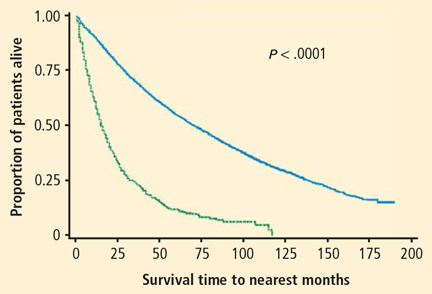
Reprinted with permission from the American College of Chest Physicians (Raz DJ, et al. Natural history of stage I non-small cell lung cancer: implications for early detection. Chest 2007; 132:193–199).
Figure 3. Survival is worse in untreated than in treated non–small cell lung cancer patients, arguing against overdiagnosis bias. Blue line: patients receiving surgery; green line: untreated patients who refused surgery.
As an argument against the significance of overdiagnosis in lung cancer screening, it has been noted that outcomes are worse for patients identified with early-stage lung cancer in screening studies who do not receive treatment. For example, the results of a study of 1,432 patients with stage I non–small cell lung cancer (NSCLC) are illustrated in Figure 3. Survival was much better in screened patients who were treated than in those who were untreated, with almost all the untreated patients dying within 10 years of diagnosis.20 However, the subjects in this study were atypical of those in most screening studies. Thirty-three percent of the patients had squamous cell carcinoma and 61% had relatively large T2 lesions, compared with a typical screening study comprised of patients with more than 50% T1 lesions and a smaller percentage of squamous cell carcinoma.
Another argument against overdiagnosis comes from gene profiling studies that have compared genetic tumor markers for tumors identified by screening with tumors identified clinically. One study found that the expression profile of 3,231 genes was similar for patients with lung cancer identified by screening or by symptoms.21 However, these investigators also found that nine genes known to be important in tumor growth differed between screened and nonscreened populations.
The significance of overdiagnosis is supported by a long-term follow-up study from the Mayo Clinic chest radiography screening trial, which found that the number of lung cancer cases remained higher in the screening group than the control group (585 vs 500 cases) for up to 28 years after screening, suggesting an overdiagnosis of lung cancer by approximately 85 cases per 500 patients screened (approximately 17%).22 Several studies have also demonstrated that screening populations may have tumors with more favorable histology or clinical characteristics, including higher levels of bronchioloalveolar carcinoma or well-differentiated adenocarcinoma.23–25 Finally, autopsy series have found undiagnosed lung tumors in as many as 1% of patients who died from natural causes, with fewer advanced tumors found in the 1970s than in the 1950s.26,27
These arguments led most to believe randomized controlled trials of CT-based screening were needed. The largest of these, the National Lung Screening Trial (NLST), has recently reported results that will clarify the impact of lung cancer screening on cancer-related mortality.28 This study enrolled 53,456 subjects between the ages of 55 and 74 years with a history of at least 30 pack-years of smoking. Patients were randomized to baseline screening followed by annual screening for 2 years using either low-dose helical CT or chest radiography and outcome follow-up 5 years after randomization. Data analysis after 6 to 8 years of follow-up found 442 lung cancer deaths in the chest radiograph arm versus 354 in the CT arm, representing a 20.3% reduction with CT.29 Screening of 320 patients using low-dose helical CT would be required to avoid each lung cancer death. Thus, after years of debate, it has been demonstrated that it is possible to reduce lung cancer-specific mortality with CT-based screening.
ARE THERE SIGNIFICANT RISKS WITH CT-BASED SCREENING?
Reprinted with kind permission from Springer Science+Business Media (Fischbach F, et al. Detection of pulmonary nodules by multislice computed tomography: improved detection rate with reduced slice thickness. Eur Radiol 2003; 13:2378–2383).
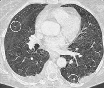
Figure 4. Benign lung nodules visualized on computed tomography.Lung cancer screening using chest CT may be associated with certain risks. The detailed high-resolution images produced by contemporary CT reveal small benign lung nodules in as many as 74% of patients(Figure 4).24,30 Although these nodules rarely represent a significant health problem, they require follow-up procedures and contribute to patient anxiety.31 In one study, every 1,000 individuals screened with CT imaging resulted in the identification of nine cases of stage I NSCLC, 235 false-positive nodules measuring at least 5 mm, and four thoracotomies for benign lesions.12
Radiation from CT tests is a potential concern, although it is difficult to quantify the importance of this risk. One estimate of CT-related radiation exposure found that annual CT screening of 50% of the eligible population between 50 and 75 years of age in the United States would result in approximately 36,000 new cancers, or a 1.8% increase in the rate of cancer over the expected rate.32 Many patients and health care professionals are already concerned about the degree of radiation exposure from medical diagnostics. A recent study that examined cumulative radiation exposure due to medical imaging in 952,420 adults aged 18 to 64 years found that approximately 57.9% of men and 78.7% of women receive at least some annual health care-related radiation exposure.33 Radiation exposure was considered moderate (> 3–20 mSv/yr) for 18.1% of men and 20.3% of women, and was considered high (> 20–50 mSv/yr) or very high (>50 mSv/hr) for 2.3% of men and 2.1% of women.
IS SCREENING COST-EFFECTIVE?
It is difficult to calculate the cost-effectiveness of CT screening because the impact of screening on mortality and the economic implications of false-positive findings are not well understood. A cost-effectiveness analysis of helical CT screening assumed that screening would result in a 50% stage shift and a 13% reduction in mortality.34 Under these assumptions, the cost-effectiveness was greater among current smokers ($116,300 per quality-adjusted life year saved by screening) than among currently quitting smokers ($558,600) or former smokers ($2,322,700). These investigators concluded that lung cancer screening is unlikely to be cost-effective, especially among those with the lowest levels of current tobacco exposure (quitting or former smokers).
Larger stage shifts or reductions in mortality would be expected to translate into greater cost-effectiveness, although the real-world effects of screening on these parameters are uncertain. Data from a US nationwide survey suggested that only about one-half of all current smokers would opt for surgery following a positive screening result, which might significantly decrease the cost-effectiveness of treatment.35
It is unclear how well the methods used in screening studies such as the NLST would translate to actual clinical practice at a national level, or how the health care system would manage the many small lung nodules that would be identified using this approach.
HOW WILL FUTURE DEVELOPMENTS AFFECT LUNG CANCER SCREENING?
Ongoing studies will continue to refine our understanding of the impact of lung cancer screening. For example, the randomized Prostate, Lung, Colorectal, and Ovarian Screening Trial is examining chest radiograph screening versus control in both smokers and never-smokers between 55 and 74 years of age.36 It is anticipated that this study will provide important information about how well chest radiographs perform for the identification of lung cancer in high- and lower-risk populations. Large randomized trials in Europe are comparing CT with no imaging for lung cancer screening.37 Efforts to better characterize specific patient populations who are at the greatest risk of lung cancer may help to improve the efficiency and cost-effectiveness of screening. Advances in molecular testing may help to identify molecular and genetic tumor biomarkers that herald increased lung cancer risk and greater need for screening. More research is needed to better understand the optimal management of patients with small lung nodules on screening tests. Professional societies are poised to publish revised screening recommendations as data from the NLST become available. Finally, insurers will need to evaluate the evidence and develop reimbursement policies.
SUMMARY AND CONCLUSIONS
Lung cancer screening efforts conducted over the last several decades have shown that it is possible to identify early lung cancer in high-risk patient populations. However, demonstrating a clear improvement in cancer-related mortality has been more difficult. Biases inherent to noncontrolled trials of screening may explain some of the beneficial effects on survival observed in some studies. Recent results from the NLST have for the first time demonstrated a significant reduction in lung cancer mortality in high-risk patients screened for lung cancer with chest CT, although there are continuing concerns about the cost of screening, the risks from radiation exposure, and the additional testing resulting from the identification of small benign lung nodules. Ongoing research will help to maximize the benefit of lung cancer screening and minimize the related risks.
Screening is the testing of an individual who is at risk for a disease, but who does not exhibit signs or symptoms of the disease. The goal of screening is to detect disease at a stage when cure or control is possible, and an effective screening program should reduce the number of disease-specific deaths in the screened population. Screening should focus on diseases that are associated with potentially serious consequences and that are detectable in the preclinical phase, yet it should avoid identifying “pseudodisease” (ie, positive test findings that would not be expected to affect the patient’s health) or causing morbidity due to the test procedure itself.1 Finally, screening is only worthwhile when treatment of the disease is more effective when administered early.
Since lung cancer screening began in the 1950s,2,3 many studies have attempted to define the medical benefits and economic impact of widespread screening. Many important unresolved issues remain, including the effectiveness of lung cancer screening for reducing disease-specific mortality, the potential harms of screening, its cost-effectiveness, and the potential impact of new research methods on the early identification of lung cancer.
DOES LUNG CANCER SCREENING REDUCE DISEASE-SPECIFIC MORTALITY?
Early studies examined the usefulness of large-scale chest radiograph programs, either with or without sputum cytology, for lung cancer screening. Although several studies reported that radiographic screening identified patients with early lung cancer and reported higher survival rates, reviews and meta-analyses of these reports concluded that screening did not significantly reduce disease-specific mortality.4,5
The utility of chest radiography for the detection of early lung cancer is limited by several factors, including poor sensitivity for the detection of small or subtle nodules and a relatively high false-positive rate.6–8 More recently, several cohort studies and randomized, controlled trials have shown that computed tomography (CT) screening is effective for the identification of early lung cancer in high-risk patients (eg, individuals with chronic, heavy tobacco use or asbestos exposure).9–11 A recent meta-analysis concluded that CT-based screening significantly increases the number of early lung cancers identified, but also increases the number of false-positive findings (nodules) and unnecessary thoracotomies for benign lesions.12
Lung cancer screening should increase the number of patients identified at early disease stages. Treatment of early-stage lung cancer should decrease the number of patients identified with late-stage cancer, resulting in a stage shift toward earlier disease for the population as a whole. Although lung cancer screening cohort studies and randomized, controlled trials have demonstrated that screening increases the number of early-stage lung cancer cases identified, these studies have generally not demonstrated decreased rates of late-stage lung cancers or stage shifting in the populations studied. In the 1970s, the National Cancer Institute began three large-scale screening trials at Mayo Clinic, Memorial Sloan-Kettering Cancer Institute, and The Johns Hopkins University, each enrolling approximately 10,000 patients. In the Mayo trial, the incidence of advanced-stage tumors was nearly identical for the screened versus unscreened patients, with 303 cancer cases detected in the screened group versus 304 cases in the control group.13 CT-based cohort studies have also reported increased rates of early recognition of lung cancer and accompanying large increases in the number of diagnostic procedures performed. However, early controlled trials of CT screening showed no differences between screened and unscreened groups in the numbers of patients with late-stage tumors or deaths due to lung cancer.14
Results such as these have led some researchers to argue that survival benefits of screening largely reflect observational biases. For example:
Figure 1. Lead-time bias. Patients identified by screening may live longer with disease than patients diagnosed clinically, although overall survival time is not improved.Lead-time bias occurs when screening results in earlier recognition of disease, but does not change the patient’s eventual lifespan, creating the appearance that the patient’s survival time with the disease is longer (Figure 1).15 Longer lead times should be observed in a successful screening program even if eventual mortality remains exactly the same, and lead time bias is therefore an expected outcome of screening.
Figure 2. Length-time bias. Indolent tumors move more gradually from the detectable stage to the onset of symptoms. These tumors are therefore more likely to be identified by intermittent screening.Length-time bias arises from the observation that any screening test that is applied intermittently is more likely to detect indolent tumors than aggressive, fast-growing tumors that would result in clinical symptoms(Figure 2).15 Indolent tumors move more gradually from the detectable state to the onset of clinical symptoms, and are therefore especially likely to be identified by screening.
Overdiagnosis bias occurs when a screening test identifies disease that never would have affected the patient’s life in the absence of screening. This type of bias might occur if screening identifies a lesion that is so indolent that it would never cause clinical disease, or if the population is otherwise in such poor health that successfully screened patients would die from other causes.
There is no question that these biases affect reports of survival in lung cancer screening, although it is unclear whether they explain the reported benefit of screening observed in cohort studies. Screening advocates have argued that the failure to screen high-risk patients for lung cancer has the potential for significant harm. In contrast, opponents of screening have argued that there was a lack of data showing a reduction in the number of patients diagnosed with late-stage cancers or in cancer-related mortality.
IS LUNG CANCER OVERDIAGNOSED IN SCREENED POPULATIONS?
Although the apparent benefit of lung cancer screening is susceptible to different sources of bias, overdiagnosis has received the greatest attention on the basis of both theoretical concerns and observations from screening studies. Estimates of lung cancer growth suggest that a typical 10-cm tumor, which is usually large enough to be fatal, has progressed through approximately 40 volume doublings during the course of its existence. In contrast, a more survivable—and clinically detectable—1-cm tumor has progressed through approximately 30 volume doublings.16,17 A lung tumor therefore spends most of its existence relatively undetectable. It has been estimated that the median doubling time is approximately 181 days, and that 22% of lung cancers have doubling times more than 465 days.18 The appearance of tumors on CT may suggest the growth rate, with 1 study showing that solid malignant nodules had a mean doubling time of 149 days, compared with 457 days for partial ground-glass–opacity nodules, and 813 days for pure ground-glass nodules.19
These estimates suggest that if a 1-cm tumor with a history of 30 volume doublings continues to grow at a typical rate (ie, a 181-day doubling time), the patient will die of cancer within 5 years. If the tumor is among the 22% of those with a 465-day doubling time, the survival time would be 12.7 years. For malignant pure ground-glass nodules, the projected time to death is 22 years. Individuals with lung cancer are often elderly, long-term cigarette smokers with emphysema or other chronic health problems—many of whom would die of other causes before their lung cancers progressed enough to cause significant health problems.
Reprinted with permission from the American College of Chest Physicians (Raz DJ, et al. Natural history of stage I non-small cell lung cancer: implications for early detection. Chest 2007; 132:193–199).
Figure 3. Survival is worse in untreated than in treated non–small cell lung cancer patients, arguing against overdiagnosis bias. Blue line: patients receiving surgery; green line: untreated patients who refused surgery.
As an argument against the significance of overdiagnosis in lung cancer screening, it has been noted that outcomes are worse for patients identified with early-stage lung cancer in screening studies who do not receive treatment. For example, the results of a study of 1,432 patients with stage I non–small cell lung cancer (NSCLC) are illustrated in Figure 3. Survival was much better in screened patients who were treated than in those who were untreated, with almost all the untreated patients dying within 10 years of diagnosis.20 However, the subjects in this study were atypical of those in most screening studies. Thirty-three percent of the patients had squamous cell carcinoma and 61% had relatively large T2 lesions, compared with a typical screening study comprised of patients with more than 50% T1 lesions and a smaller percentage of squamous cell carcinoma.
Another argument against overdiagnosis comes from gene profiling studies that have compared genetic tumor markers for tumors identified by screening with tumors identified clinically. One study found that the expression profile of 3,231 genes was similar for patients with lung cancer identified by screening or by symptoms.21 However, these investigators also found that nine genes known to be important in tumor growth differed between screened and nonscreened populations.
The significance of overdiagnosis is supported by a long-term follow-up study from the Mayo Clinic chest radiography screening trial, which found that the number of lung cancer cases remained higher in the screening group than the control group (585 vs 500 cases) for up to 28 years after screening, suggesting an overdiagnosis of lung cancer by approximately 85 cases per 500 patients screened (approximately 17%).22 Several studies have also demonstrated that screening populations may have tumors with more favorable histology or clinical characteristics, including higher levels of bronchioloalveolar carcinoma or well-differentiated adenocarcinoma.23–25 Finally, autopsy series have found undiagnosed lung tumors in as many as 1% of patients who died from natural causes, with fewer advanced tumors found in the 1970s than in the 1950s.26,27
These arguments led most to believe randomized controlled trials of CT-based screening were needed. The largest of these, the National Lung Screening Trial (NLST), has recently reported results that will clarify the impact of lung cancer screening on cancer-related mortality.28 This study enrolled 53,456 subjects between the ages of 55 and 74 years with a history of at least 30 pack-years of smoking. Patients were randomized to baseline screening followed by annual screening for 2 years using either low-dose helical CT or chest radiography and outcome follow-up 5 years after randomization. Data analysis after 6 to 8 years of follow-up found 442 lung cancer deaths in the chest radiograph arm versus 354 in the CT arm, representing a 20.3% reduction with CT.29 Screening of 320 patients using low-dose helical CT would be required to avoid each lung cancer death. Thus, after years of debate, it has been demonstrated that it is possible to reduce lung cancer-specific mortality with CT-based screening.
ARE THERE SIGNIFICANT RISKS WITH CT-BASED SCREENING?
Reprinted with kind permission from Springer Science+Business Media (Fischbach F, et al. Detection of pulmonary nodules by multislice computed tomography: improved detection rate with reduced slice thickness. Eur Radiol 2003; 13:2378–2383).
Figure 4. Benign lung nodules visualized on computed tomography.Lung cancer screening using chest CT may be associated with certain risks. The detailed high-resolution images produced by contemporary CT reveal small benign lung nodules in as many as 74% of patients(Figure 4).24,30 Although these nodules rarely represent a significant health problem, they require follow-up procedures and contribute to patient anxiety.31 In one study, every 1,000 individuals screened with CT imaging resulted in the identification of nine cases of stage I NSCLC, 235 false-positive nodules measuring at least 5 mm, and four thoracotomies for benign lesions.12
Radiation from CT tests is a potential concern, although it is difficult to quantify the importance of this risk. One estimate of CT-related radiation exposure found that annual CT screening of 50% of the eligible population between 50 and 75 years of age in the United States would result in approximately 36,000 new cancers, or a 1.8% increase in the rate of cancer over the expected rate.32 Many patients and health care professionals are already concerned about the degree of radiation exposure from medical diagnostics. A recent study that examined cumulative radiation exposure due to medical imaging in 952,420 adults aged 18 to 64 years found that approximately 57.9% of men and 78.7% of women receive at least some annual health care-related radiation exposure.33 Radiation exposure was considered moderate (> 3–20 mSv/yr) for 18.1% of men and 20.3% of women, and was considered high (> 20–50 mSv/yr) or very high (>50 mSv/hr) for 2.3% of men and 2.1% of women.
IS SCREENING COST-EFFECTIVE?
It is difficult to calculate the cost-effectiveness of CT screening because the impact of screening on mortality and the economic implications of false-positive findings are not well understood. A cost-effectiveness analysis of helical CT screening assumed that screening would result in a 50% stage shift and a 13% reduction in mortality.34 Under these assumptions, the cost-effectiveness was greater among current smokers ($116,300 per quality-adjusted life year saved by screening) than among currently quitting smokers ($558,600) or former smokers ($2,322,700). These investigators concluded that lung cancer screening is unlikely to be cost-effective, especially among those with the lowest levels of current tobacco exposure (quitting or former smokers).
Larger stage shifts or reductions in mortality would be expected to translate into greater cost-effectiveness, although the real-world effects of screening on these parameters are uncertain. Data from a US nationwide survey suggested that only about one-half of all current smokers would opt for surgery following a positive screening result, which might significantly decrease the cost-effectiveness of treatment.35
It is unclear how well the methods used in screening studies such as the NLST would translate to actual clinical practice at a national level, or how the health care system would manage the many small lung nodules that would be identified using this approach.
HOW WILL FUTURE DEVELOPMENTS AFFECT LUNG CANCER SCREENING?
Ongoing studies will continue to refine our understanding of the impact of lung cancer screening. For example, the randomized Prostate, Lung, Colorectal, and Ovarian Screening Trial is examining chest radiograph screening versus control in both smokers and never-smokers between 55 and 74 years of age.36 It is anticipated that this study will provide important information about how well chest radiographs perform for the identification of lung cancer in high- and lower-risk populations. Large randomized trials in Europe are comparing CT with no imaging for lung cancer screening.37 Efforts to better characterize specific patient populations who are at the greatest risk of lung cancer may help to improve the efficiency and cost-effectiveness of screening. Advances in molecular testing may help to identify molecular and genetic tumor biomarkers that herald increased lung cancer risk and greater need for screening. More research is needed to better understand the optimal management of patients with small lung nodules on screening tests. Professional societies are poised to publish revised screening recommendations as data from the NLST become available. Finally, insurers will need to evaluate the evidence and develop reimbursement policies.
SUMMARY AND CONCLUSIONS
Lung cancer screening efforts conducted over the last several decades have shown that it is possible to identify early lung cancer in high-risk patient populations. However, demonstrating a clear improvement in cancer-related mortality has been more difficult. Biases inherent to noncontrolled trials of screening may explain some of the beneficial effects on survival observed in some studies. Recent results from the NLST have for the first time demonstrated a significant reduction in lung cancer mortality in high-risk patients screened for lung cancer with chest CT, although there are continuing concerns about the cost of screening, the risks from radiation exposure, and the additional testing resulting from the identification of small benign lung nodules. Ongoing research will help to maximize the benefit of lung cancer screening and minimize the related risks.
References
Holin SM, Dwork RE, Glaser S, Rikli AE, Stocklen JB. Solitary pulmonary nodules found in a community-wide chest roentgenographic survey: a five-year follow-up study. Am Rev Tuberc1959; 79:427–439.
Nash FA, Morgan JM, Tomkins JG. South London Lung Cancer Study. Br Med J1968; 2:715–721.
Obuchowski NA, Graham RJ, Baker ME, Powell KA. Ten criteria for effective screening: their application to multislice CT screening for pulmonary and colorectal cancers. AJR Am J Roentgenol2001; 176:1357–1362.
Eddy DM. Screening for lung cancer. Ann Intern Med1989; 111:232–237.
Manser RL, Irving LB, Byrnes G, Abramson MJ, Stone CA, Campbell DA. Screening for lung cancer: a systematic review and meta-analysis of controlled trials. Thorax2003; 58:784–789.
Krupinski EA, Berger WG, Dallas WJ, Roehrig H. Searching for nodules: what features attract attention and influence detection?Acad Radiol2003; 10:861–868.
Yoshida H. Local contralateral subtraction based on bilateral symmetry of lung for reduction of false positives in computerized detection of pulmonary nodules. IEEE Trans Biomed Eng2004; 51:778–789.
Shiraishi J, Abe H.Engelmann R, Doi K. Effect of high sensitivity in a computerized scheme for detecting extremely subtle solitary pulmonary nodules in chest radiographs: observer performance study. Acad Radiol2003; 10:1302–1311.
Veronesi G, Bellomi M, Scanagatta P, et al. Difficulties encountered managing nodules detected during a computed tomography lung cancer screening program. J Thorac Cardiovasc Surg2008; 136:611–617.
Wilson DO, Weissfeld JL, Fuhrman CR, et al. The Pittsburgh Lung Screening Study (PLuSS): outcomes within 3 years of a first computed tomography scan [published online ahead of print July 17, 2008]. Am J Respir Crit Care Med2008; 178:956–961. doi: 10.1164/rccm.200802-336OC
Fasola G, Belvedere O, Aita M, et al. Low-dose computed tomography screening for lung cancer and pleural mesothelioma in an asbestos-exposed population: baseline results of a prospective, nonrandomized feasibility trial—an Alpe-adria Thoracic Oncology Multidisciplinary Group Study (ATOM 002). Oncologist2007; 12:1215–1224.
Gopal M, Abdullah SE, Grady JJ, Goodwin JS. Screening for lung cancer with low-dose computed tomography: a systematic review and meta-analysis of the baseline findings of randomized controlled trials. J Thorac Oncol2010; 5:1233–1239.
Fontana RS, Sanderson DR, Woolner LB, et al. Screening for lung cancer: a critique of the Mayo Lung Project. Cancer1991; 67( suppl 4):1155–1164.
Bach PB, Jett JR, Pastorino U, Tockman MS, Swensen SJ, Begg CB. Computed tomography screening and lung cancer outcomes. JAMA2007; 297:953–961.
Patz EF, Goodman PC, Bepler G. Screening for lung cancer. N Engl J Med2000; 343:1627–1633.
Weiss W. Implications of tumor growth rate for the natural history of lung cancer. J Occup Med1984; 26:345–352.
Reich JM. A critical appraisal of overdiagnosis: estimates of its magnitude and implications for lung cancer screening. Thorax2008; 63:377–383.
Winer-Muram HT, Jennings SG, Tarver RD, et al. Volumetric growth rate of stage I lung cancer prior to treatment: serial CT scanning. Radiology2002; 223:798–805.
Hasegawa M, Sone S, Takashima S, et al. Growth rate of small lung cancers detected on mass CT screening. Br J Radiol2000; 73:1252–1259.
Raz DJ, Zell JA, Ou SH, Gandara DR, Anton-Culver H, Jablons DM. Natural history of stage I non-small cell lung cancer: implications for early detection [published online ahead of print May 15, 2007]. Chest2007; 132:193–199. doi: 10.1378/chest.06-3096
Bianchi F, Hu J, Pelosi G, et al. Lung cancers detected by screening with spiral computed tomography have a malignant phenotype when analyzed by cDNA microarray. Clin Cancer Res2004; 10( 18 Pt 1):6023–6028.
Marcus PM, Bergstralh EJ, Zweig MH, Harris A, Offord KP, Fontana RS. Extended lung cancer incidence follow-up in the Mayo Lung Project and overdiagnosis. J Natl Cancer Inst2006; 98:748–756.
Sone S, Li F, Yang ZG, et al. Results of three-year mass screening programme for lung cancer using mobile low-dose spiral computed tomography scanner. Br J Cancer2001; 84:25–32.
Swensen SJ, Jett JR, Hartman TE, et al. CT screening for lung cancer: five-year prospective experience [published online ahead of print February 4, 2005]. Radiology2005; 235:259–265. doi: 10.1148/radiol.2351041662
International Early Lung Cancer Action Program Investigators, Henschke CI, Yankelevitz DF, Libby DM, et al. Survival of patients with stage I lung cancer detected on CT screening. N Engl J Med2006; 355:1763–1771.
Manser RL, Dodd M, Byrnes G, Irving LB, Campbell DA. Incidental lung cancers identified at coronial autopsy: implications for overdiagnosis of lung cancer by screening. Respir Med2005; 99:501–507.
Chan CK, Wells CK, McFarlane MJ, Feinstein AR. More lung cancer but better survival: implications of secular trends in “necropsy surprise” rates. Chest1989; 96:291–296.
National Lung Screening Trial Research Team, Aberle DR, Adams AM, Berg CD, et al. Baseline characteristics of participants in the randomized national lung screening trial [published correction appears in J Natl Cancer Inst 2011; 103:1560]. J Natl Cancer Inst2010; 102:1771–1779.
Fischbach F, Knollmann F, Griesshaber V, Freund T, Akkol E, Felix R. Detection of pulmonary nodules by multislice computed tomography: improved detection rate with reduced slice thickness [published online ahead of print May 13, 2003]. Eur Radiol2003; 13:2378–2383. doi: 10.1007/s00330-003-1915-7
van den Bergh KA, Essink-Bot ML, Borsboom GJ, et al. Short-term health-related quality of life consequences in a lung cancer CT screening trial (NELSON) [published online ahead of pring November 24, 2009]. Br J Cancer2010; 102:27–34. doi: 10.1038/sj.bjc.6605459
Brenner DJ. Radiation risks potentially associated with low-dose CT screening of adult smokers for lung cancer. Radiology2004; 231:440–445.
Fazel R, Krumholz HM, Wang Y, et al. Exposure to low-dose ionizing radiation from medical imaging procedures. N Engl J Med2009; 361:849–857.
Mahadevia PJ, Fleisher LA, Frick KD, Eng J, Goodman SN, Powe NR. Lung cancer screening with helical computed tomography in older adult smokers: a decision and cost-effectiveness analysis. JAMA2003; 289:313–322.
Silvestri GA, Nietert PJ, Zoller J, Carter C, Bradford D. Attitudes towards screening for lung cancer among smokers and their nonsmoking counterparts. [published online ahead of print November 13, 2006]Thorax2007; 62:126–130. doi: 10.1136/thx.2005.056036
Tammemagi CM, Pinsky PF, Caporaso NE, et al. Lung cancer risk prediction: Prostate, Lung, Colorectal and Ovarian Cancer Screening Trial models and validation [published online ahead of print May 23, 2011]. J Natl Cancer Inst2011; 103:1058–1068. doi: 10.1093/jnci/djr173
van Klaveren RJ, Oudkerk M, Prokop M, et al. Management of lung nodules detected by volume CT scanning. N Engl J Med2009; 361:2221–2229.
 Reprinted with permission from The New England Journal of Medicine (Patz EF, et al. Screening for lung cancer. N Engl J Med 2000; 343:1627–1633). Copyright © 2000 Massachusetts Medical Society. All rights reserved.
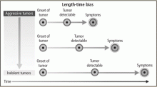 Reprinted with permission from The New England Journal of Medicine (Patz EF, et al. Screening for lung cancer. N Engl J Med 2000; 343:1627–1633). Copyright © 2000 Massachusetts Medical Society. All rights reserved.